INTRODUCTION
Sepsis is a devastating clinical condition characterized by sys-
temic inflammation occurring during a severe infection. Se- vere sepsis and septic shock are leading causes of morbidity and mortality in the intensive care unit (ICU).
1,2The reason that some patients die while others survive similar insults is partially understood, although some of this patient outcome variability may be caused by genetic variation. Several reports have con- firmed that susceptibility and outcomes from infectious disease are inheritable.
3-5Indeed, numerous studies have demonstrat- ed that innate immune responses to pathogens exhibit inter- individual variability strongly influenced by genetic factors, which may affect disease susceptibility and severity.
6-12In the pathophysiology of sepsis, the innate immune system is acti- vated prior to the acquired immune system: cells of the innate immune system, such as monocytes, macrophages, and neu-
Associations between Single Nucleotide
Polymorphisms of High Mobility Group Box 1 Protein and Clinical Outcomes in Korean Sepsis Patients
Kwangha Lee
1, Youjin Chang
2, Kyuyoung Song
3, Yun Young Park
4, Jin Won Huh
4, Sang-Bum Hong
4, Chae-Man Lim
4, and Younsuck Koh
41
Department of Internal Medicine, Pusan National University School of Medicine, Busan;
2
Division of Pulmonary and Critical Care Medicine, Department of Internal Medicine, College of Medicine, Chungbuk National University, Cheongju;
Departments of
3Biochemistry and Molecular Biology and
4Pulmonary and Critical Care Medicine, Asan Medical Center, University of Ulsan College of Medicine, Seoul, Korea.
Purpose: High mobility group box 1 (HMGB1) plays a central role in the pathogenesis of sepsis and multiple organ dysfunction syndromes. We investigated the associations of a single nucleotide polymorphism (SNP; rs1045411) in HMGB1 with various clini- cal parameters, severity, and prognosis in patients with sepsis, severe sepsis, or septic shock.
Materials and Methods: We enrolled 212 adult patients followed for 28 days. All patients were genotyped for rs1045411, and the serum levels of HMGB1 and several cytokines were measured.
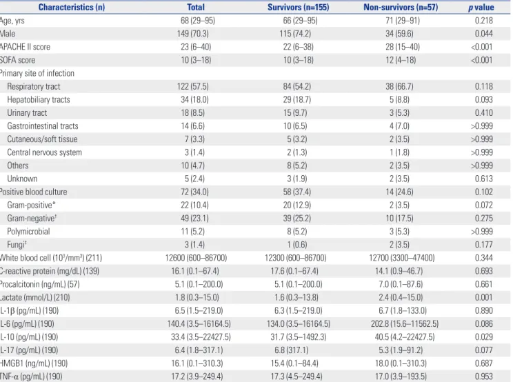
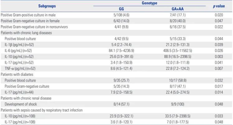
Results: The proportions of patients according to genotype were GG (71.2%), GA (26.4%), and AA (2.4%). Among patients with chronic lung disease comorbidity, patients with a variant A allele had higher positive blood culture rates and higher levels of vari- ous cytokines [interleukin (IL)-1β, IL-6, IL-10, IL-17, and tumor necrosis factor-α] than those with the GG genotype. In the analy- sis of those with diabetes as a comorbidity, patients with a variant A allele had higher blood culture and Gram-negative culture rates than those with GG genotypes; these patients also had a higher levels of IL-17. In the analysis of those with sepsis caused by a respiratory tract infection, patients with a variant A allele had higher levels of IL-10 and IL-17 (all p<0.05). This polymorphism had no significant impact on patient survival.
Conclusion: The variant A allele of rs1045411 appears to be associated with a more severe inflammatory response than the GG genotype under specific conditions.
Key Words: High mobility group box 1, single nucleotide polymorphisms, rs1045411, sepsis, the variant A allele Yonsei Med J 2016 Jan;57(1):111-117
http://dx.doi.org/10.3349/ymj.2016.57.1.111
Original Article
pISSN: 0513-5796 · eISSN: 1976-2437
Received: July 29, 2014 Revised: February 16, 2015 Accepted: March 18, 2015
Corresponding author: Dr. Younsuck Koh, Department of Pulmonary and Critical
Care Medicine, Asan Medical Center, University of Ulsan College of Medicine, 88 Olympic-ro 43-gil, Songpa-gu, Seoul 05505, Korea.
Tel: 82-2-3010-3134, Fax: 82-2-3010-6968, E-mail: [email protected]
•The authors have no financial conflicts of interest.
© Copyright: Yonsei University College of Medicine 2016
This is an Open Access article distributed under the terms of the Creative Com- mons Attribution Non-Commercial License (http://creativecommons.org/ licenses/
by-nc/3.0) which permits unrestricted non-commercial use, distribution, and repro-
duction in any medium, provided the original work is properly cited.
trophils, represent the front line of the host response to infec- tion, invasion, and injury.
13High mobility group box 1 (HMGB1) is a highly conserved, ubiquitously expressed protein, originally discovered as a non- histonal nuclear DNA binding protein.
14-16It is located on chro- mosome 13 and present in the nuclei and cytoplasm of nearly all cell types.
17In response to infection and injury, HMGB1 is secreted by activated innate immune cells and/or passively re- leased by necrotic or damaged cell.
18Some studies have dem- onstrated that HMGB1 is a late mediator of sepsis in amplifying the inflammatory response and that serum/plasma HMGB1 concentrations are elevated in patients with sepsis.
19,20Accu- mulating evidence supports a central pathogenic role for HMGB1 in the pathogenesis of sepsis and multiple organ dysfunction syndromes.
21-24The past decade has witnessed important advances in the understanding of genetic polymorphisms in sepsis; numerous studies have identified that these sepsis-related genetic poly- morphisms are associated with severity and/or outcomes.
11,12Although, several studies reported the clinical relevance of HMGB1 genetic variation,
25-27there are limited data on the re- lationship between single nucleotide polymorphisms (SNPs) of HMGB1 and clinical outcomes in patients with sepsis. More- over, the characteristics of these polymorphisms differ accord- ing to ethnicity, although few data have been reported in the Korean population. Therefore, we hypothesized that SNPs of HMGB1 could influence clinical outcomes in Korean patients with sepsis.
In this study, we genotyped a SNP of known genetic variants within HMGB1 in patients diagnosed with sepsis (including severe sepsis and septic shock), and analyzed its relationship with various clinical parameters, including disease severity and prognosis. We also investigated the relationship between this HMGB1 polymorphism and serum concentrations of HMGB1 and various cytokines.
MATERIALS AND METHODS
Study subjects
Inclusion criteria were adult patients diagnosed with sepsis, including severe sepsis and septic shock. There was no exclu- sion criterion. In total, 212 patients were enrolled from March 1, 2011 to October 31, 2012. All patients were >20 years of age [median 67.5 (range 29–95) years, M:F=149:63] and had been admitted to the ICU of a Asan Medical Center (Seoul, Korea).
Sepsis, severe sepsis, and septic shock were defined using American College of Chest Physicians/Society of Critical Care Medicine.
28,29All patients were managed according to thera- peutic recommendations based on early goal-directed thera- py and lung-protective ventilator strategy.
29,30Survivors were defined as patients who had survived for 28 days after ICU ad- mission. The study objectives and procedures were fully dis-
closed, and a case report form for this study was completed.
All data were collected from the medical records and labora- tory and radiographic findings in all patients. This study was approved by the Institutional Review Board (IRB) of the Asan Medical Center (2012-0878). Informed consent was confirmed by the IRB, and written informed consent was obtained from all study participants or their surrogates.
Data collection
The following data were gathered from the medical records of patients: age, gender, the primary cause of sepsis on initial ad- mission, underlying comorbidities, duration of mechanical ventilation, and lengths of stay in the ICU and hospital. All pa- tients were categorized as sepsis, severe sepsis, and septic shock on ICU admission. Acute Physiology and Chronic Health Eval- uation (APACHE) II and Sequential Organ Failure Assessment (SOFA) scores were calculated on the sampling day for this study.
31,32We also identified the causative pathogen for sepsis in patients with positive blood culture, and classified them ac- cordingly. In addition, we recorded laboratory data (complete blood count, lactate, C-reactive protein, procalcitonin) on sam- pling day, and surveyed for the presence of neutropenia (ab- solute neutrophil count <1500/mm
3).
SNPs genotyping
Blood samples were drawn within 24 hrs after ICU admission.
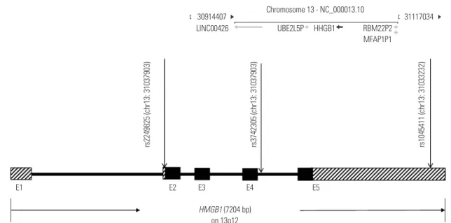
Genomic DNA was isolated from 5 mL of ethylenediaminetet- raacetic acid (EDTA)-anticoagulated venous blood by the standard method using proteinase K and phenol/chloroform extraction. SNP data for the HMGB1 gene [chromosome 13, position 29930000-29939000 (9 kb total)] was obtained from the HapMap data (version 2, release 21) for 45 unrelated Han Chinese individuals from Beijing, China (CHB) and 44 unre- lated Japanese individuals from Tokyo, Japan (JPT) samples.
From the database, a total of three SNPs with a minor allele fre-
quency >0.05 (rs1045411, rs3742305, rs2249825) were identi-
fied in HMGB1 (excluding 5’- and 3’-flanking regions) and se-
lected for genotyping; all are common SNPs with a minor allele
frequency >0.05. Among the three SNPs, rs1045411 located in
the 3’-untranslated region (Fig. 1) was chosen for further anal-
yses, because this SNP seemed to show the most significant
difference in allele frequencies between patients and normal
healthy persons (χ
2test, p<0.05) with the lowest p value and
highest odds ratio, compared to the other two SNPs (data not
shown). Genotypes for rs1045411 in all patients were deter-
mined by Sanger sequencing. The target region of HMGB1 was
amplified using forward primer 5’-TGGAAGTGGGAGGCAAT
TTA-3’ (HMGB1_1045411_F) and reverse primer 5’-TGCTGT
GCAAA GGTTGAGAG-3’ (HMGB1_1045411_R). Amplification
conditions were one cycle of 95°C for 7 min, 30 cycle of 95°C
for 30 s, 56°C for 30 s, 72°C for 1 min, plus one cycle of 72°C for
5 min. Amplification conditions were one cycle of 95°C for 7
min, 30 cycles of 95°C for 30 s, 56°C for 30 s, and 72°C for 1
min, plus one final cycle of 72°C for 5 min. Amplified products of 303 bp were confirmed by agarose gel electrophoresis and purified by treating with Exo-SAP (10:1 U ratio mixture of exo- nuclease I and shrimp alkaline phosphatase, USB corp., Cleve- land, OH, USA) at 37°C for 15 min, 80°C for 15 min, and a 4°C hold. Sequencing was carried out using an ABI 3730XL se- quencer by Cosmogenetech, Seoul, Korea. Briefly, sequencing amplifications were performed using the BigDye terminator (ver. 3.1) cycle sequencing kit (Applied Biosystems, Foster City, CA, USA). PCR products of 30–90 ng were used as tem- plates and the cycling reaction consisted of one cycle of 95°C for 90 s, 25 cycles of 95°C for 30 s, 50°C for 5 s, and 60°C for 4 min, followed by a 4°C hold with HMGB1_ 1045411_F and HMGB1_1045411_R. After purifying the cycle sequencing re- action products with magnetic beads (MagneSil GREEN, Pro- mega, Madison, WI, USA), the sequencer instrument was run according to the manufacturer’s protocol.
Serum HMGB1 and cytokines measurement
Blood samples for cytokine measurement were immediately centrifuged, and serum was frozen -80°C until the assay could be performed. We measured serum HMGB1 levels and several inflammatory [interleukin (IL)-1β, IL-6, IL-17, and tumor ne- crosis factor (TNF)-α] and anti-inflammatory (IL-10) cytokines from 190 patients, because we did not have sufficient quanti- ties of samples; we could not measure serum levels of HMGB1 or other cytokines in 22 patients. The serum HMGB1 level was measured using a sandwich enzyme-linked immunosorbent assay (HMGB1 ELISA; IBL International, Hamburg, Germa- ny). Intra-assay and inter-assay coefficients of variation (CV)
were 5.5–13.7% and 7.6–13.7%, respectively. Also, functional sensitivity was 0.2 ng/mL (lowest HMGB1 concentration with
≤20%). The serum levels of cytokines were determined by Lu- minex
®Performance Assay multiplex kits (R&D Systems, Minneapolis, MN, USA). Analyses were performed in accor- dance with manufacturer’s protocol.
Statistical analysis
The genotype frequencies of three SNPs were tested for Har- dy-Weinberg equilibrium using Haploview (v4.2) (http://
www.broadinstitute.org/haploview). No significant deviation from Hardy-Weinberg equilibrium was observed. Continuous variables are expressed as median with range. Student’s t-test or the Mann-Whitney U-test, depending on the normality of distribution, were used to compare continuous variables be- tween two groups, and the Kruskal-Wallis test was used for comparisons among three groups. Also, the χ
2and Fisher’s ex- act tests (for small numbers) were used to compare categori- cal variables. To evaluate whether these SNPs could influence clinical outcomes, univariate and multivariate Cox regression analyses were performed including all clinical data from medical records, with adjustment for age, gender. Survival curves were obtained with using the Kaplan-Meier method with the log-rank test. All statistical analyses were performed using the Statistical Package for the Social Sciences (ver. 19.0;
IBM, Armonk, NY, USA). A two-tailed p value<0.05 was con- sidered to indicate significant difference.
Fig. 1. Genetic map of the three single nucleotide polymorphism (SNP) of the HMGB1. The three long vertical arrows indicate the locations of the three SNPs. E1 to E5 show the locations of the exons. The black boxes are the protein-coding regions, and boxes including oblique lines are untrans- lated regions (UTR). rs2249825, rs3742305, and rs1045411 are located in the 5’-UTR, intron 4, and the 3’-UTR, respectively.
Location:
Sequence:
Chromosome 13 - NC_000013.10 30914407
E1 E2
HMGB1 (7204 bp)
on 13q12
E3 E4 E5
LINC00426 UBE2L5P
rs2249825 (chr13: 31037903) rs3742305 (chr13: 31037903) rs1045411 (chr13: 31033232)