Correspondence to:
Jae Gil Lee, M.D., Ph.D. Department of Surgery, Yonsei University College of Medicine, 50-1 Yonsei-ro, Seodaemun-gu, Seoul 120-752, Korea
Tel: +82-2-2228-2127 Fax: +82-2-313-8289 E-mail: [email protected]
Severe sepsis is the main cause of death in critically ill surgical patients. The mainstay of management of severe sepsis and septic shock includes eradication of infection with appropriate antibiotics and source control, and aggressive supportive care, such as fluid resuscitation, vasoactive agents or mechanical ventilation. This early goal-directed therapy is widely used. Crucial components of infection control strategy for septic shock patients are early recognition, appropriate use of antibiotics, and early effective drainage or surgical control of origins of infection. We briefly summarize the points of infection control strategy for septic shock patients. (J Acute Care Surg 2014;4:1-6)
Key Words: Sepsis, Septic shock, Intraabdominal Infections
Received April 8, 2014, Revised April 9, 2014, Accepted April 10, 2014 Copyright © 2014 by Korean Society of Acute Care Surgery
cc This is an Open Access article distributed under the terms of the Creative Commons Attribution Non-Commercial License (http://creativecommons.org/licenses/by-nc/3.0) which permits unrestricted non-commercial use, distribution, and reproduction in any medium, provided the original work is properly cited.
ISSN 2288-5862(Print), ISSN 2288-9582(Online)
외과중환자에서 중증 패혈증과
패혈성 쇼크에 대한 감염관리 전략
연세대학교 의과대학 외과학교실
이승환ㆍ김형원ㆍ이재길
Infection Control Strategy for Severe Sepsis
and Septic Shock in Critically Ill Surgical Patients
Seung Hwan Lee, M.D., Hyung Won Kim, M.D., Jae Gil Lee, M.D., Ph.D.
Department of Surgery, Yonsei University College of Medicine, Seoul, Korea
서론
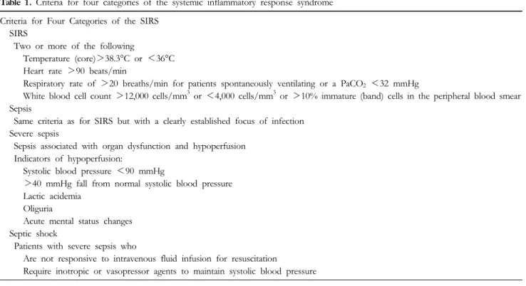
패혈증은 감염에 대한 전신 염증 반응으로 정의하며, 여기에 장기부전, 조직관류저하 등이 동반된 경우를 중증 패혈증, 적절한 수액 보충에도 불구하고 저혈압이 지속되거나, 혈압유지를 위해 혈압 상승제가 요구되는 상태를 패혈성 쇼크라 정의한다(Table 1)[1-4]. 중증 패혈증과 패혈성 쇼크는 전세계적으로 매년 백만 명 정도 발생하는 것으로 추정되며, 최근 그 빈도가 증가하는 것으로 보고 되었다[3,5-9]. 중증 패혈증과 패혈성 쇼크는 적극적인 치료에도 불구하고 각각 25∼30%, 40∼70%의 매우 높은 사망률을 보이고 [1,10,11], 치료를 위해 많은 노력과 재원이 요구되는 보건 의료 분야의 매우 중요한 문제로 인식되고 있다. 폐렴은 중증 패혈증의 가장 흔한 원인으로 약 절반 정도를 차지하며, 복강 내 감염과 요로 감염은 그 다음으로 흔한 원인으로 알려져 있다[6,7,12,13]. 한편, 전체 중증 패혈증 환자의 28.6%는 외과 환자가 차지하고 있으며[6], 수술 전 후 사망의 주된 원인 중 하나로 알려져 있다. 특히, 화상이나 외상, 대량출혈, 복강 내 감염, 장 폐쇄 및 허혈성 장 질환 등과 같이 비교적 흔한 외과적 질환에 동반되어 패혈증이 발생할 수 있다.Table 1. Criteria for four categories of the systemic inflammatory response syndrome
Criteria for Four Categories of the SIRS SIRS
Two or more of the following
Temperature (core)>38.3°C or <36°C Heart rate >90 beats/min
Respiratory rate of >20 breaths/min for patients spontaneously ventilating or a PaCO2 <32 mmHg
White blood cell count >12,000 cells/mm3 or <4,000 cells/mm3 or >10% immature (band) cells in the peripheral blood smear Sepsis
Same criteria as for SIRS but with a clearly established focus of infection Severe sepsis
Sepsis associated with organ dysfunction and hypoperfusion Indicators of hypoperfusion:
Systolic blood pressure <90 mmHg
>40 mmHg fall from normal systolic blood pressure Lactic acidemia
Oliguria
Acute mental status changes Septic shock
Patients with severe sepsis who
Are not responsive to intravenous fluid infusion for resuscitation Require inotropic or vasopressor agents to maintain systolic blood pressure SIRS: systemic inflammatory response syndrome.
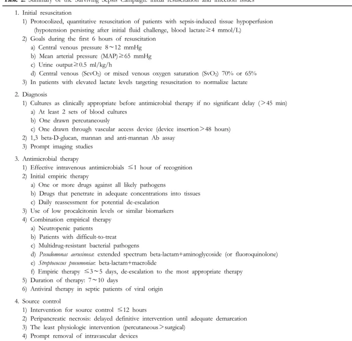
중증 패혈증 혹은 패혈성 쇼크로 진단된 25%의 환자에서 감염 부위가 다발성으로 나타나는 반면, 약 20∼30%에서는 그 원인 부위를 알 수 없는 것으로 보고되고 있다[6,14,15]. 혈액 배양은 약 30% 정도에서 양성 결과를 보인다[16]. 지금까지 알려진 중증 패혈증 및 패혈성 쇼크의 치료는 오염원 제거, 조기 항생제 치료, 보조적(adjunctive) 치료로 구성된다. 즉, 환자의 혈역학적 상태를 조기에 안정화시키고, 패혈증에 대한 염증을 제어하면서 감염에 대한 치료를 시행하는 것이다(Table 2)[2]. 최근 이러한 중증 패혈증 및 패혈성 쇼크에 대한 치료 지침 이 발표되었으며[2], 이는 중증 패혈증 및 패혈성 쇼크 환자의 치료 방향을 결정하는 데 도움을 주고 있다.
본론
패혈증 치료의 기본은 적절한 항생제의 사용이지만, 적절한 항생제를 투여하여도 일부 중증 패혈증 혹은 패혈성 쇼크의 사망 률은 감소시키지 못하는 것으로 알려져 있다. 따라서 패혈성 쇼크 의 치료는 초기 소생술과 더불어 감염의 원인을 찾아 해결하고, 숙주반응조절(스테로이드, activated protein C) 및 보조적 치료를 병행하는 것이다[10,17]. 초기 소생술은 첫 6시간 이내에 이루어지 는 것(early goal-directed therapy)이 추천된다[2,18]. 이러한 조기혈역학적 안정은 감염원을 치료하는 것만큼 중요한 처치라 할 수 있다. 물론, 감염원을 제거하지 못할 경우에는 패혈증의 치료가 실패로 돌아갈 가능성이 높아진다. 따라서 감염원을 찾기 위한 전신적인 신체 검진과 혈액 및 혈액배양 검사, 감염이 의심되는 부위에 대한 배양검사, 그리고 적절한 영상진단 검사를 체계적으 로 시행해야 한다. 본 내용에서는 초기 소생술을 기본 전제 하에 중증 패혈증 및 패혈성 쇼크에서의 감염 조절에 대해 다루고자 한다. 진단(Diagnosis) 초기 진단이 매우 중요하며, 무엇보다 패혈증의 원인으로 의심 되는 부위에 대한 검사가 필요하다. 일반적으로 항생제를 투여하 기 전에 최소 2쌍의 혈액 배양을 시행해야 한다. 최소 한 쌍은 말초 혈액에서 시행되어야 하며, 다른 한 쌍은 48시간 이상 유치되 어 있는 혈관 도관에서 확보되어야 한다[2,19]. 그 외 감염의 원인 이 될 수 있는 다른 부위, 즉 소변, 객담, 뇌척수액 및 상처 등에서 도 배양이 시행되어야 한다[2]. 또한 감염 부위가 한정되어 있고, 배양을 위한 검체를 얻을 수 있는 경우에는 이 부위에서 흡인액을 이용하여 배양검사를 시행하여야 한다. 이는 초기 광범위 항생제 사용 후에 배양 검사 및 항생제 감수성 결과에 따라 항생제의 종류를 결정하는 데 중요한 역할을 하게 된다. 그 외 전신 검사에
Table 2. Summary of the Surviving Sepsis Campaign: initial resuscitation and infection issues
1. Initial resuscitation
1) Protocolized, quantitative resuscitation of patients with sepsis-induced tissue hypoperfusion (hypotension persisting after initial fluid challenge, blood lactate≥4 mmol/L)
2) Goals during the first 6 hours of resuscitation a) Central venous pressure 8∼12 mmHg b) Mean arterial pressure (MAP)≥65 mmHg c) Urine output≥0.5 ml/kg/h
d) Central venous (ScvO2) or mixed venous oxygen saturation (SvO2) 70% or 65% 3) In patients with elevated lactate levels targeting resuscitation to normalize lactate 2. Diagnosis
1) Cultures as clinically appropriate before antimicrobial therapy if no significant delay (>45 min) a) At least 2 sets of blood cultures
b) One drawn percutaneously
c) One drawn through vascular access device (device insertion>48 hours) 2) 1,3 beta-D-glucan, mannan and anti-mannan Ab assay
3) Prompt imaging studies 3. Antimicrobial therapy
1) Effective intravenous antimicrobials ≤1 hour of recognition 2) Initial empiric therapy
a) One or more drugs against all likely pathogens
b) Drugs that penetrate in adequate concentrations into tissues c) Daily reassessment for potential de-escalation
3) Use of low procalcitonin levels or similar biomarkers 4) Combination empirical therapy
a) Neutropenic patients b) Patients with difficult-to-treat
c) Multidrug-resistant bacterial pathogens
d) Pseudomonas aerusinosa: extended spectrum beta-lactam+aminoglycoside (or fluoroquinolone) e) Streptococcus pneumoniae: beta-lactam+macrolide
f) Empiric therapy ≤3∼5 days, de-escalation to the most appropriate therapy 5) Duration of therapy: 7∼10 days
6) Antiviral therapy in septic patients of viral origin 4. Source control
1) Intervention for source control ≤12 hours
2) Peripancreatic necrosis: delayed definitive intervention until adequate demarcation 3) The least physiologic intervention (percutaneous>surgical)
4) Prompt removal of intravascular devices
서 원인 부위를 찾을 수 없는 경우에는 영상진단 검사를 통해 원인 부위를 찾도록 노력해야 한다[2].
항생제 치료(Antimicrobial Therapy)
최근 Surviving Sepsis Campaign 가이드라인에 따르면 효과적 인 항생제 투여의 지연은 사망률 증가와 연관되기 때문에 가능한 조기에 항생제 정맥 내 투여를 시작하도록 권고하고 있다[2]. 따라 서 패혈성 쇼크가 의심되는 경우, 가능성이 있는 원인 균주에 대해 광범위 항생제를 1시간 이내에 투여해야 하며, 항생제 투여 전에 혈액 배양검사를 시행해야 한다[2]. 중증 패혈증 및 패혈성 쇼크에 대한 조기 항생제 선택 전에는 fungi, methicillin-resistant
Staphylococcus aureus, vancomycin-resistant enterococcus, highly
resistant gram-negative bacilli의 유병률이 증가하고 있다는 것과 항생제 감수성의 지역적 패턴을 반드시 고려해야 한다[20]. 환자 의 전신 상태가 나쁘거나, 최근 항생제 치료를 받았거나, 다약제 내성 균주가 의심되는 경우에는 두 가지 이상의 항생제를 복합 투여하게 된다[21,22]. 부적절한 항생제가 투여되거나 항생제 사 용이 지연되는 경우 환자의 치료 결과에 악영향을 미치는 것으로
Table 5. WSES recommendations of antimicrobial agents for intra-abdominal infections
Origins Status Risk 1st choice 2nd choice
Community -acquired
Extra-biliary IAIs Stable, non-critical ESBL risk(-) Amoxicillin/clavulanate+metronidazole Ciprofloxacin+metronidazole
ESBL risk(+) Ertapenem Tigecycline
Critically ill (≥severe sepsis)
ESBL risk(-) Piperacillin/tazobactam
ESBL risk(+) Meropenem±fluconazole Imipenem±fluconazole
Biliary IAIs Stable, non-critical ESBL risk(-) Amoxicillin/clavulanate+metronidazole Ciprofloxacin+metronidazole ESBL risk(+) Tigecycline
Critically ill (≥severe sepsis)
ESBL risk(-) Piperacillin/tazobactam
ESBL risk(+) Piperacillin+tigecycline±fluconazole Hospital
-acquired
Nosocomial IAIs Stable, non-critical MDR risk(+) Piperacillin+tigecycline+fluconazole Critically ill
(≥severe sepsis)
MDR risk(+) Piperacillin+tigecycline+echinocandin Carbapenem+teicoplanin +echinocandin
WSES: World Society Emergency Surgery, IAIs: intra-abdominal infections, ESBL: extended-spectrum beta-lactamase, MDR: multidrug resistant.
Table 4. Potential bacteria responsible for intra-abdominal sepsis
and suggested antibiotics
Pathogen % Suggested antibiotics
G(-) bacilli including 60 Ertapenem (if no risk of
Pseudomonas aeruginosa) Escherichia coli 40 Piperacillin-tazobactam
P. aeruginosa 30
G(+) cocci including 30 3rd or 4th generation cephalosporin (active against P. aeruginosa) +metronidazole
Enterococcus spp. 20 Imipenem/doripenem (high-risk patients) Anaerobes including 30 ±Fluconazole
Bacteroides spp. 20 ±Aminoglycoside (if shocked)
Fungi 20
Table 3. Flora in different types of intra-abdominal infections
Primary (monomicrobial) Secondary (polymicrobial) Tertiary (polymicrobial) Escherichia coli Klebsiella spp. Streptococcus pneumoniae Enterococci Anaerobes rare Bacteroides fragilis group Other anaerobes E. coli Klebsiella spp. Other enterics Enterococci Staphylococcus epidermidis Enterococci Pseudomonas aeruginosa Candida spp. 알려져 있으므로 조기에 적절한 항생제를 선택하는 것이 필요하 다. 초기 경험적 항생제는 3∼5일 이상 투여되어서는 안되며[2], 배양검사 및 항생제 감수성 결과에 따라 가장 적절한 단일 약제로 전환하는 것이 바람직하겠다[23]. 항생제 치료 기간은 다소 논란 이 있으나, 최근의 권고 기간은 7∼10일 정도 유지하는 것으로 되어 있다[2]. 물론 환자의 상태 또는 배양된 균에 따라 그 기간이 달라질 수 있는데, 임상 반응이 늦거나, 감염원이 제거되지 않을 때, 혹은 호중구 감소 등의 면역결핍이 있을 때 항생제 사용 기간 이 다소 길어질 수 있다[2]. 최근에는 항생제 사용 여부를 procalc-itonin 농도에 따라 정하기도 한다[24,25]. 배양검사 결과 및 항생제 내성에 대한 검사 결과가 확인되면 적절한 항생제를 다시 선택하여야 한다. 또한 여러 약제를 사용하 는 경우에는 항생제의 부작용을 고려하여야 하며, 환자 상태를 적절히 판단하여 항생제의 사용 및 선택을 수시로 확인하고, 필요 시에는 항생제의 중단 등을 고려하게 된다. 일반적으로 감염 발생 부위에 따라 원인균의 차이가 있으며 (Table 3)[26], 복강 내 감염의 경우에 흔한 원인균과 사용할 수 있는 항생제는 Table 4와 같다[27]. 최근 복강 내 감염이 있는 패혈증 환자에서 항생제의 선택에 대한 World Society Emergency Surgery의 권고안은 Table 5와 같이 정리하였다[28]. 오염원 제거(Source Control) 중증 패혈증과 패혈성 쇼크에서 오염원 제거에 대한 치료의 시기와 적절성은 매우 중요하다. 왜냐하면 오염원의 제거가 불완 전하거나 지연되는 경우 성공적인 치료 결과를 기대하기 힘들기 때문이다. 감염의 원인을 제거하고, 세균이 퍼지는 것을 차단하며, 해부학적인 손상을 해결하여 생리적인 기능을 정상화시키는 것 이 오염원 제거의 주된 목적이 된다. 오염원을 제거하는 방법으로 는 농양이나 감염된 체액을 배액하고, 괴사되거나 감염된 조직을
제거하며, 감염된 기구를 제거함으로써 지속되는 오염의 근원을 완전히 조절하는 것이다[29,30]. 오염원에 대한 조절은 수술이나 비수술적인 방법을 모두 사용할 수 있으며, 환자의 상태가 불안정 한 경우에는 조직의 손상을 최소화할 수 있는 덜 침습적인 방법을 고려해야 한다[31]. 또한 정맥관 또는 체내 유치관의 감염이 의심 되는 경우에는 가능한 빨리 이들을 제거하여야 한다[32]. 만일 수술이 필요한 환자에서는 수술 전에 충분한 소생술을 시행하여 혈역학적으로 안정화시키는 것이 더 중요하며, 이와 함께 가능한 빠른 시간 내에 수술적 처치를 통해 감염원을 제거해 야 한다[33].
결론
중증 패혈증 및 패혈성 쇼크 환자에서 감염에 대한 성공적인 치료의 핵심은 조기 진단, 적절한 항생제 치료 및 감염원 제거가 종합적으로 이루어져야 한다. 따라서 감염이 의심되는 환자에서 진단을 위한 적극적인 검사, 초기의 광범위 항생제 사용, 그리고 감염원을 제거하는 방법 등을 충분히 이해하는 것이 중요하다.References
1. Martin GS. Sepsis, severe sepsis and septic shock: changes in incidence, pathogens and outcomes. Expert Rev Anti Infect Ther 2012;10:701-6.
2. Dellinger RP, Levy MM, Rhodes A, Annane D, Gerlach H, Opal SM, et al; Surviving Sepsis Campaign Guidelines Com-mittee including The Pediatric Subgroup. Surviving Sepsis Campaign: international guidelines for management of severe sepsis and septic shock, 2012. Intensive Care Med 2013;39: 165-228.
3. Angus DC, van der Poll T. Severe sepsis and septic shock. N Engl J Med 2013;369:840-51.
4. Bone RC, Balk RA, Cerra FB, Dellinger RP, Fein AM, Kna-us WA, et al. Definitions for sepsis and organ failure and guidelines for the use of innovative therapies in sepsis. The ACCP/SCCM Consensus Conference Committee. American College of Chest Physicians/Society of Critical Care Medi-cine. Chest 1992;101:1644-55.
5. Morrell MR, Micek ST, Kollef MH. The management of se-vere sepsis and septic shock. Infect Dis Clin North Am 2009;23:485-501.
6. Angus DC, Linde-Zwirble WT, Lidicker J, Clermont G, Carcillo J, Pinsky MR. Epidemiology of severe sepsis in the United States: analysis of incidence, outcome, and associated costs of care. Crit Care Med 2001;29:1303-10.
7. Lagu T, Rothberg MB, Shieh MS, Pekow PS, Steingrub JS, Lindenauer PK. Hospitalizations, costs, and outcomes of
se-vere sepsis in the United States 2003 to 2007. Crit Care Med 2012;40:754-61.
8. Martin GS, Mannino DM, Eaton S, Moss M. The epidemiol-ogy of sepsis in the United States from 1979 through 2000. N Engl J Med 2003;348:1546-54.
9. Sihler KC, Nathens AB. Management of severe sepsis in the surgical patient. Surg Clin North Am 2006;86:1457-81. 10. Bernard GR, Vincent JL, Laterre PF, LaRosa SP, Dhainaut
JF, Lopez-Rodriguez A, et al; Recombinant Human Protein C Worldwide Evaluation in Severe Sepsis (PROWESS) Study Group. Efficacy and safety of recombinant human activated protein C for severe sepsis. N Engl J Med 2001;344:699-709. 11. Annane D, Aegerter P, Jars-Guincestre MC, Guidet B; CUB-
Réa Network. Current epidemiology of septic shock: the CUB-Réa Network. Am J Respir Crit Care Med 2003;168: 165-72.
12. Ranieri VM, Thompson BT, Barie PS, Dhainaut JF, Douglas IS, Finfer S, et al; PROWESS-SHOCK Study Group. Drotrecogin alfa (activated) in adults with septic shock. N Engl J Med 2012;366:2055-64.
13. Vincent JL, Rello J, Marshall J, Silva E, Anzueto A, Martin CD, et al; EPIC II Group of Investigators. International study of the prevalence and outcomes of infection in in-tensive care units. JAMA 2009;302:2323-9.
14. Fisher CJ Jr, Agosti JM, Opal SM, Lowry SF, Balk RA, Sadoff JC, et al. Treatment of septic shock with the tumor necrosis factor receptor:Fc fusion protein. The Soluble TNF Receptor Sepsis Study Group. N Engl J Med 1996;334: 1697-702.
15. Annane D, Bellissant E, Cavaillon JM. Septic shock. Lancet 2005;365:63-78.
16. Abraham E, Reinhart K, Opal S, Demeyer I, Doig C, Rodriguez AL, et al; OPTIMIST Trial Study Group. Efficacy and safety of tifacogin (recombinant tissue factor pathway in-hibitor) in severe sepsis: a randomized controlled trial. JAMA 2003;290:238-47.
17. Annane D, Sébille V, Charpentier C, Bollaert PE, François B, Korach JM, et al. Effect of treatment with low doses of hydrocortisone and fludrocortisone on mortality in patients with septic shock. JAMA 2002;288:862-71.
18. Rivers E, Nguyen B, Havstad S, Ressler J, Muzzin A, Kno-blich B, et al; Early Goal-Directed Therapy Collaborative Gr-oup. Early goal-directed therapy in the treatment of severe sepsis and septic shock. N Engl J Med 2001;345:1368-77. 19. Weinstein MP, Reller LB, Murphy JR, Lichtenstein KA. The
clinical significance of positive blood cultures: a comprehen-sive analysis of 500 episodes of bacteremia and fungemia in adults. I. Laboratory and epidemiologic observations. Rev In-fect Dis 1983;5:35-53.
20. Rosenthal VD, Bijie H, Maki DG, Mehta Y, Apisarnthanarak A, Medeiros EA, et al; INICC members. International Noso-comial Infection Control Consortium (INICC) report, data summary of 36 countries, for 2004-2009. Am J Infect Con-trol 2012;40:396-407.
benefit of combination antibiotic therapy for serious infec-tions associated with sepsis and septic shock is contingent only on the risk of death: a meta-analytic/meta-regression study. Crit Care Med 2010;38:1651-64.
22. Klastersky J. Management of fever in neutropenic patients with different risks of complications. Clin Infect Dis 2004;39 (Suppl 1):S32-7.
23. Garnacho-Montero J, Gutiérrez-Pizarraya A, Escoresca-Ortega A, Corcia-Palomo Y, Fernández-Delgado E, Herrera-Melero I, et al. De-escalation of empirical therapy is associated with lower mortality in patients with severe sepsis and septic shock. Intensive Care Med 2014;40:32-40.
24. Bouadma L, Luyt CE, Tubach F, Cracco C, Alvarez A, Schwebel C, et al; PRORATA trial group. Use of procalcito-nin to reduce patients' exposure to antibiotics in intensive care units (PRORATA trial): a multicentre randomised con-trolled trial. Lancet 2010;375:463-74.
25. Hohn A, Schroeder S, Gehrt A, Bernhardt K, Bein B, We-gscheider K, et al. Procalcitonin-guided algorithm to reduce length of antibiotic therapy in patients with severe sepsis and septic shock. BMC Infectious Diseases 2013;13:158.
26. Mazuski JE, Solomkin JS. Intra-abdominal infections. Surg Clin North Am 2009;89:421-37, ix.
27. Textoris J, Wiramus S, Martin C, Leone M. Antibiotic ther-apy in patients with septic shock. Eur J Anaesthesiol 2011;
28:318-24.
28. Sartelli M, Viale P, Catena F, Ansaloni L, Moore E, Malan-goni M, et al. 2013 WSES guidelines for management of in-tra-abdominal infections. World J Emerg Surg 2013;8:3. 29. Jimenez MF, Marshall JC; International Sepsis Forum. Source
control in the management of sepsis. Intensive Care Med 2001;27(Suppl 1):S49-62.
30. Marshall JC, Maier RV, Jimenez M, Dellinger EP. Source control in the management of severe sepsis and septic shock: an evidence-based review. Crit Care Med 2004;32(11 Suppl): S513-26.
31. Sartelli M, Catena F, Di Saverio S, Ansaloni L, Malangoni M, Moore EE, et al. Current concept of abdominal sepsis: WSES position paper. World J Emerg Surg 2014;9:22. 32. O'Grady NP, Alexander M, Burns LA, Dellinger EP, Garland
J, Heard SO, et al; Healthcare Infection Control Practices Advisory Committee (HICPAC). Guidelines for the preven-tion of intravascular catheter-related infecpreven-tions. Clin Infect Dis 2011;52:e162-93.
33. Boyer A, Vargas F, Coste F, Saubusse E, Castaing Y, Gbi-kpi-Benissan G, et al. Influence of surgical treatment timing on mortality from necrotizing soft tissue infections requiring intensive care management. Intensive Care Med 2009;35: 847-53.