The worldwide incidence of pulmonary tuberculosis has been increasing in recent years, a fact which is large- ly attributable to human immunodeficiency virus (HIV) infection (1, 2). It has been suggested that pulmonary tu- berculosis in immunocompromised patients often has
an atypical pattern and distribution, and this has been well documented particularly in patients with acquired immunodeficiency syndrome (AIDS) (1, 2).
End-stage renal disease (ESRD) is synonymous with the late stages of chronic renal failure, eventually requir- ing maintenance dialysis or kidney transplantation (3).
Patients with ESRD belong to the moderately immuno- compromised sector of the population and are thus sus- ceptible to tuberculous infections (4-6). The purpose of our study was to describe the clinical and radiological manifestations of tuberculosis in patients with ESRD.
Tuberculosis in Patients with End-Stage Renal Disease
1Hyo-Cheol Kim, M.D., Jin Mo Goo, M.D., Myung Jin Chung, M.D., Min Hoan Moon, M.D., Young Hwan Koh, M.D., and Jung-Gi Im, M.D.
Purpose: The purpose of our study was to describe the clinical and radiological mani- festations of tuberculosis in patients with end-stage renal disease.
Materials and Methods: The medical records, chest radiographs, and CT scans of 42 patients with tuberculosis among 871 consecutive patients with end-stage renal dis- ease were reviewed. Patterns of initial chest radiographs were categorized as primary, postprimary, miliary, or atypical, according to the predominant radiologic findings.
Results: Chest radiographs and CT scans revealed pulmonary tuberculosis in 28 pa- tients and extrapulmonary tuberculosis in 15. The pattern of chest radiographs indica- tive of pulmonary tuberculosis was primary in 12 cases, postprimary in 11, miliary in one, demonstrated atypical infiltrates in three, and was normal in one. Tuberculosis in- volved the extrathoracic lymph nodes in six cases, the peritoneum in four, the spine in three, and the bone marrow in two. The primary pattern, seen in 12 patients, manifest- ed as pleural effusion or segmental consolidation, and in ten of the twelve the former was dominant.
Conclusion: The radiological pattern of pulmonary tuberculosis in end-stage renal dis- ease is often primary, and extrapulmonary involvement is frequent.
Index words : Tuberculosis Kidney, failure Radiograph
Computed Tomography (CT)
1Department of Radiology, Seoul National University College of Medicine and the Institute of Radiation Medicine, SNUMRC
This study was supported in part by 2000 BK 21 Project for Medicine, Dentistry and Pharmacy.
Received October 6, 2000; Accepted January 16, 2001
Address reprint requests to : Jung-Gi Im, M.D., Department of Radiology, Seoul National University Hospital,
28 Yongon-dong, Chongno-gu, Seoul, 110-744, Korea.
Tel. 82-2-760-2584 Fax. 82-2-743-6385 E-mail: [email protected]
Materials and Methods
Retrospective analysis of the medical records at our in- stitution relating to the period January 1995 to December 1999 revealed 871 cases in which mainte- nance dialysis had been required. Patients who had re- ceived kidney transplantation or had renal tuberculosis were excluded from the analysis. In 42 (4.8%) of the 871 patients [26 men and 16 women, aged 21-74 (mean, 53.2) years] active tuberculosis was diagnosed. The medical records of all patients were reviewed to identify those for whom the cause of renal failure, as well as clinical signs and symptoms, was stated. All patients were HIV-seronegative, as indicated by the negative re- sults of enzyme-linked immunosorbent assay tests.
In 33 patients, the diagnosis of tuberculosis was con- firmed bacteriologically and/or histopathologically (Table 1). Clinical and radiological diagnosis (n=9) was based on relevant radiographic findings, which showed radiological and clinical improvement with antitubercu- lous chemotherapy. In all patients, culture demonstrat- ed that no other organisms existed concurrently with tu- berculosis.
Patients fulfilling at least one of the following criteria were considered to have extrapulmonary infection: (1) biopsy specimen revealing caseating granuloma with acid-fast bacilli (n=12); (2) decreased size of lesion(s) at an extrapulmonary site following antituberculous chemotherapy (n=3).
In 12 patients chest CT images were obtained with continuous 7 to 10-mm collimation and intravenous in- jection of contrast material, and in two, thin-section CT images (with 1-mm collimation at 10-mm intervals and a high spatial frequency reconstruction algorithm, but without injection of contrast material) were obtained.
One patient underwent both thick- and thin-section chest CT scanning. Abdominal CT scans and neck CT scans were obtained in two and three patients, respec- tively.
Chest radiographs and CT scans were retrospectively reviewed by two experienced observers; conclusions were reached by consensus, with radiographs always in- terpreted before scans. Both sets of images were used to determine the presence and location of lymphadenopa- thy, consolidation, nodules, cavitation, and pleural effu- sion.
Initial chest radiographs were categorized on the basis of the predominance of findings (2, 7). Hilar or mediasti-
nal lymphadenopathy, pleural effusion, air-space con- solidation without cavitation, or any combination of the above were considered typical findings of the primary pattern of tuberculosis. Our operational definition of features that suggested a postprimary infection was fo- cal consolidation and/or cavitary lesions in the apical and posterior segments of the upper lobe and superior segment of the lower lobe with occasional evidence of endobronchial spread to other segments of the lungs.
Bilateral, symmetric and diffuse micronodules of 1 to 3 mm were considered typical of the miliary pattern of pulmonary tuberculosis. Radiographic findings that were abnormal but did not fit either of the above cate- gories were arbitrarily classified as atypical.
Results
Twenty-seven of the 42 patients had pulmonary tuber- culosis only, 14 had extrapulmonary tuberculosis only, and in one there was both pulmonary and extrapul- monary involvement (Fig. 1) (Table 2). The clinical find- ings are summarized in Table 3. Seven patients had a history of previous pulmonary tuberculosis and one had previously suffered tuberculous spondylitis. In 26 pa- tients tuberculosis was diagnosed 1 to 108 months after the initiation of maintenance dialysis, and in the remain- ing 16, diagnosis was 1 to 24 months before the initia- tion of dialysis. Pleural tapping was performed in eight patients, and in all cases laboratory testing demonstrat- ed that the exudate contained mainly lymphocytes.
The radiographic characteristics of the 28 patients with pulmonary involvement are summarized in Table
─ 346 ─
Table 1. Basis for Diagnosis of Tuberculosis in Patients with End- Stage Renal Disease
Method Number of patients
Bacteriology
Culture positive Sputum 6
Bronchial washing 1 Spine 1
AFB positive Sputum 10
Pleural effusion 2 Ascites 2 Histology Lymph node biopsy 6
Spine surgery 3
Pleural biopsy 2
Bone marrow biopsy 2 Peritoneum biopsy 1 Therapeutic trial Response to
antituberculous drugs 9 AFB: acid-fast bacilli
4. In twelve of these (43%), a primary pattern of pul- monary tuberculosis was observed (Fig. 2), and within this subgroup, ten patients demonstrated pleural effu- sion (bilateral 5, right-sided 3, left-sided 2), five had mid- dle or lower lung zone consolidation, and one manifest- ed intrathoracic lymphadenopathy. Five of the 28 had a cavitary lung lesion.
Chest CT scanning was performed in 13 patients whose chest radiographs showed a primary (n=9), post- primary (n=1), or atypical (n=3) pattern (Fig. 3). The re- sults demonstrated mediastinal lymphadenopathy in
Table 2. Involved Sites of Tuberculosis in 42 Patients with End- Stage Renal Disease
Number of patients (n=42)
Pulmonary tuberculosis 28 (67)
Extrapulmonary tuberculosis 15 (36) Extrathoracic lymphadenopathy 06 (14)
Cervical lymphadenopathy 04 Axillary lymphadenopathy 01 Abdominal lymphadenopathy 01
Peritoneum 04 (10)
Spine 03 (7)
Bone marrow 02 (5)
Note.─ one patient had both pulmonary and extrapulmonary in- volvement. Numbers in parentheses are percentages.
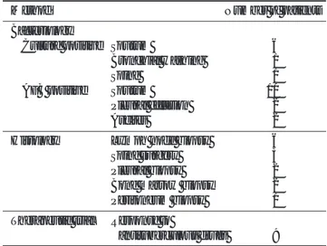
Fig. 1. 55-year-old woman with who presented with malaise.
CT scan shows portocaval and retroperitoneal lymphadenopa- thy with central low-density areas and an enhancing wall (ar- rows). CT guided biopsy revealed granulomatous inflamma- tion with caseation necrosis. She underwent hemodialysis one month after the initiation of antituberculous medication.
Table 3. Clinical Findings of Patients with End-Stage Renal Disease
Number of patients Causes of ESRD Diabetes Mellitus 22
Glomerulonephritis 3
Systemic lupus nephritis 2 Recurrent urinary infection 1
Unknown etiology 14
Symptoms and Signs Dyspnea 11
Cough 9
Fever 8
Abdominal pain 6
Malaise 5
Chest pain 4
Low back pain 3
Palpable mass 3
Clinical outcome Cure 30
Aggravation 3
Expire because of other causes 3
Follow-up loss 6
ESRD: end-stage renal disease
A B
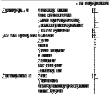
Fig. 2. 58-year-old man undergoing hemodialysis during 14 months who presented with dyspnea.
A. Chest radiograph shows bilateral pleural effusions, which was considered a primary pattern.
B. CT scan shows bilateral pleural effusions with thickening and enhancement of the parietal pleura (arrowheads).
three (23%) of these 13 patients. Enlargement of the me- diastinal lymph nodes involved the right paratracheal region in one case and the subcarinal lymph nodes in two. Parenchymal consolidation was observed in six pa-
tients (46%), and nodular opacity with centrilobular dis- tribution in seven (54%). Pleural effusion, identified in nine patients (69%), was left-sided in three cases and bi- lateral in six.
Discussion
The major host defense against the tubercle bacillus is cell-mediated immunity, effected primarily through macrophages and T-lymphocytes (8). A wide range of immunological derangements in ESRD have been postu- lated as the cause of the increased susceptibility of dialy- sis patients to tuberculosis. Tuberculosis has been re- ported to be more common in the dialysis population than in the general population (5, 6), and in our series, the prevalence of tuberculosis associated with ESRD
─ 348 ─ Table 4. Radiographic Characteristics of Pulmonary Tuberculosis in Patients with End-Stage Renal Disease
Radiographic Finding Number of patinets (n=28)
Postprimary pattern 11 (39)
Primary pattern 12 (43)
Pleural effusion 10 (36)
Consolidation 05 (18)
Thoracic lymphadenopathy 01 (4)
Atypical opacification 03 (11)
Miliary pattern 01 (4)
Normal radiographs 01 (4)
Note.─Numbers in parentheses are percentages.
A
C
B
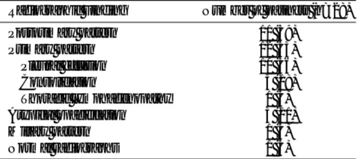
Fig. 3. 61-year-old man undergoing hemodialysis dur- ing 29 months who presented with fever.
A. Chest radiograph shows left-sided pleural effusion and multiple nodular opacities, which was catego- rized a atypical pattern.
B. CT scan obtained with mediastinal window set- tings shows subcarinal lymphadenopathy and lobar lymphadenopathy.
C. CT scan obtained with lung window settings at the same level as figure 2B shows multiple centrilobular nodules and linear opacities.
was 4.8%, about five times that of the general popula- tion of our country (9).
The inordinately high percentage of patients with AIDS whose radiographic findings are more typical of primary than of postprimary tuberculosis has been well documented (1, 2). In our series, tuberculosis with ES- RD showed a pattern similar to that of AIDS. The prima- ry pattern, manifesting as pleural effusion or segmental consolidation, was seen in twelve patients (43%), in ten of whom pleural effusion dominated. Pleural effusion in uremic patients may result from heart failure, uremic pleurisy, parapneumonic effusion, or atelectasis (10), but tuberculous pleural effusion can usually be distin- guished from these other types by microscopic and chemical examination of pleural fluid prior to the result of pleural fluid acid-fast bacilli smear and culture. In some patients pleural biopsy may confirm the diagnosis, as in two of our cases.
It has been reported that cavities had formed in 46%
of patients with pulmonary tuberculosis who were dia- betic or otherwise immunocompromised [diabetes (n=31), malignancy (n=6), or steroid therapy (n=2)], and about half of them had multiple cavities (11). In our series, five patients showed evidence of cavitary lung le- sions, a proportion similar to that observed in relation to the rare phenomenon of cavity formation in patients with AIDS (1, 2) and in contrast to the high prevalence of cavitation seen in patients with intact immunity (12).
Prior to the epidemic of HIV infection, approximately 13% of newly reported cases of tuberculosis involved extrapulmonary sites (13). In HIV-infected patients, however, both absolute and relative rates of extrapul- monary involvement have increased by up to two thirds (14), in our series, extrapulmonary tuberculosis account- ed for 36% of all cases. Multiorgan involvement is prob- ably a much more common than is usually recognized:
once M. tuberculosis is identified in any specimen, oth- er sites are generally not evaluated.
Since presenting symptoms may be non-specific and protean, the diagnosis of tuberculosis in patients with ESRD can be difficult (5, 6). In many patients tuberculo- sis is diagnosed at a time when symptoms such as dysp- nea, malaise, and fever could be readily attributed to uremia, volume overload, pyogenic reaction to dialysate or the relatively common bacterial infections of vascular access sites that occur in patients undergoing dialysis.
The failure of the radiologist to alert the physician to the possibility of pulmonary tuberculosis frequently leads to long delays in the institution of therapy. Unfortunately,
there are no radiologic findings, alone or in combina- tion, that are pathognomonic of tuberculosis, and for correct diagnosis, especially in those patients at in- creased risk, it is important that a degree of suspicion is maintained.
Our study has several limitations. First, CD4+ T-lym- phocyte counts were not checked in any patient. We made no objective measurements reflecting the immune status of patients with ESRD. Second, radiologic find- ings such as pleural effusion may result partly from heart failure, a common complication of ESRD, though the pleural fluid encountered was exudate consisting principally of lymphocytes. Third, only 13 patients, in nine of whom the radiographic pattern was primary, un- derwent chest CT scanning.
In conclusion, the results of the present study indicate that there is a high incidence of tuberculosis in patients with ESRD, and that the radiologic appearance of pul- monary tuberculosis in ESRD commonly shows a pri- mary pattern, with frequent extrapulmonary involve- ment. Tuberculosis should be considered in the differ- ential diagnosis of any patient with ESRD presenting with pleural effusion, any kind of pulmonary infiltrate not responding to common antibiotics, or extrapul- monary manifestations.
References
1. Pitchenik AE, Rubinson HA. The radiographic appearance of tu- berculosis in patients with AIDS and pre-AIDS. Am Rev Respir Dis 1985;131:393-396
2. Greenberg SD, Frager D, Suster B, Walker S, Stavropoulos C, Rothpearl A. Active pulmonary tuberculosis in patients with AIDS: spectrum of radiographic findings (including a normal ap- pearance). Radiology 1994;193:115-119
3. Bennett JC, Plum F. Cecil textbook of medicine, 20th ed. W.B.
Saunders Company, 1996:556-563
4. Newberry WM, Sanford JP. Defective cellular immunity in renal failure: depression of reactivity of lymphocytes to phytohemagglu- tinin by renal failure serum. J Clin Invest 1971;50:1262-1271 5. Andrew OT, Schoenfeld PY, Hopewell PC, Humphreys MH.
Tuberculosis in patients with end-stage renal disease. Am J Med 1980;68:59-65
6. Cengiz K. Increased incidence of tuberculosis in patients undergo- ing hemodialysis. Nephron 1996;73:421-424
7. Miller WT, Macgregor RR. Tuberculosis: frequency of unusual ra- diographic findings. AJR Am J Roentgenol 1978;130:867-875 8. Barnes PF, Bloch AB, Davidson PT, Snider DE. Tuberculosis in pa-
tients with human immunodeficiency virus infection. N Engl J Med 1991;324:1644-1650
9. Hong YP, Kim SJ, Lew WJ, Lee EK, Han YC. The seventh nation- wide tuberculosis prevalance survey in Korea. 1995. Int J Tuberc Lung Dis 1998;2:27-38
10. Jarratt MJ, Sahn SA. Pleural effusions in hospitalized patients re- ceiving long-term hemodialysis. Chest 1995;108:470-474
─ 350 ─ 11. Ikezoe J, Takeuchi N, Johkoh T, et al. CT appearance of pul-
monary tuberculosis in diabetic and immunocompromised pa- tients: comparison with patients who had no underlying disease.
AJR Am J Roentgenol 1992;159:1175-1179
12. Im J-G, Itoh H, Shim YS, et al. Pulmonary tuberculosis: CT find- ings-early active disease and sequential change with antitubercu- lous therapy. Radiology 1993;186:653-660
13. Farer LS, Lowell AM, Meador MP. Extrapulmonary tuberculosis in the United States. Am J Epidemiol 1979;109:205-217
14. Small PM, Schecter GF, Goodman PC, Sande MA, Chaisson RE, Hopewell PC. Treatment of tuberculosis in patients with advanced human immunodeficiency virus infection. N Engl J Med 1991;324:
289-294
대한방사선의학회지 2001;44:345-350
말기신부전 환자에서의 결핵1
1서울대학교 의과대학 방사선과학교실, 서울대학교 의학연구원 방사선의학연구소
김효철・구진모・정명진・문민환・고영환・임정기
목적: 말기신부전 환자에서 결핵의 임상적, 방사선학적 소견을 알아보고자 하였다.
대상과 방법: 말기신부전 환자 871명 중 활동성 결핵으로 진단된 42명의 의무기록과 방사선 사진을 후향적으로 분
석하였다. 흉부방사선 사진을 주된 소견에 따라 일차 폐결핵, 이차 폐결핵, 속립성 폐결핵, 비전형 폐결핵으로 나누 었다.
결과: 폐결핵이 28명, 폐외결핵이 15명에서 있었다. 흉부방사선 사진의 소견상 일차 폐결핵이 12명, 이차 폐결핵이 11명, 속립성 폐결핵이 1명, 비전형 폐결핵이 3명, 정상 소견이 1명 있었다. 폐외결핵에서는 흉부외림프절결핵이 6 명, 복막결핵이 4명, 척추결핵이 3명, 골수결핵이 2명에서 있었다. 흉수나 폐경결로 나타나는 12명의 일차 폐결핵 환자 중 10명에서 흉수가 두드러진 소견이었다.
결론: 말기신부전 환자에서의 결핵은 일차 폐결핵이 흔하고 폐외결핵도 자주 보이는 소견이다.