Background: Mortality rates associated with sepsis have increased progressively in Korea, but domestic epidemiologic data remain limited. The objective of this study was to investigate the characteristics, management and clinical outcomes of sepsis patients in Korea.
Methods: This study is a multicenter retrospective cohort study. A total of 64,021 adult pa- tients who visited an emergency department (ED) within one of the 19 participating hospitals during a 1-month period were screened for eligibility. Among these, patients diagnosed with sepsis based on the third International Consensus Definitions for Sepsis and Septic Shock (Sepsis-3) were included in the study.
Results: Using the Sepsis-3 criteria, 977 sepsis patients were identified, among which 36.5%
presented with septic shock. The respiratory system (61.8%) was the most common site of in- fection. The pathogen involved was identified in 444 patients (45.5%) and multi-drug resis- tance (MDR) pathogens were isolated in 171 patients. Empiric antibiotic therapy was appro- priate in 68.6% of patients, but the appropriateness was significantly reduced in infections associated with MDR pathogens as compared with non-MDR pathogens (58.8% vs. 76.0%, P <0.001). Hospital mortality was 43.2% and 18.5% in sepsis patients with and without shock, respectively. Of the 703 patients who survived to discharge, 61.5% were discharged to home and 38.6% were transferred to other hospitals or facilities.
Conclusions: This study found the prevalence of sepsis in adult patients visiting an ED in Ko- rea was 1.5% (15.2/1,000 patients). Patients with sepsis, especially septic shock, had a high mortality and were often referred to step-down centers after acute and critical care.
Key Words: epidemiology; Korea; mortality; prevalence; sepsis; septic shock
Kyeongman Jeon
1, Soo Jin Na
1, Dong Kyu Oh
2, Sunghoon Park
3, Eun Young Choi
4, Seok Chan Kim
5, Gil Myeong Seong
6, Jeongwon Heo
7, Youjin Chang
8, Won Gun Kwack
9, Byung Ju Kang
10, Won-Il Choi
11, Kyung Chan Kim
12, So Young Park
13*, Sang Hyun Kwak
14, Yoon Mi Shin
15, Heung Bum Lee
16, So Hee Park
17, Jae Hwa Cho
18, Beongki Kim
19, Chae-Man Lim
2; Korean Sepsis Alliance (KSA) study group
1Department of Critical Care Medicine, Samsung Medical Center, Sungkyunkwan University School of Medicine, Seoul; 2Department of Pulmonary and Critical Care Medicine, Asan Medical Center, University of Ulsan College of Medicine, Seoul; 3Department of Pulmonary, Allergy and Critical Care Medicine, Hallym University Sacred Heart Hospital, Anyang; 4Division of Respiratory and Critical Care Medicine, Department of Internal Medicine, Yeungnam University Medical Center, Daegu; 5Division of Pulmonary and Critical Care Medicine, Department of Medicine, Seoul St. Mary’s Hospital, College of Medicine, The Catholic University of Korea, Seoul; 6Department of Internal Medicine, Jeju National University Hospital, Jeju National University School of Medicine, Jeju;
7Department of Internal Medicine, Kangwon National University Hospital, Chuncheon; 8Division of Pulmonary and Critical Care Medicine, Department of Internal Medicine, Inje University Sanggye Paik Hospital, Seoul; 9Division of Pulmonary, Allergy and Critical Care Medicine, Department of Internal Medicine, Kyung Hee University Hospital, Seoul; 10Department of Internal Medicine, Ulsan University Hospital, University of Ulsan College of Medicine, Ulsan; 11Department of Medicine, Keimyung University Dongsan Medical Center, Daegu; 12Department of Internal Medicine, Daegu Catholic University Medical Center, Daegu Catholic University College of Medicine, Daegu; 13Division of Pulmonary and Critical Care Medicine, Department of Internal Medicine, Chungnam National University Hospital, Daejeon; 14Department of Anesthesiology, Chonnam National University Hospital, Gwangju; 15Division of Pulmonary and Critical Care Medicine, Department of Internal Medicine, Chungbuk National University Hospital, Cheongju; 16Department of Internal Medicine, Research Center for Pulmonary Disorders, Chonbuk National University Medical School, Jeonju; 17Division of Pulmonary and Critical Care Medicine, Department of Internal Medicine, Inje University Ilsan Paik Hospital, Inje University College of Medicine, Goyang; 18Division of Pulmonology, Department of Internal Medicine, Gang- nam Severance Hospital, Yonsei University College of Medicine, Seoul; 19Division of Pulmonology, Department of Internal Medicine, Korea University Ansan Hospital, Ansan, Korea
Characteristics, management and clinical outcomes of patients with sepsis: a multicenter cohort study in Korea
Original Article
Received: April 19, 2019 Revised: May 21, 2019 Accepted: May 30, 2019 Corresponding author Chae-Man Lim
Department of Pulmonary and Critical Care Medicine, Asan Medical Center, University of Ulsan College of Medicine, 88 Olympic-ro 43-gil, Songpa-gu, Seoul 05505, Korea
Tel: +82-2-3010-4710 Fax: +82-2-2045-4039 E-mail: [email protected]
* Current affiliation: Division of Pulmonary and Critical Care Medicine, Department of Internal Medicine, Ewha Womans University Seoul Hospital, Seoul, Korea Copyright © 2019 The Korean Society of Critical Care Medicine
This is an Open Access article distributed under the terms of Creative Attributions Non-Commercial License (http://
creativecommons.org/li-censes/by-nc/4.0/) which permits unrestricted noncommercial use, distribution, and reproduction in any medium, provided the original work is properly cited.
| pISSN 2586-6052 | eISSN 2586-6060
INTRODUCTION
Sepsis is an important global health concern. A recent sys- tematic review of study data from high-income countries yielded estimates of 31.5 million sepsis cases and 5.3 million deaths worldwide each year [1]. In addition, many patients who survive sepsis subsequently suffer from substantial cog- nitive impairment and functional disability [2]. To reduce the burden of sepsis, the World Health Organization has recom- mended implementing epidemiologic surveillance systems and monitoring the incidence and outcomes from sepsis, to- gether with concerted efforts to reduce antimicrobial resis- tance [3]. Given the incidence, etiology, treatment and out- comes of sepsis vary by geographical region and change over time, national data must be continually updated to guide each country’s healthcare policy and to allocate appropriate health- care resources to manage sepsis [4-6].
The mortality rates associated with sepsis have increased progressively in Korea [7]. But domestic epidemiologic data, especially regarding sepsis cases that are recognized outside the intensive care unit (ICU) remain limited. This study aimed to investigate the incidence, characteristics, treatment and outcomes of sepsis in Korea.
MATERIALS AND METHODS
Study Design and Population
This was a nationwide multicenter retrospective cohort study conducted by the Korean Sepsis Alliance. Nineteen tertiary or university-affiliated hospitals in Korea agreed to participate in the study. The steering committee developed the study proto- col, periodically reviewed the progress, and provided overall supervision of the study. The present study was approved by the Institutional Review Boards of each participating hospital, and the requirement for informed consent was waived be- cause of the non-interventional observational nature of the study.
We screened all consecutive patients who presented to the emergency department (ED) in one of the participating hos- pitals during a 1-month period (from January 1 through Janu- ary 31, 2018) for eligibility. Patients who were over 19 years of age and had sepsis as defined by clinical criteria from the third International Consensus Definitions for Sepsis and Septic Shock (Sepsis-3) were included in the study and followed up until death or hospital discharge. We considered sepsis to be the diagnosis if the patient satisfied the following two condi- tions: (1) a probable or confirmed diagnosis of infection, and
KEY MESSAGES
■ The prevalence of sepsis in adult patients visiting an emer- gency department during a 1-month period in Korea was 1.5% (15.2/1,000 patients).
■ Overall in-hospital mortality was 27.5% in adult patients with sepsis who were admitted to hospitals through emer- gency departments in Korea.
■ Patients with sepsis were more commonly referred to step-down facilities rather than discharged to home, even after acute sepsis care.
(2) an acute change in total Sequential Organ Failure Assess- ment (SOFA) score of 2 or more consequent to the infection [8]. The baseline SOFA score was assumed to be zero in pa- tients not known to have pre-existing organ dysfunction.
Data Collection
Trained study coordinators in each participating center used the hospital records for each patient to prepare a standardized Excel spreadsheet-based case report form. The following in- formation was collected retrospectively: (1) demographic data, including age, sex, comorbidities, SOFA score, physio- logical and laboratory measurements at the time of ED visit;
(2) infection data, including source and type of infection, and presence of multi-drug resistance (MDR) pathogens in the case of culture-positive-infected patients; (3) treatment data, including choice and appropriateness of empiric therapy, im- plementation of nonsurgical or surgical source control, imple- mentation of the 1-hour Surviving Sepsis Campaign bundle, use of adjunctive steroids, and decisions regarding limitation of life-sustaining treatments during the hospitalization; and (4) clinical outcomes, including in-hospital death and dis- charge destination for patients who survived to discharge. For patients admitted to the ICU for sepsis, data regarding SOFA scores at ICU admission and at the first 48 hours after ICU ad- mission, resource use and medical events during ICU stay, and need for organ support treatment at the time of ICU dis- charge were also collected. All participating centers were asked to complete data entry and email the data to the coordi- nating center at the Samsung Medical Center, where the qual- ity of data were assessed for completeness and logical errors.
Definitions
Infection was defined as the presence of a clinical or radiolog- ical infectious focus, or both, plus the administration of anti- biotics, and was classified into one of three categories: (1) mi-
Table 1. Baseline characteristics of patients with sepsis who were admitted to hospitals through emergency departments in Korea
Variable Overall (n=977) Sepsis (n=620) Septic shock (n=357) P-value
Age (yr) 75 (64–81) 75 (65–82) 73 (63–80) 0.017
Male sex 559 (57.2) 348 (56.1) 211 (59.1) 0.366
Body mass index (kg/m2) 22.3 (19.7–24.6) 22.2 (19.3–24.9) 22.4 (20.0–24.3) 0.961
Comorbidity
Diabetes 284 (29.1) 168 (27.1) 116 (32.5) 0.074
Cardiovascular disease 270 (27.6) 160 (25.8) 110 (30.8) 0.092
Chronic neurological disease 213 (21.8) 136 (21.9) 77 (21.6) 0.894
Chronic lung disease 172 (17.6) 122 (19.7) 50 (14.0) 0.025
Chronic liver disease 107 (11.0) 65 (10.5) 42 (11.8) 0.537
Chronic kidney disease 162 (16.6) 105 (17.0) 57 (16.0) 0.679
Connective tissue disease 24 (2.5) 16 (2.6) 8 (2.2) 0.741
Solid malignant tumors 258 (26.4) 160 (25.8) 98 (27.5) 0.574
Hematological malignancies 56 (5.7) 36 (5.8) 20 (5.6) 0.895
Immunocompromised 43 (4.4) 24 (3.9) 19 (5.3) 0.287
Charlson comorbidity index 5 (4–7) 5 (4–7) 5 (4–8) 0.218
SOFA score 5 (3–7) 4 (3–6) 8 (5–10) <0.001
Respiration 2 (1–2) 2 (1–2) 2 (1–3) 0.006
Coagulation 0 (0–1) 0 (0–1) 0 (0–2) 0.001
Liver 0 (0–1) 0 (0–1) 0 (0–1) 0.030
Cardiovascular 0 (0–1) 0 (0–1) 2 (0–3) <0.001
Central nervous system 0 (0–2) 0 (0–1) 0 (0–3) <0.001
Renal 1 (0–2) 0 (0–1) 1 (0–2) <0.001
Vital sign
Systolic blood pressure (mm Hg) 110 (89–137) 120 (100–141) 90 (75–113) <0.001
Diastolic blood pressure (mm Hg) 65 (52–80) 70 (60–84) 54 (43–68) <0.001
Mean blood pressure (mm Hg) 80 (65–99) 87 (73–105) 67 (54–83) <0.001
Heart rate (/min) 104 (89–120) 102 (88–118) 108 (90–122) 0.043
Temperature (ºC) 37.2 (36.5–38.2) 37.3 (36.6–38.2) 37.1 (36.4–38.0) 0.004
Laboratory finding
Lactate (mmol/L) 2.4 (1.5–4.1) 1.70 (1.20–2.67) 3.90 (2.50–6.26) <0.001
White blood cell (103/L) 11.1 (6.8–16.5) 11.2 (7.3–16.2) 10.9 (5.5–17.0) 0.298
Hemoglobin (g/dl) 11.2 (9.7–13.0) 11.4 (9.9–13.0) 11.0 (9.2–12.7) 0.003
Platelet count (103/L) 178 (108–257) 185 (118–266) 150 (96–237) <0.001
Sodium (mmol/L) 136 (132–140) 136 (132–139) 136 (132–140) 0.746
Potassium (mmol/L) 4.3 (3.7–4.7) 4.3 (3.8–4.7) 4.3 (3.6–4.8) 0.305
Chloride (mmol/L) 101 (97–106) 101 (97–106) 101 (97–106) 0.892
Blood urea nitrogen (mg/dl) 26.6 (17.1–44.0) 25 (16–39) 31 (20–55) <0.001
Creatinine (mg/dl) 1.30 (0.85–2.26) 1.18 (0.79–1.98) 1.61 (1.05–2.63) <0.001
AST (U/L) 36 (24–67) 33 (23–58) 41 (26–90) <0.001
ALT (U/L) 22 (14–44) 21 (13–41) 24 (15–49) 0.002
Albumin (g/dl) 3.0 (2.7–3.5) 3.1 (2.8–3.6) 2.9 (2.5–3.3) <0.001
Prothrombin time (INR) 1.20 (1.08–1.38) 1.17 (1.06–1.29) 1.27 (1.13–1.52) <0.001
(Continued to the next page)
crobiologically documented infection (infection with patho- gen identification), (2) clinically documented infection (infec- tion without causative pathogen identification), and (3) possi- ble infection (all other situations). We also divided the infec- tions into community-acquired infection, which was present upon admission or developed within 48 hours of hospital ad- mission, and hospital-acquired infection, which occurred
>48 hours after hospital admission. Cultured pathogens were defined as the presence of any etiologic microorganism re- covered from cultures collected within 2 days before and 2 days after admission. MDR was defined as acquired nonsus- ceptibility to at least one agent in three or more antimicrobial categories [9]. Initial antimicrobial therapy begun in the ab- sence of definitive microbiologic pathogen identification was considered an empiric therapy, and the appropriateness of empiric therapy was determined according to the results of the drug susceptibility test or the guideline recommendations [10].
Septic shock was characterized by persistent arterial hypo- tension requiring vasopressors to maintain mean arterial pres- sure ≥65 mm Hg and a serum lactate level >2 mmol/L de- spite adequate volume resuscitation [8]. The 1-hour Surviving Sepsis Campaign bundle consisted of the following elements:
measure lactate level, obtain blood cultures prior to adminis- tration of antibiotics, administer broad spectrum antibiotics, begin rapid administration of 30 ml/kg of intravenous crystal- loid fluid for hypotension or lactate level ≥4 mmol/L, apply vasopressors to maintain a mean arterial pressure ≥65 mm Hg, and remeasure lactate level if initial lactate is >2 mmol/L [11].
Statistical Analysis
The data were summarized using descriptive statistics: medi- an and interquartile range (IQR; 25th and 75th percentiles)
were calculated for continuous variables, while categorical variables were summarized as numbers and percentages. In order to assess differences between subgroups, data were compared using the Mann-Whitney U-test for continuous variables and the chi-square or Fisher’s exact test for categori- cal variables, when applicable. For all analyses, a two-tailed test with a P-value less than 0.05 was considered statistically significant. Statistical analyses were performed using STATA ver. 14.0 (Stata Corp., College Station, TX, USA).
RESULTS
Patient Characteristics
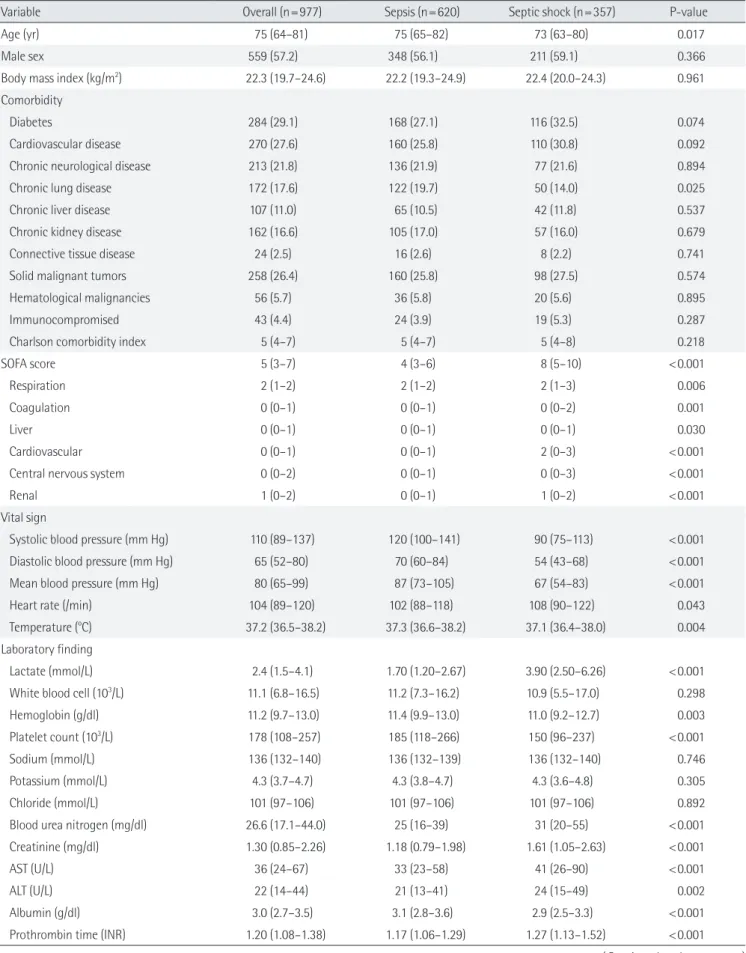
During the 1-month study period, 64,021 patients visited the EDs of the participating hospitals: 977 (1.5%) were identified as having sepsis through the medical records review and were included in the study. Among these, 357 patients (36.5%) met the clinical criteria for septic shock. Patient characteristics at the time of the ED visit are summarized in Table 1. The medi- an age was 75 years (IQR, 64–81 years) and 57.2% were male.
Diabetes (29.1%), cardiovascular diseases (27.6%), and solid malignant tumors (26.4%) were the most frequent comorbidi- ties. The median SOFA score was 5 (IQR, 3–7).
The comorbidities of patients with sepsis were similar to those of patients with shock, except for chronic pulmonary diseases (Table 1). The SOFA score was higher in patients with septic shock (4 vs. 8, P<0.001). The scores of each organ sys- tem were also different according to the presence of septic shock and the site of infection. In addition, myocardial de- pression was identified in 40 (46.5%) of 88 sepsis patients and 48 (62.3%) of 78 septic shock patients who underwent echo- cardiography (P=0.043), and the proportion of severe left ven- tricular systolic dysfunction tended to be higher in patients
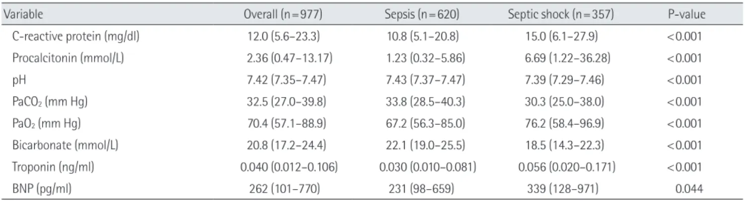
Variable Overall (n=977) Sepsis (n=620) Septic shock (n=357) P-value
C-reactive protein (mg/dl) 12.0 (5.6–23.3) 10.8 (5.1–20.8) 15.0 (6.1–27.9) <0.001
Procalcitonin (mmol/L) 2.36 (0.47–13.17) 1.23 (0.32–5.86) 6.69 (1.22–36.28) <0.001
pH 7.42 (7.35–7.47) 7.43 (7.37–7.47) 7.39 (7.29–7.46) <0.001
PaCO2 (mm Hg) 32.5 (27.0–39.8) 33.8 (28.5–40.3) 30.3 (25.0–38.0) <0.001
PaO2 (mm Hg) 70.4 (57.1–88.9) 67.2 (56.3–85.0) 76.2 (58.4–96.9) <0.001
Bicarbonate (mmol/L) 20.8 (17.2–24.4) 22.1 (19.0–25.5) 18.5 (14.3–22.3) <0.001
Troponin (ng/ml) 0.040 (0.012–0.106) 0.030 (0.010–0.081) 0.056 (0.020–0.171) <0.001
BNP (pg/ml) 262 (101–770) 231 (98–659) 339 (128–971) 0.044
Values are presented as median (interquartile range) or number (%).
SOFA: Sequential Organ Failure Assessment; AST: aspartate aminotransferase; ALT: alanine aminotransferase; INR: international normalized ratio;
PaCO2: partial pressure of carbon dioxide in arterial blood; PaO2: partial pressure of oxygen in arterial blood; BNP: brain natriuretic peptide.
Table 1. Continued
with septic shock (35.1% vs. 56.3%, P=0.053).
Characteristics of Infection
Most of the infections were community-acquired infections (80.9%) (Table 2). The most common primary site of infection was the respiratory system (61.8%), followed by the abdomi- nal cavity (16.5%) and genitourinary system (12.5%). Although respiratory system infections were the most common cause of both sepsis and septic shock, the percentages of patients with respiratory system infections (54.1% vs. 66.3%, P<0.001) were lower and intra-abdominal infections (21.9% vs. 13.4%, P<0.001) were higher in patients with septic shock than in patients with-
out shock.
Microbiologic pathogens were identified in 444 patients (45.5%) and bacteremia developed in 186 (42.3%) of these pa- tients. Infection due to MDR pathogens occurred in 171 (38.5%) of patients with microbiologically documented infection. En- terobacteriaceae accounted for about half of the MDR patho- gens, and Staphylococcus aureus was the next most common (21.1%). Pathogens were more frequently identified (52.1% vs.
41.6%, P=0.002), and the percentages of patients with bacte- remia (54.5% vs. 35.2%, P<0.001) and MDR pathogens (21.1%
vs. 15.8%, P=0.039) were greater in patients with septic shock than in sepsis patients without shock.
Table 2. Characteristics of infection in patients with sepsis who were admitted to hospitals through emergency departments in Korea
Variable Overall (n=977) Sepsis (n=620) Septic shock (n=357) P-value
Classification of infection
Microbiologically documented infection 444 (45.5) 258 (41.6) 186 (52.1) 0.002
Clinically documented infection 430 (44.0) 293 (47.3) 137 (38.4) 0.007
Possible infection 103 (10.5) 69 (11.1) 34 (9.5) 0.431
Site of infection
Respiratory tract 604 (61.8) 411 (66.3) 193 (54.1) <0.001
Abdominal cavity 161 (16.5) 83 (13.4) 78 (21.9) <0.001
Urinary tract 122 (12.5) 85 (13.7) 37 (10.4) 0.128
Skin/soft tissue 27 (2.8) 17 (2.7) 10 (2.8) 0.957
Catheter-related 7 (0.7) 3 (0.5) 4 (1.1) 0.266
Neurological 7 (0.7) 5 (0.8) 2 (0.6) >0.999
Infections without a clear primary site of infection 49 (5.0) 16 (2.6) 33 (9.2) <0.001
Type of infection 0.338
Community-acquired infection 790 (80.9) 507 (81.8) 283 (79.3)
Hospital-acquired infection 187 (19.1) 113 (18.2) 74 (20.7)
Cultured pathogena
Respiratory 147 (33.4) 93 (37.7) 52 (27.2) 0.021
Blood 186 (42.3) 87 (35.2) 104 (54.5) <0.001
Urine 66 (15.0) 48 (19.4) 18 (9.4) 0.004
Catheter 2 (0.5) 0 2 (1.1) 0.190
Others 34 (7.7) 19 (7.7) 15 (7.9) 0.950
Multi-drug resistance pathogen 171 (17.8) 96 (15.8) 75 (21.1) 0.039
Staphylococcus aureus 36 (21.1) 17 (17.7) 19 (25.3)
Enterococcus species 13 (7.6) 6 (6.3) 7 (9.3)
Enterobacteriaceae 86 (50.3) 52 (54.2) 34 (45.3)
Pseudomonas aeruginosa 18 (10.5) 15 (15.6) 3 (4.0)
Acinetobacter species 9 (5.3) 2 (2.1) 7 (9.3)
Clostridium perfringens 1 (0.6) 0 1 (1.3)
No data 8 (4.7) 4 (4.2) 4 (5.3)
Values are presented as number (%).
aData were available for 438 patients (247 patients with sepsis and 191 patients with septic shock).
Treatment Characteristics
Empiric combination therapy was used in a total of 484 pa- tients (50.5%) (Table 3). Beta-lactam antibiotics (87.5%) and fluoroquinolones (30.8%) were frequently chosen for empiric therapy. The percentages of patients administered glycopep- tides (13.5% vs. 6.3%, P <0.001) and carbapenem (13.0% vs.
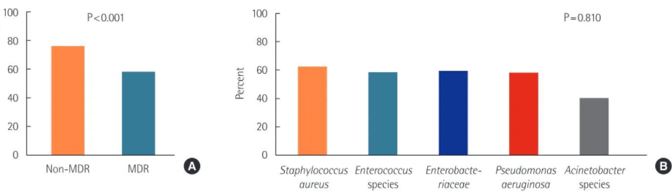
4.5%, P<0.001) were significantly greater in patients with sep- tic shock than in patients without shock. The selection of em- piric antibiotic regimen was appropriate in 68.6% of patients.
The appropriateness of empiric antibiotic selection was sig- nificantly lower in infections by MDR pathogen than in infec-
tions by non-MDR pathogen (58.1% vs. 76.0%, P<0.001) (Fig- ure 1). In infections by MDR pathogen, the appropriateness of empiric antibiotic selection was observed in 62.2% for Staphy- lococcus aureus, 58.3% for Enterococcus species, 59.3% for En- terobacteriaceae, 57.9% for Pseudomonas aeruginosa and 40.0%
for Acinetobacter species (P=0.810). Nonsurgical source con- trol measures were implemented in 132 patients (13.5%), in- cluding intravascular or other catheter removal in 35 cases and drainage catheter insertion of 86 cases, and surgical source control was performed in 20 patients (2.1%).
In terms of compliance rates for the 1-hour sepsis bundle Table 3. Characteristics of treatments for sepsis
Variable Overall (n=977) Sepsis (n=620) Septic shock (n=357) P-value
Initial empirical antibiotics
Beta-lactam 837 (87.5) 534 (88.7) 303 (85.4) 0.130
Fluroquinolone 295 (30.8) 189 (31.4) 106 (29.9) 0.619
Aminoglycoside 12 (1.3) 7 (1.2) 5 (1.4) 0.769
Glycopeptide 86 (9.0) 38 (6.3) 48 (13.5) <0.001
Colistin 1 (0.1) 0 1 (0.3) 0.371
Carbapenem 73 (7.6) 27 (4.5) 46 (13.0) <0.001
Macrolide 96 (10.0) 75 (12.5) 21 (5.9) 0.001
Others 75 (15.2) 35 (11.2) 40 (22.9) 0.001
Combination antibiotic therapy 484 (50.5) 293 (48.5) 191 (53.8) 0.113
Appropriateness of initial antibiotics
Appropriate 670 (68.6) 420 (67.9) 250 (70.0) 0.480
Inappropriate 172 (17.6) 103 (16.6) 69 (19.3) 0.288
Not available 134 (13.7) 96 (15.5) 38 (10.6) 0.033
Nonsurgical source control measure implemented 132 (13.5) 71 (11.5) 61 (17.1) 0.013
Removal of infected intravascular or other catheters 35 (4.2) 11 (2.1) 24 (7.7) <0.001
Insertion of percutaneous drain catheters 86 (10.3) 49 (9.3) 37 (11.9) 0.227
Pleural 33 (38.4) 16 (32.7) 17 (46.0)
Hepatobiliary 29 (33.7) 18 (36.7) 11 (29.7)
Peritoneum 10 (11.6) 6 (12.2) 4 (10.8)
Others 14 (16.3) 9 (18.4) 5 (13.5)
Other nonsurgical source control measure 20 (2.4) 16 (3.0) 4 (1.3) 0.109
Surgical source control 20 (2.1) 9 (1.5) 11 (3.2) 0.075
Intensive care unit admission 294 (33.9) 111 (20.6) 183 (55.5) <0.001
Treatment during intensive care unit staya
Mechanical ventilation 182 (63.9) 68 (58.6) 114 (67.5) 0.127
Renal replacement therapy 70 (24.6) 25 (21.6) 45 (26.6) 0.328
Extracorporeal membrane oxygenation 12 (4.2) 5 (4.3) 7 (4.1) >0.999
Hemoperfusion 22 (7.7) 4 (3.5) 18 (10.7) 0.025
Values are presented as number (%).
aData were available for 286 patients (116 patients with sepsis and 170 patients with septic shock).
Figure 2. One-hour Surviving Sepsis Campaign bundle compliance between sepsis and septic shock. IV: intravenous.
Sepsis Septic shock Measure lactate level 100
80 60 40 20 0
Percent
P<0.001
Sepsis Septic shock Obtain blood cultures 100
80 60 40 20 0
Percent
P=0.769
Sepsis Septic shock Broad spectrum antibiotics 100
80 60 40 20 0
Percent
P<0.001
Sepsis Septic shock 30 ml/kg of IV crystalloid fluid 100
80 60 40 20 0
Percent
P<0.001
Sepsis Septic shock Apply vasopressors 100
80 60 40 20 0
Percent
P<0.001
Sepsis Septic shock Remeasure lactate 100
80 60 40 20 0
Percent
P<0.001
Figure 1. Appropriateness of initial antibiotics. (A) Comparison of multi-drug resistance (MDR) and non-MDR pathogen. (B) Comparison among specific MDR pathogens.
Staphylococcus aureus
Enterococcus species
Enterobacte- riaceae
Pseudomonas aeruginosa
Acinetobacter species 100
80 60 40 20 0
Percent
P=0.810
Non-MDR MDR
100 80 60 40 20 0
Percent
P<0.001
A B
(Figure 2), lactate was measured in 80.5% of patients, and it was remeasured in 254 (67.0%) of the 379 patients with initial lactate >2 mmol/L. Blood cultures were obtained within 1 hour in 91.8% of patients, but antibiotics were administered to only 69.7% of patients in the same interval. Intravenous fluid administration and vasopressor infusion were performed in 38.9% and 35.0% of the patients, respectively. Compliance with survival bundle components was significantly greater in patients with septic shock than patients without shock, except with respect to obtaining blood cultures.
A total of 294 patients (33.9%) were admitted to an intensive
care unit, and data regarding treatment during the ICU stay were available for 286 of them: 182 (63.9%) and 70 (24.6%) pa- tients received mechanical ventilation and renal replacement therapy during ICU stay, respectively. Extracorporeal mem- brane oxygenation for circulatory support was performed in 12 patients (4.2%). There was no significant difference in the need for organ support treatment in patients with and without shock. However, patients with septic shock were more often treated with hemoperfusion (10.7% vs. 3.5%, P=0.025).
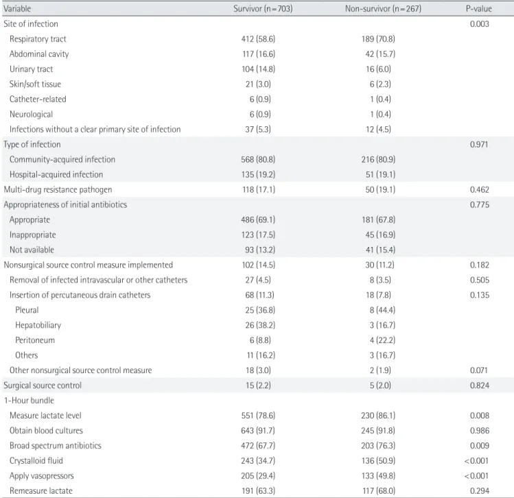
Table 4. Comparison of survivors and non-survivors
Variable Survivor (n=703) Non-survivor (n=267) P-value
Age (yr) 74 (63–81) 75 (67–82) 0.049
Male sex 404 (57.5) 152 (56.9) 0.879
Body mass index (kg/m2) 22.3 (19.8–24.9) 22.2 (19.5–24.2) 0.185
Comorbidity
Diabetes 207 (29.5) 71 (26.6) 0.380
Cardiovascular disease 187 (26.6) 78 (29.2) 0.415
Chronic neurological disease 155 (22.1) 57 (21.4) 0.814
Chronic lung disease 126 (17.9) 45 (16.9) 0.696
Chronic liver disease 81 (11.5) 25 (9.4) 0.336
Chronic kidney disease 120 (17.1) 40 (15.0) 0.442
Connective tissue disease 17 (2.4) 7 (2.6) 0.855
Solid malignant tumor 173 (24.6) 84 (31.5) 0.031
Hematological malignancy 41 (5.8) 15 (5.6) 0.898
Immunocompromised 30 (4.3) 13 (4.9) 0.684
Charlson comorbidity index 5 (4–7) 6 (4–8) 0.085
SOFA score 4 (3–7) 6 (4–10) <0.001
Vital sign
Systolic blood pressure (mm Hg) 110 (90–140) 104 (84–129) 0.005
Diastolic blood pressure (mm Hg) 67 (54–81) 60 (50–77) 0.004
Mean blood pressure (mm Hg) 83 (67–101) 77 (62–93) 0.007
Heart rate (/min) 104 (88–119) 106 (90–122) 0.148
Temperature (°C) 37.5 (36.6–38.4) 36.9 (36.3–37.7) <0.001
Laboratory finding
Lactate (mmol/L) 2.2 (1.3–3.5) 3.3 (1.9–6.0) <0.001
Hemoglobin (g/dl) 11.4 (9.8–13.0) 10.8 (9.1–12.8) 0.002
Platelet count (103/L) 179 (112–263) 170 (98–250) 0.073
Blood urea nitrogen (mg/dl) 24 (16–37) 37 (21–59) <0.001
Creatinine (mg/dl) 1.23 (0.81–2.03) 1.63 (0.92–2.63) <0.001
AST (U/L) 34 (23–59) 44 (26–91) <0.001
ALT (U/L) 22 (14–43) 24 (16–48) 0.197
Albumin (g/dl) 3.1 (2.8–3.6) 2.8 (2.3–3.2) <0.001
Prothrombin time (INR) 1.17 (1.06–1.32) 1.29 (1.16–1.58) <0.001
C-reactive protein (mg/dl) 10.9 (4.9–21.9) 15.9 (6.7–27.9) <0.001
Procalcitonin (mmol/L) 1.85 (0.39–10.95) 4.93 (0.87–20.49) <0.001
pH 7.42 (7.37–7.47) 7.39 (7.28–7.47) <0.001
PaCO2 (mm Hg) 33.2 (27.8–39.7) 31.8 (25.0–39.8) 0.076
PaO2 (mm Hg) 70.0 (57.5–88.3) 72.3 (56.0–93.0) 0.736
Bicarbonate (mmol/L) 21.3 (18.0–24.6) 19.5 (14.6–23.2) <0.001
Troponin (ng/ml) 0.034 (0.010–0.096) 0.051 (0.020–0.180) <0.001
BNP (pg/ml) 222 (89–592) 352 (142–1,445) 0.002
(Continued to the next page)
Variable Survivor (n=703) Non-survivor (n=267) P-value
Site of infection 0.003
Respiratory tract 412 (58.6) 189 (70.8)
Abdominal cavity 117 (16.6) 42 (15.7)
Urinary tract 104 (14.8) 16 (6.0)
Skin/soft tissue 21 (3.0) 6 (2.3)
Catheter-related 6 (0.9) 1 (0.4)
Neurological 6 (0.9) 1 (0.4)
Infections without a clear primary site of infection 37 (5.3) 12 (4.5)
Type of infection 0.971
Community-acquired infection 568 (80.8) 216 (80.9)
Hospital-acquired infection 135 (19.2) 51 (19.1)
Multi-drug resistance pathogen 118 (17.1) 50 (19.1) 0.462
Appropriateness of initial antibiotics 0.775
Appropriate 486 (69.1) 181 (67.8)
Inappropriate 123 (17.5) 45 (16.9)
Not available 93 (13.2) 41 (15.4)
Nonsurgical source control measure implemented 102 (14.5) 30 (11.2) 0.182
Removal of infected intravascular or other catheters 27 (4.5) 8 (3.5) 0.505
Insertion of percutaneous drain catheters 68 (11.3) 18 (7.8) 0.135
Pleural 25 (36.8) 8 (44.4)
Hepatobiliary 26 (38.2) 3 (16.7)
Peritoneum 6 (8.8) 4 (22.2)
Others 11 (16.2) 3 (16.7)
Other nonsurgical source control measure 18 (3.0) 2 (1.9) 0.071
Surgical source control 15 (2.2) 5 (2.0) 0.824
1-Hour bundle
Measure lactate level 551 (78.6) 230 (86.1) 0.008
Obtain blood cultures 643 (91.7) 245 (91.8) 0.986
Broad spectrum antibiotics 472 (67.7) 203 (76.3) 0.009
Crystalloid fluid 243 (34.7) 136 (50.9) <0.001
Apply vasopressors 205 (29.4) 133 (49.8) <0.001
Remeasure lactate 191 (63.3) 117 (68.0) 0.294
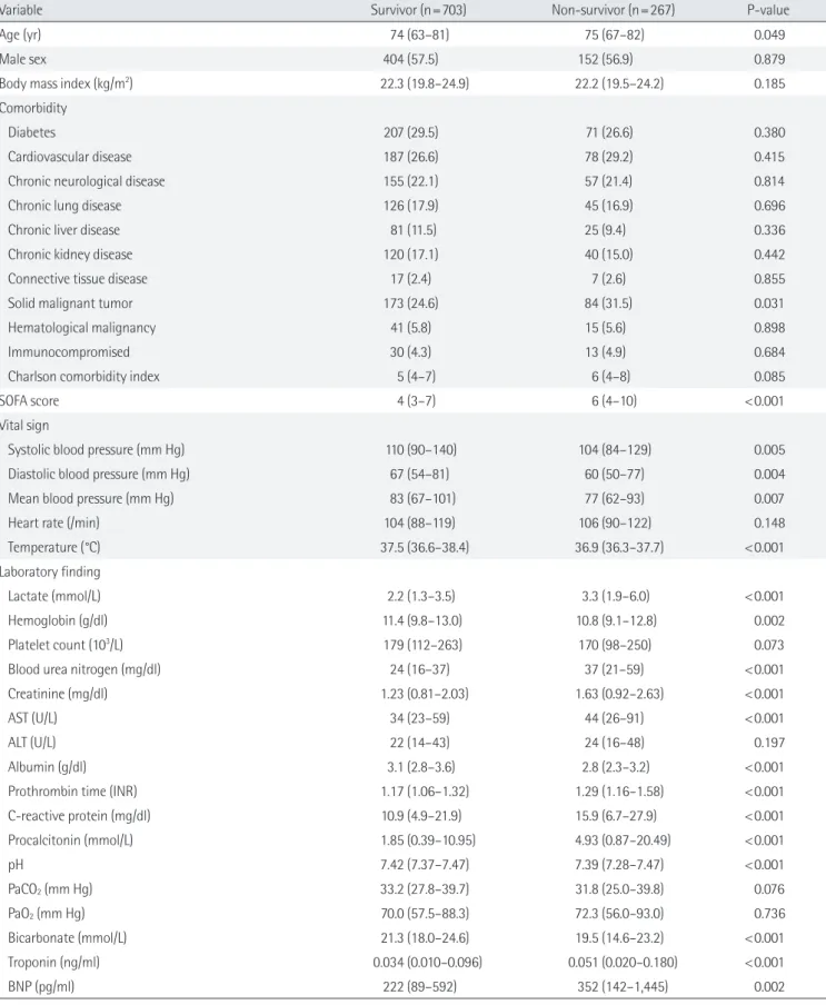
Values are presented as median (interquartile range) or number (%).
SOFA: Sequential Organ Failure Assessment; AST: aspartate aminotransferase; ALT: alanine aminotransferase; INR: international normalized ratio;
PaCO2: partial pressure of carbon dioxide in arterial blood; PaO2: partial pressure of oxygen in arterial blood; BNP: brain natriuretic peptide.
Table 4. Continued
Clinical Outcomes
Overall, 267 patients (27.5%) died in the hospital, and hospital mortality was significantly greater in patients with septic shock than in patients without shock (43.2% vs. 18.5%, P<0.001). Pa- tients who died in the hospital were older and had a higher proportion of solid malignant tumor than survivors (Table 4).
In addition, non-survivors were more frequently associated
with organ dysfunction at the time of visit to the ED and respi- ratory tract infection. However, there was no significant differ- ence in the proportion of MDR pathogen, appropriateness of initial antibiotics, and source control measures. In a multivari- able analysis, age, initial SOFA scores, solid or hematological malignancies, and site of infection were significant prognostic predictors for hospital mortality (Table 5).
Of the patients who survived to discharge from hospital, 61.5% were discharged to home and 38.6% were transferred to hospitals or facilities (Table 6). Most of the referral cases were transferred to step-down care. Patients with septic shock were more likely to be referred than sepsis patients without shock (48.3% vs. 34.7%, P =0.002). Patients with septic shock had more frequent limitation of life–sustaining treatments (52.0%
vs. 23.6%, P<0.001).
DISCUSSION
In the present study, we described the clinical and microbio-
logical characteristics and outcomes of patients with sepsis who visited an ED in Korea. We found that sepsis accounted for 1.5% (15.2/1,000 patients) of the population of adult pa- tients visiting the ED during a 1-month period, and 36.5% of the sepsis patients presented with septic shock. The reported incidence of sepsis varies widely in different studies [12], al- though direct comparison is difficult because of differences in screening methods and criteria for defining sepsis between studies. Our study showed comparable results when com- pared with reported incidence of severe sepsis (by Sepsis-2 criteria) or sepsis (by Sepsis-3 criteria) from neighboring coun- tries or high-income countries. Recent observational studies Table 5. Multivariable logistic regression analysis for probability of hospital mortality in patients with sepsis who were admitted to hospi- tals through emergency departments in Korea
Variable Univariable Multivariable
OR (95% CI) P-value Adjusted OR (95% CI) P-value
Age (yr) 1.01 (1.00–1.02) 0.061 1.02 (1.01–1.03) 0.004
Initial SOFA score 1.17 (1.13–1.22) <0.001 1.14 (1.08–1.19) <0.001
Septic shock 3.35 (2.50–4.49) <0.001 2.56 (1.80–3.62) <0.001
Solid or hematological malignancy 1.34 (1.00–1.80) 0.054 1.84 (1.31–2.58) <0.001
Site of infectiona
Abdominal 0.78 (0.53–1.16) 0.221 0.66 (0.43–1.02) 0.060
Urinary 0.34 (0.19–0.58) <0.001 0.30 (0.17–0.54) <0.001
Othersb 0.62 (0.37–1.05) 0.078 0.37 (0.20–0.67) 0.001
Measure lactate level 1.69 (1.14–2.50) 0.008 1.11 (0.72–1.71) 0.649
Obtain blood cultures 1.00 (0.60–1.68) 0.986 0.89 (0.50–1.57) 0.677
Broad spectrum antibiotics 1.54 (1.11–2.12) 0.009 1.17 (0.81–1.69) 0.413
Crystalloid fluid 1.95 (1.47–2.60) <0.001 1.02 (0.71–1.46) 0.905
OR: odds ratio; CI: confidence interval; SOFA: Sequential Organ Failure Assessment.
aThe reference group is pulmonary infection; bOthers include skin/soft tissue, catheter-related, neurological, and systemic infections refer to infections without a clear primary site of infection.
Table 6. Clinical outcomes of patients with sepsis who were admitted to hospitals through emergency departments in Korea
Variable Overall (n=977) Sepsis (n=620) Septic shock (n=357) P-value
Hospital mortality 267 (27.5) 114 (18.5) 153 (43.2) <0.001
Discharge destination
Home 432 (61.5) 328 (65.3) 104 (51.7) 0.001
Transfer 271 (38.6) 174 (34.7) 97 (48.3) 0.002
Step-down referral 227 (83.8) 146 (83.9) 81 (83.5)
Step-up referral 18 (6.6) 6 (3.5) 12 (12.4)
Unknown 26 (9.6) 22 (12.6) 4 (4.1)
Hospital length of stay (day) 9 (3–19) 10 (4–19) 8 (2–19) 0.013
Limitation of life–sustaining treatments at any time during the current admission
330 (33.9) 146 (23.6) 184 (52.0) <0.001
Values are presented as number (%) or median (interquartile range).
from East Asian countries demonstrated an incidence of 0.4%–1.2% or 461–639 cases/100,000 person-years, and Fleis- chmann et al. [1] estimated the worldwide incidence of sepsis to be 437 cases per 100,000 person-years [13,14].
The mortality rate was also similar to that reported in stud- ies of sepsis patients with organ dysfunction [1]. In our study, patients with sepsis showed a high mortality rate: one quarter of sepsis patients—and more than 40% of patients with septic shock—died in the hospital. The patients in our cohort were older than sepsis patients in studies from Western countries, reflecting the rapidly aging Korean population. Increasing age has been suggested to be an independent factor associated with poor outcomes in sepsis [15,16]. Hospital mortality among the patients in our study was consistent with reported mortal- ity for sepsis patients, but approximately 40% of our patients were transferred to step-down care facilities after recovery from sepsis. The high rates of underlying comorbidities and limitation of life-sustaining treatment in elderly patients may have influenced these outcomes. A recent study from Japan also showed that elderly patients with various comorbidities were major population of patients receiving ICU treatment due to sepsis and only one-third of sepsis patients were dis- charged home [16].
The respiratory system was the most common route of in- fection, similar to other studies [15,17,18]. In addition, the pro- portion of infections of respiratory system in our study was greater than in other studies, including that from another study from Korea [19]. Increasing age is considered a risk factor for community-acquired pneumonia in high-income countries, and death rates due to pneumonia in elderly patients have in- creased over the past 30 years in Korea, in association with so- cioeconomic improvements and aging of the population [20].
In addition to the relatively older population, having collected patient data during the winter may be also a reason for the higher proportion of respiratory system infections in our study [21]. Among the infections where the causative microorgan- ism was identified as the cause of sepsis, 38% were associated with MDR pathogens, which was greater than in the Extended Prevalence of Infection in Intensive Care (EPIC II) study de- scribing the prevalence of infections in ICUs in Western coun- tries [6]. MDR is associated with initially inappropriate antibi- otic therapy, and it results in an increased risk of in-hospital mortality [22]. While consistent with prior studies, a recent retrospective observational study from India shows that high- er odds for mortality of MDR infection in the non-ICU popu- lation, but this relationship was not statistically significant in the ICU population [23]. In our study, the inappropriate anti-
biotic therapy in infections associated with MDR pathogens was high, but did not affect mortality. This result might be at- tributed to the development of organ failure at the time of presentation, although there was no difference in the initial SOFA scores between the two groups.
We characterized the patterns of empiric antibiotic therapy and the 1-hour sepsis bundle approach in clinical practice, as well as the epidemiology of sepsis, and identified consider- able differences in the initial resuscitation and treatment of sepsis depending on presence of shock. In patients with septic shock, the use of glycopeptides and carbapenem and admin- istration of antibiotics and measurement of lactate within one hour were significantly more frequent than in sepsis patients without shock. This difference might reflect the lack of com- pliance with the international guidelines for management of sepsis [10], but could also be interpreted as a result of failure of early recognition of sepsis in patients without hemodynam- ic instability. The rates of fluid and vasopressor infusion were also significantly greater in patients with septic shock. How- ever, it is difficult to distinguish whether these results were due to lower adherence to the bundle approach or to lower rates fluid or vasopressor therapy in patients without shock, because we did not assess indicators of hydration status (hy- potension or lactate ≥4 mmol/L) or the use of vasopressor therapy (hypotension during or after fluid resuscitation) [11].
Although our study provided information regarding the prevalence, patient and infection characteristics, and out- comes in sepsis patients diagnosed using Sepsis-3 definitions, and regarding compliance with the 1-hour sepsis bundle in Korea, there are several limitations that should be considered.
First, because of the retrospective nature of the study, our find- ings remain prone to various biases. We used a national mul- ticenter design to improve the generalizability of our findings, but there is a potential risk of selection bias, because only pa- tients visiting tertiary or university-affiliated hospitals were included in the study. Next, the incidence of sepsis might have been underestimated in this study, as we did not evaluate sep- sis identified on general wards or in ICUs during hospitaliza- tion, or sepsis caused by viral or fungal infections. In addition, given the incidence and characteristics of infection may vary depending on the season, we could not exclude the possibility that the incidence of sepsis might have been different if the study had been conducted during a different (non-winter) season. Finally, the causal relationship between sepsis and death could not be identified in this study. Additional pro- spective studies of sepsis, addressing a larger number of pa- tients followed for a longer period of time, are needed to bet-
ter define the public health and economic burden of sepsis.
Further prospective and nation-wide studies should deal with evaluation for viral and fungal infection in order to compre- hensively understand the microbiological characteristics and improve the appropriateness of antimicrobial use, and analy- sis of detailed cost and economic burden as well as clinical outcomes.
In Korea, the incidence and mortality of patients with sep- sis were comparable to those reported in other high-income countries. Patients with sepsis, in addition to having a high mortality, were more commonly referred to step-down facili- ties rather than discharged home even after acute sepsis care.
Our study found differences between the participating hospi- tals with regard to compliance with current recommendations for initial resuscitation and treatment of sepsis and septic shock, which suggests that knowledge of and experience with early recognition and treatment of sepsis may still be lacking. Fur- ther epidemiologic studies and development of healthcare policies aimed at improving awareness of sepsis and promot- ing implementation of the recommended sepsis care proto- cols are needed to improve the outcomes of sepsis.
CONFLICT OF INTEREST
No potential conflict of interest relevant to this article was re- ported.
ACKNOWLEDGMENTS
The Korean Sepsis Alliance (KSA) Study Group Steering and Writing Committee: Kyeongman Jeon, Soo Jin Na, Dong Kyu Oh, Sunghoon Park, Sang-Bum Hong, Gee Young Suh, Chae‐
Man Lim.
ORCID
Kyeongman Jeon https://orcid.org/0000-0002-4822-1772 Soo Jin Na https://orcid.org/0000-0002-8551-2472 Dong Kyu Oh https://orcid.org/0000-0002-7511-9634 Sunghoon Park https://orcid.org/0000-0001-7004-6985 Eun Young Choi https://orcid.org/0000-0003-2974-5447 Seok Chan Kim https://orcid.org/0000-0002-0054-5962 Gil Myeong Seong https://orcid.org/0000-0002-0765-8273 Jeongwon Heo https://orcid.org/0000-0001-7694-6730 Youjin Chang https://orcid.org/0000-0002-4838-466X Won Gun Kwack https://orcid.org/0000-0001-9611-4341 Byung Ju Kang https://orcid.org/0000-0002-1396-7398
Won-Il Choi https://orcid.org/0000-0001-7705-0098 Kyung Chan Kim https://orcid.org/0000-0001-5697-9674 So Young Park https://orcid.org/0000-0003-2718-0518 Sang Hyun Kwak https://orcid.org/0000-0001-6077-2086 Yoon Mi Shin https://orcid.org/0000-0002-1909-5148 Heung Bum Lee https://orcid.org/0000-0002-8267-8434 So Hee Park https://orcid.org/0000-0002-3320-9949 Jae Hwa Cho https://orcid.org/0000-0002-3432-3997 Beongki Kim https://orcid.org/0000-0003-2009-7658 Chae‐Man Lim https://orcid.org/0000-0001-5400-6588
AUTHOR CONTRIBUTIONS
Conceptualization: KJ, SHP, CML. Methodology: KJ, SHP, CML. Investigation: KJ, DKO, SP, EYC, SCK, GMS, JH, YC, WGK, BJK, WIC, KCK, SYP, SHK, YMS, HBL, SHP, JHC, BK.
Formal analysis: KJ, SJN. Writing – original draft: KJ, SJN. Writ- ing – review and editing: all authors.
REFERENCES
1. Fleischmann C, Scherag A, Adhikari NK, Hartog CS, Tsaganos T, Schlattmann P, et al. Assessment of global incidence and mortality of hospital-treated sepsis: current estimates and limitations. Am J Respir Crit Care Med 2016;193:259-72.
2. Iwashyna TJ, Ely EW, Smith DM, Langa KM. Long-term cog- nitive impairment and functional disability among survivors of severe sepsis. JAMA 2010;304:1787-94.
3. Reinhart K, Daniels R, Kissoon N, Machado FR, Schachter RD, Finfer S. Recognizing sepsis as a global health priority: a WHO resolution. N Engl J Med 2017;377:414-7.
4. Vincent JL, Marshall JC, Namendys-Silva SA, François B, Mar- tin-Loeches I, Lipman J, et al. Assessment of the worldwide burden of critical illness: the intensive care over nations (ICON) audit. Lancet Respir Med 2014;2:380-6.
5. Kaukonen KM, Bailey M, Suzuki S, Pilcher D, Bellomo R. Mor- tality related to severe sepsis and septic shock among critical- ly ill patients in Australia and New Zealand, 2000-2012. JAMA 2014;311:1308-16.
6. Vincent JL, Rello J, Marshall J, Silva E, Anzueto A, Martin CD, et al. International study of the prevalence and outcomes of infection in intensive care units. JAMA 2009;302:2323-9.
7. Choe YJ, Choe SA, Cho SI. Trends in infectious disease mor- tality, South Korea, 1983-2015. Emerg Infect Dis 2018;24:320-7.
8. Singer M, Deutschman CS, Seymour CW, Shankar-Hari M, Annane D, Bauer M, et al. The third international consensus definitions for sepsis and septic shock (Sepsis-3). JAMA 2016;
315:801-10.
9. Magiorakos AP, Srinivasan A, Carey RB, Carmeli Y, Falagas ME, Giske CG, et al. Multidrug-resistant, extensively drug-re- sistant and pandrug-resistant bacteria: an international ex- pert proposal for interim standard definitions for acquired resistance. Clin Microbiol Infect 2012;18:268-81.
10. Rhodes A, Evans LE, Alhazzani W, Levy MM, Antonelli M, Ferrer R, et al. Surviving sepsis campaign: international guide- lines for management of sepsis and septic shock: 2016. Inten- sive Care Med 2017;43:304-77.
11. Levy MM, Evans LE, Rhodes A. The Surviving Sepsis Campaign Bundle: 2018 update. Intensive Care Med 2018;44:925-8.
12. Sakr Y, Jaschinski U, Wittebole X, Szakmany T, Lipman J, Ña- mendys-Silva SA, et al. Sepsis in intensive care unit patients:
worldwide data from the intensive care over nations audit.
Open Forum Infect Dis 2018;5:ofy313.
13. Lee CC, Yo CH, Lee MG, Tsai KC, Lee SH, Chen YS, et al. Adult sepsis: a nationwide study of trends and outcomes in a popu- lation of 23 million people. J Infect 2017;75:409-19.
14. Zhou J, Tian H, Du X, Xi X, An Y, Duan M, et al. Population- based epidemiology of sepsis in a subdistrict of Beijing. Crit Care Med 2017;45:1168-76.
15. Angus DC, Linde-Zwirble WT, Lidicker J, Clermont G, Carcillo J, Pinsky MR. Epidemiology of severe sepsis in the United States: analysis of incidence, outcome, and associated costs of care. Crit Care Med 2001;29:1303-10.
16. Abe T, Ogura H, Shiraishi A, Kushimoto S, Saitoh D, Fujishima S, et al. Characteristics, management, and in-hospital mortal- ity among patients with severe sepsis in intensive care units
in Japan: the FORECAST study. Crit Care 2018;22:322.
17. Wang HE, Jones AR, Donnelly JP. Revised national estimates of emergency department visits for sepsis in the United States.
Crit Care Med 2017;45:1443-9.
18. Phua J, Koh Y, Du B, Tang YQ, Divatia JV, Tan CC, et al. Man- agement of severe sepsis in patients admitted to Asian inten- sive care units: prospective cohort study. BMJ 2011;342:d3245.
19. Park DW, Chun BC, Kim JM, Sohn JW, Peck KR, Kim YS, et al.
Epidemiological and clinical characteristics of community- acquired severe sepsis and septic shock: a prospective obser- vational study in 12 university hospitals in Korea. J Korean Med Sci 2012;27:1308-14.
20. Lee JY, Yoo CG, Kim HJ, Jung KS, Yoo KH. Disease burden of pneumonia in Korean adults aged over 50 years stratified by age and underlying diseases. Korean J Intern Med 2014;29:
764-73.
21. Kim HJ, Choi SM, Lee J, Park YS, Lee CH, Yim JJ, et al. Respira- tory virus of severe pneumonia in South Korea: prevalence and clinical implications. PLoS One 2018;13:e0198902.
22. Zilberberg MD, Shorr AF, Micek ST, Vazquez-Guillamet C, Kollef MH. Multi-drug resistance, inappropriate initial antibiotic therapy and mortality in Gram-negative severe sepsis and septic shock: a retrospective cohort study. Crit Care 2014;18:
596.
23. Gandra S, Tseng KK, Arora A, Bhowmik B, Robinson ML, Pani- grahi B, et al. The mortality burden of multidrug-resistant pathogens in India: a retrospective, observational study. Clin Infect Dis 2019;69:563-70.