Tuberc Respir Dis 2012;73:266-272
CopyrightⒸ2012. The Korean Academy of Tuberculosis and Respiratory Diseases. All rights reserved.
A Multicenter Study of Pertussis Infection in Adults with Coughing in Korea: PCR-Based Study
Sunghoon Park, M.D.1, Myung-Gu Lee, M.D.2, Kwan Ho Lee, M.D.3, Yong Bum Park, M.D.4, Kwang Ha Yoo, M.D.5, Jeong-Woong Park, M.D.6, Changhwan Kim, M.D.7, Yong Chul Lee, M.D.8, Jae Seuk Park, M.D.9, Yong Soo Kwon, M.D.10, Ki-Hyun Seo, M.D.11, Hui Jung Kim, M.D.12, Seung Min Kwak, M.D.13, Ju-Ock Kim, M.D.14, Seong Yong Lim, M.D.15, Hwa-Young Sung, M.D.16, Sang-Oun Jung, M.S.16, Ki-Suck Jung, M.D.1
1Division of Pulmonary, Allergy and Critical Care Medicine, Hallym University Sacred Heart Hospital, Hallym University College of Medicine, Anyang, 2Division of Pulmonary, Allergy and Critical Care Medicine, Chuncheon Sacred Heart Hospital, Lung Research Institute of Hallym University College of Medicine, Chuncheon, 3Division of Pulmonary, Allergy and Critical Care Medicine, Yeungnam University Medical Center, Yeungnam University College of Medicine, Daegu, 4Division of Pulmonary, Allergy and Critical Care Medicine, Kangdong Sacred Heart Hospital, Hallym University College of Medicine, Seoul, 5Division of Pulmonary, Allergy and Critical Care Medicine, Konkuk University Hospital, Konkuk University School of Medicine, Seoul,
6Division of Pulmonary and Critical Care Medicine, Gachon University Gil Hospital, Gachon University of Medicine and Science, Incheon, 7Division of Pulmonary and Critical Care Medicine, Sejong General Hospital, Bucheon, 8Division of Pulmonary, Allergy and Critical Care Medicine, Chonbuk National University Hospital, Chonbuk National University Medical School, Jeonju, 9Division of Pulmonary, Allergy and Critical Care Medicine, Dankook University Hospital, Dankook University College of Medicine, Cheonan, 10Division of Pulmonary and Critical Care Medicine, Chonnam National University Hospital, Chonnam National University Medical School, Gwangju, 11Division of Pulmonary, Allergy and Critical Care Medicine, Soonchunhyang University Hospital, Soonchunhyang University College of Medicine, Cheonan, 12Division of Pulmonary and Critical Care Medicine, Wonkwnag University Sanbon Hospital, Wonkwang University School of Medicine, Gunpo, 13Division of Pulmonary and Critical Care Medicine, Inha University Hospital, Inha University School of Medicine, Incheon, 14Division of Pulmonary and Critical Care Medicine, Chungnam National University Hospital, Chungnam National University School of Medicine, Daejeon, 15Division of Pulmonary and Critical Care Medicine, Kangbuk Samsung Hospital, Sungkyunkwan University School of Medicine, Seoul,
16Division of Bacterial Respiratory Infection, Korea National Institute of Health, Korea Centers for Disease Control and Prevention, Seoul, Korea
Background: Limited data on the incidence and clinical characteristics of adult pertussis infections are available in Korea.
Methods: Thirty-one hospitals and the Korean Centers for Disease Control and Prevention collaborated to investigate the incidence and clinical characteristics of pertussis infections among adults with a bothersome cough in non-outbreak, ordinary outpatient settings. Nasopharyngeal aspirates or nasopharyngeal swabs were collected for polymerase chain reaction (PCR) and culture tests.
Results: The study enrolled 934 patients between September 2009 and April 2011. Five patients were diagnosed as confirmed cases, satisfying both clinical and laboratory criteria (five positive PCR and one concurrent positive culture). Among 607 patients with cough duration of at least 2 weeks, 504 satisfied the clinical criteria of the US Centers for Disease Control and Prevention (i.e., probable case). The clinical pertussis cases (i.e., both probable and confirmed cases) had a wide age distribution (45.7±15.5 years) and cough duration (median, 30 days;
interquartile range, 18.0~50.0 days). In addition, sputum, rhinorrhea, and myalgia were less common and dyspnea was more common in the clinical cases, compared to the others (p=0.037, p=0.006, p=0.005, and p=0.030, respectively).
Conclusion: The positive rate of pertussis infection may be low in non-outbreak, ordinary clinical settings if a PCR-based method is used. However, further prospective, well-designed, multicenter studies are needed.
Key Words: Adult; Cough; Incidence; Whooping Cough; Signs and Symptoms
Address for correspondence: Ki-Suck Jung, M.D.
Division of Pulmonary, Allergy and Critical Care Medicine, Department of Internal Medicine, Hallym University Sacred Heart Hospital, Hallym University College of Medicine, 22 Gwanpyeong-ro 170beon-gil, Dongan-gu, Anyang 431-796, Korea Phone: 82-31-380-3715, Fax: 82-31-380-3973, E-mail: [email protected]
Received: Mar. 19, 2012 Revised: Apr. 9, 2012 Accepted: Oct. 26, 2012
CCIt is identical to the Creative Commons Attribution Non-Commercial License (http://creativecommons.org/licenses/by-nc/3.0/).
Introduction
Pertussis infection is characterized by paroxysmal coughing, inspiratory whooping, and post-tussive vomit- ing and is also a bacterial cause of acute bronchitis re- quiring antibiotic treatment
1. Although the incidence of pertussis infection in the United States has declined dra- matically since the introduction of the inactivated whole-cell pertussis vaccine in the 1940s, the number of reported cases is again increasing, with a striking in- crease in adolescents and adults since 1976
2. This re- surgence has also been observed in countries such as Canada and Argentina since the 1980s
3-7. In a large European study (1998∼2002), the incidence of pertus- sis infection did not decrease in children, but rather in- creased in adults
3.
Multiple mechanisms may have caused this increase in the rate of pertussis infection: development of more sensitive diagnostic methods; changes in nationwide surveillance systems for communicable diseases; and waning immunity among adolescents and adults
2,8. In particular, the clinical presentation of adults with pertus- sis infection is not typical, and symptoms are frequently less severe in this population. Therefore, pertussis can go undiagnosed in adults, and adults can be the primary source of pertussis infection in non-vaccinated infants, in whom infection is potentially fatal
2.
In terms of vaccination schedule, a dose of tetanus and reduced dose of diphtheria and acellular pertussis (Tdap) is recommended for adolescents and adults by the US Advisory Committee on Immunization Practice and the Canada National Advisory Committee on Im- munization. Tdap was introduced recently in Korea.
However, contrary to the seriousness of adult pertussis infection in Western countries, data for Korea are lim- ited; most reported pertussis cases have been in child- ren. Therefore, a study of adult pertussis infection is ur- gently required to estimate the current burden of pertus- sis infection and facilitate a future booster vaccination program. This study investigated the incidence and clin- ical characteristics of adult pertussis infection in non- outbreak, ordinary clinical settings.
Materials and Methods 1. Sites and subjects
This study was conducted from September 2009 to April 2011 at 31 hospitals (14 primary care clinics, six secondary referral hospitals, and 11 tertiary referral hos- pitals) in eight South Korean provinces.
We enrolled only outpatients (≥18 years old) who presented with bothersome coughs. Exclusion criteria were a history of antibiotic treatment within 7 days; ac- tive lesions on the chest or paranasal sinus radiographs, if available; immunocompromised (e.g., acquired im- mune deficiency syndrome, leukemia, aplastic anemia, organ transplant, autoimmune diseases, or chemothera- py); or cough illness with a confirmed alternative cause (e.g., drugs [angiotensin-converting enzyme inhibitors], pneumonia, allergic rhinitis, sinusitis, or gastroesopha- geal reflux).
2. Clinical data and specimen collection
For all enrolled patients, the participating inves- tigators at outpatient departments collected clinical in- formation, including data on age, gender, chronic respi- ratory diseases, co-morbid illnesses, smoking status, cough duration, classic pertussis symptoms, other respi- ratory symptoms, history of diphtheria, tetanus, and per- tussis or diphtheria, tetanus, and acellular pertussis (DTP or DTaP) vaccination; and a history of hospital visits.
We collected laboratory specimens via nasophar-
yngeal aspiration (NPA, bulb aspiration kit prefilled with
saline, N-Pak; M-Pro, Annandale, MN, USA) or naso-
pharyngeal swabbing (NPS, liquid Amies medium on
flocked swabs; Copan Diagnostic, Murrieta, CA, USA),
and the specimens were transferred at room temper-
ature to the Department of Bacterial Respiratory Infec-
tion at the Korean Centers for Disease Control and
Prevention (KCDC) within 24 hours. To ensure con-
sistent specimen quality, independent personnel, usu-
ally a nurse, performed the sampling procedure in each
hospital during the study period, and all microbiological
tests were performed and validated by the KCDC.
Table 1. Baseline characteristics (n=934)
Characteristics No. (%)
Age, yr 45.9±15.2
Female/Male 623/311
Smoker, never/ex-/current 805/31/98
Diabetes 41 (4.4)
Hypertension 85 (9.1)
Bronchial asthma 70 (7.5)
COPD 4 (0.4)
Bronchiectasis 5 (0.5)
Interstitial lung disease 1 (0.1)
Heart disease 24 (2.6)
Renal disease 2 (0.2)
Liver disease 10 (1.1)
Cerebrovascular disease 4 (0.4)
History of DTaP vaccination 123 (13.2)
Prior hospital visit 362 (38.7)
Prior antibiotics 72 (7.7)
Cough duration (range), days 17 (9∼30) COPD: chronic obstructive pulmonary disease; DTaP: diph- theria, tetanus, and acellular pertussis.
3. Case definition
The US Centers for Disease Control and Prevention (CDC) clinical criteria for pertussis infection were de- fined as when patients presented with a cough illness for ≥2 weeks and had one of the following classical symptoms: 1) paroxysmal coughing, 2) inspiratory whooping, or 3) post-tussive vomiting
9. The laboratory criteria for diagnosis were isolation of Bordetella pertus- sis or a positive polymerase chain reaction (PCR) assay.
A "confirmed case" was diagnosed when a patient sat- isfied both the clinical and laboratory criteria and a
"probable case" was diagnosed when a patient satisfied only the clinical criteria
9.
4. Specimens and microbiologic tests
All specimens were subject to PCR and culture tests.
Regan and Lowe agar medium (charcoal agar supple- mented with 10% horse blood) with 40 mg/mL cepha- lexin was used for culture tests
10. After inoculation, the plates were incubated for at least 7 days under humid conditions (35∼36
oC). Identification was based on both biological characteristics and PCR
11.
A portion of each specimen was boiled for 5 minutes for PCR. After centrifugation, 1∼2μL of the super- natant was used as the PCR template. Although no standard PCR method exists, the "repeated-insertion se- quence" and "pertussis toxin promoter region" have been used most frequently as target regions
12,13. We used the repeated-insertion sequence and primers BP1 (5'-GATTCAATAGGTTGTATGCATGGTT-3') and BP2 (5'- TTCAGGCACACAAACTTGATGGGCG-3'). In-house PCR was performed using a commercial pre-mixed Taq poly- merase (AccuPower PCR PreMix; Bioneer, Daejeon, Korea).
The PCR conditions were 95
oC for 5 minutes, fol- lowed by 40 cycles at 95
oC for 5 seconds and 55
oC for 10 seconds. The PCR products were resolved by electro- phoresis on 2% agarose gels, and identification of a 180-bp band was considered positive.
5. Data analyses
We investigated the incidence of confirmed cases and compared the clinical characteristics between the clin- ical cases of pertussis infection and other cases, among patients with a cough duration≥2 weeks. Data are ex- pressed as the mean±standard deviation (or median and interquartile range) for continuous variables and as percentages for categorical variables, unless otherwise indicated. Student's t-tests or Mann-Whitney U-tests were performed for continuous data; whereas chi- square or Fisher's exact tests were used for categorical data. A p<0.05 was considered statistically significant, and all analyses were conducted using SAS statistical software, EG version (SAS Institute, Cary, NC, USA).
Results
1. Demographics and clinical symptoms
In total, 938 patients were initially enrolled, and four
with incomplete clinical data were excluded (primary
care clinics, 157 patients; secondary referral hospitals,
214 patients; and tertiary referral hospitals, 563 pa-
Table 2. Symptoms and signs (n=934)
Symptoms No. (%)
Sputum 565 (60.4)
Rhinorrhea 329 (35.2)
Febrile sense 111 (11.9)
Chillness 84 (9.0)
Malaise 46 (4.9)
Myalgia 106 (11.3)
Chest pain 119 (12.7)
Dyspnea 134 (14.3)
Hemoptysis 27 (2.9)
Wheezing 72 (7.7)
Hemoptysis 16 (1.7)
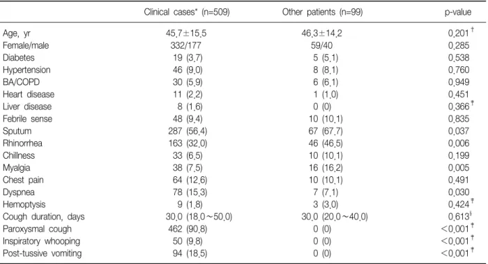
Table 3. Comparisons of the clinical characteristics between clinical cases and other patients
Clinical cases* (n=509) Other patients (n=99) p-value
Age, yr 45.7±15.5 46.3±14.2 0.201†
Female/male 332/177 59/40 0.285
Diabetes 19 (3.7) 5 (5.1) 0.538
Hypertension 46 (9.0) 8 (8.1) 0.760
BA/COPD 30 (5.9) 6 (6.1) 0.949
Heart disease 11 (2.2) 1 (1.0) 0.451
Liver disease 8 (1.6) 0 (0) 0.366‡
Febrile sense 48 (9.4) 10 (10.1) 0.835
Sputum 287 (56.4) 67 (67.7) 0.037
Rhinorrhea 163 (32.0) 46 (46.5) 0.006
Chillness 33 (6.5) 10 (10.1) 0.199
Myalgia 38 (7.5) 16 (16.2) 0.005
Chest pain 64 (12.6) 10 (10.1) 0.491
Dyspnea 78 (15.3) 7 (7.1) 0.030
Hemoptysis 9 (1.8) 3 (3.0) 0.424‡
Cough duration, days 30.0 (18.0∼50.0) 30.0 (20.0∼40.0) 0.613§
Paroxysmal cough 462 (90.8) 0 (0) <0.001‡
Inspiratory whooping 50 (9.8) 0 (0) <0.001‡
Post-tussive vomiting 94 (18.5) 0 (0) <0.001‡
Values are represented as number (% or range).
*Confirmed (n=5) and probable (n=504) cases defined by the US Centers for Disease Control and Prevention's criteria. †Student's t-tests. ‡Fisher's exact test. §Mann-Whitney U-tests.
BA: bronchial asthma; COPD: chronic obstructive pulmonary disease.