INTRODUCTION
Pulmonary alveolar proteinosis (PAP) was first described by Rosen et al. in 1958 (1). It is a rare disorder in which lipo- proteinaceous material accumulates within alveoli. The clin- ical course of the disease is variable, ranging from spontaneous
resolution to death due to pneumonia or respiratory failure.
Pulmonary alveolar proteinosis occurs in three clinically dis- tinct forms: congenital, secondary, and idiopathic (acquired).
Congenital PAP is a heterogeneous group of disorders (2) caused by mutations in the genes encoding surfactant pro- tein B, C or bcchain of the receptor for granulocyte-macro-
393
Min Kwang Byun1, Dong Soon Kim2, Young Whan Kim3, Man Pyo Chung4, Jae Jeong Shim5, Seung Ick Cha6, Soo-Taek Uh7, Choon Sik Park8, Sung Hwan Jeong9, Yong Bum Park10, Hong Lyeol Lee11, and Moo Suk Park1
Division of Pulmonary Medicine1, Department of Internal Medicine, Yonsei University, College of Medicine, Yonsei University Health System, Seoul;
Department of Pulmonary and Critical Care Medicine2, Asan Medical Center, University of Ulsan College of Medicine, Seoul; Division of Pulmonary and Critical Care Medicine3, Department of Internal Medicine and Lung Institute, Seoul National University College of Medicine, Seoul; Division of Pulmonary and Critical Care Medicine4, Samsung Medical Center, Sungkyunkwan University School of Medicine, Seoul;
Division of Pulmonary5, Allergy and Critical Care Medicine, Department of Internal Medicine, Korea University Anam Hospital, Seoul; Division of Pulmonary and Critical Care Medicine6, Department of Internal Medicine, Kyungpook National University Hospital, Daegu; Division of Allergy and Respiratory Medicine7, Department of Internal Medicine, Soonchunhyang University Hospital, Seoul; Division of Allergy and Respiratory Medicine8, Department of Internal Medicine, Soonchunhyang University Bucheon Hospital, Bucheon; Division of Pulmonology9, Department of Internal Medicine, Gachon University Gil Medical Center, Incheon; Division of Pulmonary10, Allergy & Critical Care Medicine, Department of Internal Medicine, Hallym University Kangdong Sacred Heart Hospital, Seoul; Pulmonary Division11, Department of Internal Medicine, Inha University Hospital, Incheon, Korea
Address for Correspondence Moo Suk Park, M.D.
Division of Pulmonary Medicine, Department of Internal Medicine, Yonsei University, College of Medicine, Yonsei University Health System, 250 Seongsan-ro, Seodaemun-gu, Seoul 120-752, Korea Tel : +82.2-2228-1974, Fax : +82.2-393-6884 E-mail : [email protected]
Clinical Features and Outcomes of Idiopathic Pulmonary Alveolar Proteinosis in Korean Population
Idiopathic pulmonary alveolar proteinosis (PAP) is a rare disorder in which lipopro- teinaceous material accumulates within alveoli. There were few reports on Asian populations with idiopathic PAP. We retrospectively reviewed 38 patients with idio- pathic PAP in Korea. We assessed clinical features, therapeutic efficacy and out- comes of whole lung lavage in patients with idiopathic PAP. The mean age at diag- nosis was 52 yr. Eighty six percent of patients were symptomatic at diagnosis. Dys- pnea and cough were the most common symptoms. Crackles were the most com- mon physical examination finding. On pulmonary function test, a mild restrictive ven- tilatory defect was common, with a predicted mean forced vital capacity (FVC) of 77% and forced expiratory volume in one second (FEV1) of 84.6%. Diffusing capaci- ty was disproportionately reduced at 67.7%. Arterial blood gas analysis revealed hypoxemia with a decreased PaO2of 69.0 mmHg and an increased D(A-a)O2of 34.2 mmHg. After whole lung lavage, PaO2, D(A-a)O2and DLCOwere significantly improved, but FVC and total lung capacity (TLC) were not different. This is the first multicenter study to analyze 38 Korean patients with idiopathic PAP. The clinical features and pulmonary parameters of Korean patients with idiopathic PAP are con- sistent with reports in other published studies. Whole lung lavage appears to be the most effective form of treatment.
Key Words : Pulmonary Alveolar Proteinosis; Irrigation; Treatment Outcome; Koreans
Received : 13 March 2009 Accepted : 1 June 2009
ⓒ 2010 The Korean Academy of Medical Sciences.
This is an Open Access article distributed under the terms of the Creative Commons Attribution Non-Commercial License (http://creativecommons.org/licenses/by-nc/3.0) which permits unrestricted non-commercial use, distribution, and reproduction in any medium, provided the original work is properly cited.
phage colony stimulating factor (GM-CSF) (3, 4). Secondary PAP occurs as a consequence of any one of a heterogeneous group of underlying clinical conditions (hematologic cancers, pharmacologic immunosuppression, inhalation of inorganic dust or toxic fumes, and certain infections) that impair alve- olar macrophage function, resulting in surfactant accumula- tion (5). Idiopathic (acquired) PAP is a disorder of unknown etiology that is thought to represent approximately 90% of PAP cases. The prevalence of idiopathic pulmonary alveolar proteinosis has been estimated to be 0.37 per 100,000 per- sons (6-9). The median age at the time of diagnosis is 39 yr;
most patients are men, and 72% have a history of smoking (10). Idiopathic pulmonary alveolar proteinosis is an enigmat- ic and fascinating disorder (1). Recent observations in trans- genic mice and humans, however, have provided important clues about its pathogenesis. The first real clue was provid- ed by the discovery that mice deficient in GM-CSF develop a lung phenotype that is biochemically, histologically, phys- iologically, and ultrastructurally indistinguishable from idio- pathic PAP (11, 12).
The observation of PAP in GM-CSF-deficient mice was quickly followed by the evaluation of GM-CSF therapy, first in a single patient and then in several small series of patients (13-16). GM-CSF deficiency has not been reported in humans (17, 18). However, a second vital pathogenic clue was the observation that primary PAP is specifically and strongly associated with very high levels of GM-CSF autoantibodies (19).
So far, most reports have focused on PAP patients from western countries, but reports on Asian populations are ext- remely rare, with only one report based on a Japanese popu- lation (20). The first case in Korea was reported in 1987 (21) and Kim and colleagues reviewed twelve cases in 1999 (22).
We reviewed the clinical features, treatment outcomes, and prognosis of patients with idiopathic PAP in Korea.
MATERIALS AND METHODS
We retrospectively reviewed patients with idiopathic PAP that were diagnosed between 1993 and 2007. The data were collected from 10 clinical research centers in Korea (College of Medicine in University of Ulsan, Seoul National Univer- sity, Yonsei University, Sungkyunkwan University, Korea University, Kyungpook National University, Soonchunhyang University, Gachon University, Hallym University, and Inha University). Patients with PAP were identified by review of the Korean Interstitial Lung Disease Study Group. The medi- cal records of eligible patients were reviewed by one investi- gator of each institution, and data were extracted using a stan- dard data collection instrument. The diagnosis of PAP was confirmed by a physician diagnosis and pathologic specimens were obtained by video-assisted thoracoscopic surgery or trans- bronchial lung biopsy with bronchoalveolar lavage (BAL).
The symptoms and signs present at the initial assessment at each institution were used as data. We contacted patients by telephone to ascertain follow-up information and outcomes.
Arterial blood gas analysis, spirometry, and lung volume mea- surements were estimated at diagnosis and if possible, esti- mated again after whole lung lavage. We assessed the effica- cy of whole lung lavage based on these parameters.
RESULTS
We identified 38 patients (24 males and 14 females) with PAP. The mean age at diagnosis was 52 yr (Table 1). There were no cases of PAP due to occupational exposure.
Eighty six percent of patients were symptomatic at diag- nosis and the most common symptom at presentation was dyspnea, and followed by cough (Table 2). The most promi- nent finding on physical examination was crackles in 50%
of patients, with cyanosis present in one patient. The chest radiographs for all but one patient showed bilateral air-space
Parameters No. Mean±SD Range
Age (yr) 38 51.6±12.8 15-81
Male (%) 24 63.2%
WBC (×103/mL) 37 6.7±2.2 3.5-13.9
Hb (g/dL) 37 14.2±2.3 8.8-19.7
LDH (IU/L) 21 455±183 153-834
CEA (ng/mL) 11 6.8±6.8 1.8-25.6
PaO2(mmHg) 31 69.0±14.2 39.7-93.7
D(A-a)O2(mmHg) 28 34.2±15.9 6.8-67.4
FEV1(% pred) 32 84.6±20.8 30-119
FVC (% pred 32 77.3±17.7 45-107
TLC (% pred) 25 81.9±16.0 46-110
DLCO(% pred) 28 67.7±31.6 17-131
Table 1. Demographic and clinical features upon diagnosis of idiopathic pulmonary alveolar proteinosis in Korean patients
Symptoms No. %
Dyspnea 25 50
Cough 12 24
Sputum 5 10
Fatigue 1 2
Chest pain 1 2
Epigastric pain 1 2
Asymptomatic 5 10
Table 2. Symptoms of Korean patients with idiopathic pulmonary alveolar proteinosis at presentation
WBC, white blood cell; Hb, hemoglobin; LDH, lactate dehydrogenase;
CEA, carcinoembryogenic antigen; PaO2, partial pressure of oxygen in arterial blood; D(A-a)O2, alveolar-arterial O2gradient; FEV1, forced expi- ratory volume in one second; FVC, forced vital capacity; TLC, total lung
capacity; DLCO, diffusing capacity of the lung for carbon monoxide. *Some patients presented with more than one symptom.
disease with an ill-defined nodular or confluent pattern. High resolution computed tomography showed patchy ground-glass opacifications with superimposed interlobular septal thick- ening in all patients.
On pulmonary function testing, a mild restrictive ventila- tory defect was common, the mean percent predicted forced vital capacity (FVC) was slightly decreased at 77%, the mean percent predicted forced expiratory volume in one second (FEV1) was normal at 84.6%, but the mean percent predict- ed total lung capacity (TLC) of 81% was not decreased. How- ever, the percent predicted diffusing capacity of the lung for carbon monoxide (DLCO) was disproportionately reduced (mean
±SD; 67.7±31.6) among 29 patients. Arterial blood gas analysis showed hypoxemia with a mean room air partial pres- sure of oxygen (PaO2) of 69.0±14.2 mmHg and a mean alve- olar arterial O2gradient; D(A-a)O2of 36.0±4.4 mmHg. The serum lactate dehydrogenase (LDH) level was elevated with a mean value of 455.4±183.1 IU/L (range 153 to 834 IU/
L). Transbronchial lung biopsy was performed in 35 patients (92%) and video-assisted thoracoscopic surgery was perform- ed in 16 patients (42%). Diagnosis was established by trans- bronchial lung biopsy with BAL or surgical lung biopsy. On light microscopic examination, we observed the characteristic finding of alveoli filled with a granular, eosinophilic material that stained with periodic acid-Schiff, and a well-preserved alveolar architecture. Whole lung lavage was indicated for patients who presented with dyspnea or hypoxemia and dete- riorated chest radiography. Whole lung lavage was performed in 26 patients (68%), of whom 11 patients underwent another lavage. We defined the recurrence as significant progression of respiratory symptoms attributable to PAP or the applica- tion of further therapeutic interventions such as repeated lavage.
Five patients (13.2%) showed spontaneous resolution after unspecified periods of observation. We analyzed oxygenation/
hypoxemia, spirometric measurements, lung volume param- eters, and radiologic findings before and after whole lung lavage (Table 3). Oxygenation improved dramatically after therapeu-
tic lavage, and hypoxemia was corrected in 16 of 17 patients for whom data were available. D(A-a)O2also improved after lung lavage. Spirometric results before and after whole lung lavage were available for 12 patients. FVC improved in eight patients and worsened in four patients. There was no signif- icant difference in the mean FVC before and after whole lung lavage (P=0.388), as analyzed using the Wilcoxon signed rank test. FEV1was increased in six patients and decreased in six patients. The difference in FEV1before and after whole lung lavage was not statistically significant (P=0.609). Eight of nine patients showed improvement in lung volume measurements following therapeutic lavage. DLCOafter whole lung lavage showed significant improvement (P=0.015). TLC showed a slight improvement after lavage, but the difference before and after lavage was not significant (P=0.089). Using radiologic evaluation with chest radiography or chest computed tomog- raphy, we found that 22 of 26 patients (85%) had improved after whole lung lavage therapy. Radiologic studies after lavage was unavailable in 2 patients, no changes in 2 patients. The mean follow-up period was 3 yr (range, 0.04 to 11.5 yr). At the end of the study, 20 patients were alive, 2 patients had died, and 16 patients were no longer available for follow-up.
One patient died soon after the whole lung lavage and the other patient died of advanced gastric cancer. The definition of disease recurrence varied between reports, but significant progression of respiratory symptoms due to PAP and the appli- cation of further therapeutic lavage were considered as recur- rences. PAP recurred in 4 out of 24 patients (16.7%) during the follow-up period. Of the four patients with a recurrence of PAP, three underwent another whole lung lavage and the fourth patient was closely observed without lavage.
DISCUSSION
PAP was first recognized in 1958 by Rosen and colleagues (1). Since then, there have been several reports on the clini- cal respects of idiopathic PAP. Seymour and Presneill (10) ana- lyzed 410 identifiable separate cases of PAP in 241 separate initial publications; most of these cases involved patients from western countries. Asamoto and colleagues (20) reported on the clinical manifestations of PAP in a Japanese population.
The Japanese report was unique because it focused on an Asian population with idiopathic PAP, but it was different from the retrospective meta-analysis by Seymour and colleagues (10) in several respects. First, two-thirds of the pathologic diagnoses were established by bronchoscopic biopsy instead of surgical lung biopsy. Further, it reported that the therapeu- tic outcomes of whole lung lavage were superior to those of other studies (Table 4). Before our study, however, this was the only data available on idiopathic PAP in Asian popula- tion. In this study, we retrospectively reviewed 38 patients with idiopathic PAP in Korea. All the Korean patients were born and had grown up in Korea. The median age at diag-
Variables No. Pre-lavage* Post-lavage* P value� PaO2, mmHg 17 64.9±11.1 77.3±15.7 0.005 D(A-a)O2, mmHg 16 35.3±12.4 20.1±13.2 0.001 FEV1, % pred 12 75.1±22.1 76.3±17.9 0.609 FVC, % pred 12 66.3±17.3 68.2±11.9 0.367 TLC, % pred 7 73.6±9.5 80.6±13.2 0.089 DLCO, % pred 9 53.9±22.3 65.2±17.6 0.024 Table 3. Pre-lavage and post-lavage pulmonary parameters for patients with idiopathic PAP
*Mean±SD (standard deviation); �P value is from the Wilcoxon signed rank test for comparison of pre-lavage versus post-lavage data for each parameter from each patient for whom data were available.
PaO2, partial pressure of oxygen in arterial blood; D(A-a)O2, alveolar-arte- rial O2gradient; FEV1, forced expiratory volume in one second; FVC, forced vital capacity; TLC, total lung capacity; DLCO, diffusing capacity of the lung for carbon monoxide.
nosis was 52 yr for both genders, which is in contrast to the distribution of patients from the report by Seymour and Pres- neill (10) who reported a median age of 39 yr and a different median age in men and women (Table 4). The median age in our study was close to that of the recent report on autoim- mune PAP in Japan by Inoue and colleagues (23).
Recently, the use of surgical lung biopsy has decreased (24, 25) because a diagnosis of PAP can be established in approx- imately 75% of clinically suspected cases based on the typi- cal findings of a milky fluid from the BAL (25) that contains large and foamy alveolar macrophages or monocyte-like alve- olar macrophage and an increased numbers of lymphocytes (26, 27). There are relatively few inflammatory cells of other cell types, unless superinfection is present. There is also a large
amount of amorphous, lipoproteinaceous material that is char- acteristically eosinophilic, granular, and positive with a peri- odic acid-Schiff stain (25, 27). In this study, pathologic diag- nosis was established by surgical lung biopsy (42%) or trans- bronchial biopsy with BAL (58%). Milky fluid on BAL and consistent transbronchial lung biopsy specimens provided diagnosis, such as surgical lung biopsy.
Pulmonary function test at diagnosis showed results that were similar to those of previous studies: a restrictive defect, with a disproportionate reduction in diffusing capacity, a slight- ly decreased mean FVC of 77.3% and a moderately decreased DLCOof 67.7%. Arterial blood gas analysis at the initial pre- sentation also showed the same results; PaO2was decreased to 69.0 mmHg and D(A-a)O2was increased to 35.3 mmHg
Fig. 1. Paired pre-lavage and post-lavage arterial blood gas analysis data from patients with idiopathic PAP. The P value is for the com- parison of pre-lavage versus post-lavage data for individual patients for each parameter for only those patients with available data using a Wilcoxon signed rank test. The number of evaluable patients for each parameter: PaO2(n=17), D(A-a)O2(n=16).
Fig. 2. Paired pre-lavage and post-lavage pulmonary function data from patients with idiopathic PAP. The P value is for the comparison of pre-lavage versus post-lavage data for individual patients for each parameter for only those patients with available data using a Wilcoxon signed rank test. The number of evaluable patients for each parameter: FEV1(n=12), FVC (n=12), TLC (n=7), DLCO(n=9).
Pre Post
P=0.005 P=0.001
PaO2(mmHg)
100
80
60
40
20
0 Pre Post
D(A-a) O2(mmHg)
100
80
60
40
20
0
P=0.609
FEV1(% pred)
150 100 50
0
Pre Post
P=0.367
FVC (% pred)
150 100 50
0
Pre Post
P=0.089
TLC (% pred)
150 100 50
0
Pre Post
P=0.024
DLco(% pred)
150 100 50
0
Pre Post
*Cases establishing diagnosis by autopsy, not shown.
NA=not available.
Diagnostic procedure (%)*
Surgical lung
biopsy Autopsy Broncho-
scopy
No. Mean
age (yr)
Whole lung lavage
(%)
Mortality (%) Male
(%) Study
(Reference)
Rosen et al. (1958) (1) 27 34 78 0 78 19 NA 30
Prakash et al. (1987) (9) 34 41 71 12 76 6 62 9
Asamoto et al. (1995) (20) 68 44 75 90 10 0 82 0
Seymour and Presneill (2002) (10) 410 39 71 10 71 11 54 16
Current study 38 52 63 58 42 0 68 5
Table 4. Comparison of published pulmonary alveolar proteinosis studies
at the initial presentation. The efficacy of whole lung lavage was evaluated with radiologic findings and pulmonary func- tion tests. After the therapeutic lavage, oxygenation param- eters such as PaO2and D(A-a)O2improved significantly in comparison to the initial examinations (Fig. 1). DLCO also increased dramatically. FVC and TLC improved slightly, but this improvement was not statistically significant (Fig. 2).
These results are similar to those from previous studies on idiopathic PAP.
The possibility of spontaneous resolution was initially pro- posed in the Rosen et al. report (1). They described the pati- ents as either having stable persistent symptoms, progressive deterioration in their process, or spontaneous improvement.
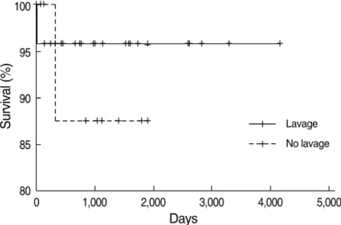
Other authors also have made similar observations. Kariman and colleagues (28) reported that spontaneous resolution oc- curred in 24% of a series of 23 patients. Seymour and Presneill (10) reported that 24 of 303 patients (7.9%) showed a signif- icant degree of spontaneous improvement. The reason for the variation in the amount of spontaneous resolution reported by different studies and authors may be because authors used different individual criteria. In our study, we defined spon- taneous resolution as radiographic and symptomatic improve- ment until follow-up. Based on these criteria, 5 of 38 pati- ents (13%) showed spontaneous resolution. In the survival analysis, there was no difference between the lavage and no lavage groups (Fig. 3). The mean follow-up period from the time of whole lung lavage of 3 yr was too short to compare the survival between the lavage and no lavage groups. More- over, one patient died soon after therapeutic lavage in the lavage group and one patient died of stomach cancer in the no lavage group, but neither death was directly related to PAP.
This study has several limitations; it was a retrospective study, the follow-up period was too short to analyze survival outcomes, and the smoking history of the patients was una- vailable.
This is the first multicenter study to analyze 38 Korean
patients with idiopathic PAP. The clinical features and pul- monary parameters of Korean patients with idiopathic PAP are consistent with reports in other published studies. Whole lung lavage appears to be the most effective form of treatment.
REFERENCES
1. Rosen SH, Castleman B, Liebow AA. Pulmonary alveolar proteino- sis. N Engl J Med 1958; 258: 1123-42.
2. Teja K, Cooper PH, Squires JE, Schnatterly PT. Pulmonary alveo- lar proteinosis in four siblings. N Engl J Med 1981; 305: 1390-2.
3. Nogee LM, de Mello DE, Dehner LP, Colten HR. Brief report: defi- ciency of pulmonary surfactant protein B in congenital alveolar pro- teinosis. N Engl J Med 1993; 328: 406-10.
4. Nogee LM, Dunbar AE 3rd, Wert SE, Askin F, Hamvas A, Whitsett JA. A mutation in the surfactant protein C gene associated with famil- ial interstitial lung disease. N Engl J Med 2001; 344: 573-9.
5. Kavuru MS, Bonfield TL, Thompson MJ. Pulmonary alveolar pro- teinosis. In: Mason RJ, Broaddus VC, Murray JF, Nadel JA, editors.
Textbook of pulmonary medicine, 4th ed. Philadelphia, PA: Elsevier 2006; 1716-34.
6. Ben-Dov I, Kishinevski Y, Roznman J, Soliman A, Bishara H, Zel- ligson E, Grief J, Mazar A, Perelman M, Vishnizer R, Weiler-Ravel D. Pulmonary alveolar proteinosis in Israel: ethnic clustering. Isr Med Assoc J 1999; 1: 75-8.
7. deMello DE, Lin Z. Pulmonary alveolar proteinosis: a review. Pedi- atr Pathol Mol Med 2001; 20: 413-32.
8. Goldstein LS, Kavuru MS, Curtis-McCarthy P, Christie HA, Farver C, Stoller JK. Pulmonary alveolar proteinosis: clinical features and outcomes. Chest 1998; 114: 1357-62.
9. Prakash UB, Barham SS, Carpenter HA, Dines DE, Marsh HM. Pul- monary alveolar phospholipoproteinosis: experience with 34 cases and a review. Mayo Clin Proc 1987; 62: 499-518.
10. Seymour JF, Presneill JJ. Pulmonary alveolar proteinosis: progress in the first 44 years. Am J Respir Crit Care Med 2002; 166: 215-35.
11. Dranoff G, Crawford AD, Sadelain M, Ream B, Rashid A, Bronson RT, Dickersin GR, Bachurski CJ, Mark EL, Whitsett JA, Mulligan RC. Involvement of granulocyte-macrophage colony-stimulating fac- tor in pulmonary homeostasis. Science 1994; 264: 713-6.
12. Stanley E, Lieschke GJ, Grail D, Metcalf D, Hodgson G, Gall JA, Maher DW, Cebon J, Sinickas V, Dunn AR. Granulocyte/macro- phage colony-stimulating factor-deficient mice show no major per- turbation of hematopoiesis but develop a characteristic pulmonary pathology. Proc Natl Acad Sci USA 1994; 91: 5592-6.
13. Seymour JF, Dunn AR, Vincent JM, Presneill JJ, Pain MC. Efficacy of granulocyte-macrophage colony-stimulating factor in acquired alveolar proteinosis. N Engl J Med 1996; 335: 1924-5.
14. Bonfield TL, Kavuru MS, Thomassen MJ. Anti-GM-CSF titer pre- dicts response to GM-CSF therapy in pulmonary alveolar proteinosis.
Clin Immunol 2002; 105: 342-50.
15. Seymour JF, Presneill JJ, Schoch OD, Downie GH, Moore PE, Doyle IR, Vincent JM, Nakata K, Kitamura T, Langton D, Pain MC, Dunn AR. Therapeutic efficacy of granulocyte-macrophage colony-stimu-
Survival (%)
100
95
90
85
80
0 1,000 2,000 3,000 4,000 5,000
Days
Lavage
Fig. 3. Overall survival from the time of diagnosis of idiopathic PAP.
There was no survival difference between two groups (lavage, n=26; no lavage, n=12, P=0.524).
No lavage
lating factor in patients with idiopathic idiopathic alveolar protei- nosis. Am J Respir Crit Care Med 2001; 163: 524-31.
16. Tazawa R, Hamano E, Arai T, Ohta H, Ishimoto O, Uchida K, Watan- abe M, Saito J, Takeshita M, Hirabayashi Y, Ishige I, Eishi Y, Hagi- wara K, Ebina M, Inoue Y, Nakata K, Nukiwa T. Granulocyte-mac- rophage colony-stimulating factor and lung immunity in pulmonary alveolar proteinosis. Am J Respir Crit Care Med 2005; 171: 1142-9.
17. Trapnell BC, Whitsett JA, Nakata K. Pulmonary alveolar proteinosis.
N Engl J Med 2003; 349: 2527-39.
18. Carraway MS, Ghio AJ, Carter JD, Piantadosi CA. Detection of gran- ulocyte-macrophage colony-stimulating factor in patients with pul- monary alveolar proteinosis. Am J Respir Crit Care Med 2000; 161:
1294-9.
19. Kitamura T, Tanaka N, Watanabe J, Uchida, Kanegasaki S, Yamada Y, Nakata K. Idiopathic pulmonary alveolar proteinosis as an auto- immune disease with neutralizing antibody against granulocyte/macro- phage colony-stimulating factor. J Exp Med 1999; 190: 875-80.
20. Asamoto H, Kitaichi M, Nishimura K, Itoh H, Izumi T. Primary pulmonary alveolar proteinosis: clinical observations of 68 patients in Japan. Nihon Kyobu Shikkan Gakkai Zasshi 1995; 33: 835-45.
21. Kim HT, Chung HS, Han SK, Shim Y, Kim KY, Han YC. A case of pulmonary alveolar proteinosis. Korean J Intern Med 1987; 33:
668-74.
22. Kim G, Lee SJ, Lee HP, Yoo CG, Han SK, Shim YS, Kim YW. The
clinical characteristics of pulmonary alveolar proteinosis: experi- ence at Seoul National University Hospital, and review of the litera- ture. J Korean Med Sci 1999; 14: 159-64.
23. Inoue Y, Trapnell BC, Tazawa R, Arai T, Takada T, Hizawa N, Kasa- hara Y, Tatsumi K, Hojo M, Ichiwata T, Tanaka N, Yamaguchi E, Eda R, Oishi K, Tsuchihashi Y, Kaneko C, Nukiwa T, Sakatani M, Krischer JP, Nakata K. Characteristics of a large cohort of patients with autoimmune pulmonary alveolar proteinosis in Japan. Am J Respir Crit Care Med 2008; 177: 752-62.
24. Shah PL, Hansell D, Lawson PR, Reid KB, Morgan C. Pulmonary alveolar proteinosis: clinical aspects and current concepts on patho- genesis. Thorax 2000; 55: 67-77.
25. Wang BM, Stern EJ, Schmidt RA, Pierson DJ. Diagnosing pulmonary alveolar proteinosis. A review and an update. Chest 1997; 111: 460-6.
26. Iyonaga K, Suga M, Yamamoto T, Ichiyasu H, Miyakawa H, Ando M. Elevated bronchoalveolar concentrations of MCP-1 in patients with pulmonary alveolar proteinosis. Eur Respir J 1999; 14: 383-9.
27. Schoch OD, Schanz U, Koller M, Nakata K, Seymour JF, Russi EW, Boehler A. BAL findings in a patient with pulmonary alveolar pro- teinosis successfully treated with GM-CSF. Thorax 2002; 57: 277- 80.
28. Kariman K, Kylstra JA, Spock A. Pulmonary alveolar proteinosis:
prospective clinical experience in 23 patients for 15 years. Lung 1984;
162: 223-31.