• 검색 결과가 없습니다.

Pedunculated Hepatocellular Carcinoma Mimicking Adrenal Cortical Carcinoma

N/A
N/A
Protected

Academic year: 2021

Share "Pedunculated Hepatocellular Carcinoma Mimicking Adrenal Cortical Carcinoma"

Copied!
6
0
0

로드 중.... (전체 텍스트 보기)

전체 글

(1)

Corresponding Author: Yu Na Kang, M.D., Department of Pathology, Keimyung University School of Medicine 56 Dalseong-ro, Jung-gu, Daegu 700-712, Korea

Tel: +82-53-250-7290, +82-53-580-3814

E-mail: yunakang@dsmc.or.kr, swmpath@gmail.com

Introduction

Pedunculated hepatocellular carcinoma (HCC) is a rare form of cancer, which protrudes from the liver as a massive tumor with or without a pedicle and was first described by Roux in 1897. It is rare variant of HCC, reported most commonly from

Japan and rarely from other parts of the world.

Because of anatomical adjacency and similarity of histopathologic findings, differential diagnosis from adrenal gland tumor was very difficult. And incidence of pedunculated hepatocellular carcinoma of 0.24~3.0% has been reported in Japan [1]. Here, we report a case that had much help in differential

Abstract

Hepatocellular carcinoma (HCC) is the most common primary liver tumor. However extrahepatic growing hepatocellular carcinoma has rarely been reported and is characteristically large and encapsulated.

Extrahepatic growing HCC which invaded right adrenal gland is very difficult to differential diagnose between hepatocelluar carcinoma and adrenal gland tumor. We report a 56-year-old male patient with almost one-year- history of right flank pain. Dynamic computerized tomography (CT) showed the tumor grew so large that invaded directly right adrenal gland. For this reason, we had to rule out hepatocelluar carcinoma, adrenal cortical carcinoma, and pheochromocytoma. In diagnosis process, we got much help from immunohistochemistry. Therefore, we presented a case of pedunculated hepatocellular carcinoma mimicking adrenal cortical carcinoma, diagnosed by immunohistochemical stains.

Key Words : Adrenal cortical carcinoma, Hepatocellular carcinoma, Immunohistochemistry, Pedunculated HCC

Undergraduate Student, Department of pathology

1

, Keimyung University School of Medicine, Daegu, Korea

Seong Wook Shin, Yu Na Kang

1

, M.D.

Pedunculated Hepatocellular Carcinoma Mimicking Adrenal Cortical

Carcinoma

(2)

d i a g n o s i n g f r o m t h e a d r e n a l t u m o r s b y immunohistochemistry.

Case Report

A 56-year-old male patient was referred to our hospital for further evaluation of intra-abdominal mass, which was found by computed tomography (CT) at other local hospital. Hypertension and diabetes mellitus are not found, but he has chronic hepatitis. Intermittent right flank pain was felt one year before admission but followed without treatment. On admission, blood pressure was 100/80 mmHg and other vital sign was stable. Physical examination showed normal appearance. Level of alpha-fetoprotein was 61.50 mg/ml (normal : <9.6 mg/ml), 24-hr urine vanillyl mandelic acid (VMA) was 23.96 mg (normal: <8.0 mg), 24-hr urine metanephrine was 1.720 mg (normal: <1.3 mg), plasma epinephrine was 0.020 mg (normal: <0.30 mg) and plasma norepinephrine was 0.177 mg (normal: <0.80 mg).

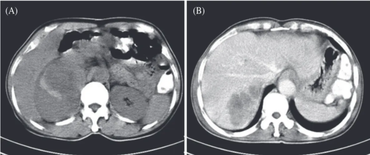
In contrast enhancement phase of CT, the inhomogeneous mass of right adrenal gland had

internal low attenuation area that represents areas of hemorrhage and necrosis. In delayed phase, the intrahepatic mass showed irregular heterogeneous low-attenuated areas, and is located in hepatic segment 6 (Fig. 1).

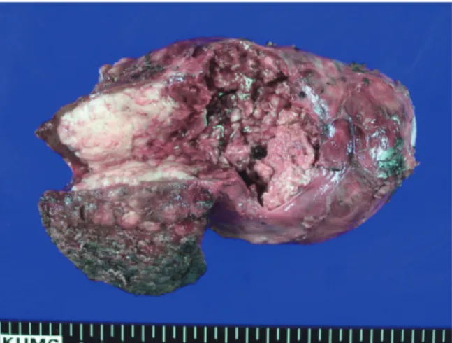
The patient underwent right unilateral adrenalectomy and segmentectomy of hepatic segment 6. Clinically, adrenal gland origin tumor, such as pheochromocytoma or adrenal cortical carcinoma, and primary hepatic carcinoma should be considered. Grossly, tumor mass is composed of intrahepatic lesion and protruding mass encasing the right adrenal gland, measuring 17.0 cm in maximum length. The cut surface of the mass showed pale tan solid and variegated mass with extensive areas of necrosis and hemorrhage (Fig. 2).

Microscopically, variable differentiation, from well dif ferentiated sinusoidal pattern to undifferentiated solid sheet growth, within intrahepatic tumor mass was seen. Undifferentiated tumor portion was composed of large, bizarre, hyperchromatic nuclei, markedly increasing mitotic rate (more than 20 per 10 high power fields), atypical mitoses, and extensive invasion into blood vessels and bile duct. In contrast, sections of the

Fig. 1. (A) Cotrast enhanced CT shows ill-defined inhomogeneous lesion in the right adrenal gland. (B) Delayed phase CT shows irregular heterogeneous low-attenuated mass in hepatic segment 6.

(A) (B)

(3)

adrenal gland was replaced by extensive undifferentiated tumor growth and showed thin collapsed normal architecture, such as zona glomerulosa, fasciculate, and reticularis, beneath the thin capsule (Fig. 3).

In immunohistochemical stains, the tumor cells were diffusely stained for cytokeratin, glutamine synthetase, HSP-70, hepatocyte, and the tumor mass showed thick sinusoidal capillarization for CD34.

But the immunohistochemical staining for the adrenal gland tumor markers, such as calretinin, chromogramin, synaptophysin, and inhibin, were all negative at anaplastic tumor cells except collapsed adrenal tissue (Fig. 4).

Fig. 2. Bisected gross finding of intrahepatic and protruding tumor mass, measuring 17.0 cm in maximum length.

Fig. 3. (A) Mixed patterns of sinusoidal growth (left) and diffuse sheet-like growth (right) are seen in the

intrahepatic and intraadrenal tumor mass (H&E, ×100). (B) Diffuse sheet-like growth of tumor cells is seen

with numerous atypical mitoses (H&E, ×400). (C) Thick sinusoidal growth pattern of tumor cells is seen

(H&E, ×200). (D) Collapsed adrenal cortex beneath the adrenal capsule is seen with expansile tumor mass

(H&E, ×40).

(4)

After surgical resection of tumor, patient’s symptom was improved. And he discharged with regular follow up. But 3 month later, local tumor recurrence with multiple hepatic metastasis was found on follow up computerized tomography (CT). Recurrent masses were localized around previous operation site, 3.0 cm in maximum length, and right hepatic lobe, 5.0 cm in maximum length.

After that, he was lost for follow-up evaluation.

Discussion

Primary hepatic cellular carcinoma in male is the fifth most frequently diagnosed cancer worldwide, and is the second leading cause of cancer-related death in the world [2]. Hepatic tumors generally progress in the patterns of local expansion, intrahepatic spread, and distant metastases.

However HCC occasionally grows outside the liver

in pedunculated form. Macroscopically, common hepatic tumors may present as nodular or diffuse type. The nodular type of hepatic tumors may be solitary or multiple. Tumor nodules are round to oval, grey or green (if the tumor produces bile), well circumscribed but not encapsulated. The diffuse type is poorly circumscribed and infiltrates t h e p o r t a l v e i n s, o r t h e h e p a t i c v e i n s.

Microscopically, there are four architectural and cytological types of common hepatocellular carcinoma: hepatocellular, pseudoglandular (adenoid), pleomorphic (giant cell) and clear cell types. The most common type is hepatocellular type, formation of thick trabecular hepatic cords lined by endothelial cells. When the tumor is well- differentiated, the neoplastic cells often show features that can be seen in normal hepatocytes such as fat and bile; however, in the poorly differentiated tumors, the cells may also be quite pleomorphic with giant cells and may be difficult to

Fig. 4. (A) Immunohistochemical stains show positive reactivity for cytokeratin (×400), (B) Hepatocyte (×200), (C)

Glutamine synthetase (×200), (D) HSP-70 (×200), (E) Negative reactivity for calretinin at tumor cells

(×100). (F) Also the endothelial cells of sinusoid pattern shows positive for CD34 (×200).

(5)

differentiate from certain metastatic tumors.

Hepatocellular carcinoma needs to be differentiated from poorly differentiated cholangiocarcinoma, regenerative nodules of cirrhosis, atypical hepatocellular adenoma, and other adrenal gland tumors. Especially pedunculated HCC variants are characteristically large and encapsulated, and classified as nodular, diffuse, massive [3].

Histologically, most of these carcinomas are poorly differentiated and show features of rapid growth including numerous mitoses and a Ki-67 labeling index. The mechanism for extrahepatic growth remains unknown.

Adrenal cortical carcinoma is a rare endocrine malignancy with a reported incidence of 0.5~2 cases per million individuals per year [4]. Although adrenal cortical carcinoma most commonly arises sporadically, its pathogenesis is not understood.

Adrenal cortical carcinoma are classified as functional tumor that are related to excess of corticosteroid, androgen, estrogen, and rarely mineralocorticoids or non-functional tumor that are related to mass effect. Non-functional tumor is related to ominous signs and symptoms including fever, anemia, pain, weight loss, and anorexia. Most common metastatic spread site is lungs (45%), liver (42%), and lymph nodes (24%).

Grossly, adrenocortical carcinomas are often large, with a tan-yellow cut surface, and areas of hemorrhage and necrosis. On microscopic examination, wide range of differentiation from tumor that is so well differentiated as to be almost impossible to distinguish from adenoma to totally undifferentiated neoplasms composed of giant cell with abundant acidophilic cytoplasm and bizarre hyperchromatic nuclei, sometimes multiple.

Adrenal cortical carcinomas may show evidence of neuroendocrine differentiation, as manifested by immunoreactivity for synaptophysin and typically negative for chromogranin, in contrast to

pheochromocytoma. Positivity for immunohisto- chemical markers such as Bcl-2 and calretinin, also assist the diagnosis of adrenal cortical carcinoma.

Bcl-2 is typically present in all cell layers of the normal adrenal cortex, but is consistently absent from the medulla [5]. Calretinin, a calcium binding protein, is expressed in 73% of adrenal cortical c a r c i n o m a [ 6 ] . I n t h i s c a s e , t h e immunohistochemical staining for the adrenal gland t u m o r m a r k e r s, s u c h a s c h r o m o g r a m i n, synaptophysin, and inhibin, were all negative at anaplastic tumor cells except collapsed adrenal tissue. Endothelial markers, such as CD31, CD34, and factor Ⅷ helps differentiate HCC from other malignant neoplasm and may accentuate trabecular growth pattern. Because one of the hallmark histopathologic features of HCC is the presence of endothelial cells lining the sinusoids that surround enlarged hepatocellular plates. Almost all HCC show positive staining for cytokeratin antibody, particularly low-molecular-weight cytokeratins (CK8, CK18). Hep Par-1 (Hepatocyte antigen) is an antigen reflecting hepatocytic differentiation and yields a diffuse cytoplasmic granular staing pattern in normal and neoplastic hepatocytes including approximately 80% to 90% of HCC cases [7].

Reticulin fibers are thin fibers composed of collagen

Ⅲ which form delicate stromal network in many organs. The reticulin network is particularly rich in the liver and can be seen along hepatic trabeculae.

Therefore reticulin stain provides important information about the architecture of the liver.

When hepatocytes are damaged and undergo

necrosis, the reticulin fibers surrounding the

collapse in the empty space left behind. Areas of

reticulin crowding thus indicate focal hepatocytes

loss. Therefore a reticulin stain is useful for

demonstrating liver architecture; hepatocyte necrosis

and hepatocyte regeneration. In our patient, positive

stain for cytokeratin (CK) shows that this tumor is

(6)

carcinoma. Furthermore, positive stains for hepatocyte, glutamine synthetase, HSP-70 and negative for calretinin demonstrate that this tumor originate not in adrenal cortical cell but in hepatocellular carcinoma. Finally CD34 positive stain for endothelial cells of sinusoid means endothelial cells capillarization typical found in hepatocellular carcinoma (HCC). After all, we finally diagnosed hepatocellular carcinoma.

Pedunculated HCC has been considered a poor- prognosis HCC variant. Most investigators support that tumor size plays a role in the prognosis and significantly affects survival. Furthermore, large tumors are associated with a significantly higher risk of recurrence. However, some patients benefit from surgical resection because both the capsule and the pedicle may prevent vascular invasion, therefore improving prognosis. In our case, Pedunculated tumor resection was primary because the tumor was still suspicious of adrenal cortical carcinoma. So there was no necessary for liver lobectomy.

In conclusion, in case of very ambiguous tumor as our case especially pedunculated hepatocellular carcinoma invading adrenal gland or tumor of unknown origin because of poorly differentiated tumor or tumor which is not found typical findings of hepatocellular carcinoma (HCC) in radiologically, the panel of immunohistochemical stains can be helpful for diagnosis of hepatocellular carcinoma (HCC).

Summary

A 56-year-old male patient was referred to our hospital for further evaluation of abdominal large mass involving segment 6 of liver and right adrenal gland. The patient underwent right unilateral

adrenalectomy and liver segmentectomy 6. At last, histological and immunohistochemical studies of intrahepatic and adrenal gland tumor confirmed hepatocellular carcinoma, invading adrenal gland.

When clinical and histopathologic differential diagnosis from adjacent organ neoplasms is difficult, the panel of immunohistochemistry give much help for confirming tools as our case.

References

1. Horie Y, Katoh S, Yoshida H, Imaoka T, Suou T, Hirayama C. Pedunculated hepatocellular carcinoma.

Report of three cases and review of literature. Cancer 1983;51:746-51.

2. Jemal A, Bray F, Center MM, Ferlay J, Ward E, Forman D. Global cancer statistics. CA Cancer J Clin 2011;61:69-90.

3. Yeh CN, Lee WC, Jeng LB, Chen MF. Pedunculated hepatocellular carcinoma: clinicopathologic study of 18 surgically resected cases. World J Surg 2002;26:1133-8.

4. Brennan MF. Adrenocortical carcinoma. CA Cancer J Clin 1987;37:348-65.

5. Laforga JB, Aranda FI. Giant adrenal cortical carcinoma, clinically "nonfunctional": report of a case containing cytoplasmic hyaline globules of vimentin. Diagn Cytopathol 1999;21:394-7.

6. Sangoi AR, Fujiwara M, West RB, Montgomery KD, Bonventre JV, Higgins JP, et al. Immunohisto-chemical distinction of primary adrenal cortical lesions from metastatic clear cell renal cell carcinoma: a study of 248 cases. Am J Surg Pathol 2011;35:678-86.

7. Kakar S, Gown AM, Goodman ZD, Ferrell LD. Best practices in diagnostic immunohistochemistry:

hepatocellular carcinoma versus metastatic neoplasms.

Arch Pathol Lab Med 2007;131:1648-54.

수치

Fig. 1.   (A) Cotrast enhanced CT shows ill-defined inhomogeneous lesion in the right adrenal gland
Fig. 2.  Bisected gross finding of intrahepatic and  protruding tumor mass, measuring 17.0 cm in  maximum length.

참조

관련 문서

A retrospective analysis of invasive tumor percentages of GSS tumor samples showed that samples with high tumor content correlated well with FNAB samples

Effects of Tetrapanax papyriferum Extracts on the KB Human Oral

Especially, in the case of DPRK(Democratic People's Republic of Korea) of which energy consumption have been decreased by economic depression, decomposition

Polypoid lesions of the gallbladder; Report of 111 cases with surgical reference to operative indication.. Polypoid lesion of GB: Indication of carcinoma

1 John Owen, Justification by Faith Alone, in The Works of John Owen, ed. John Bolt, trans. Scott Clark, &#34;Do This and Live: Christ's Active Obedience as the

Concentration-Dependent inhibition of cell viability by adenosine in human oral fibroblast and FaDu human head and neck squamous cell carcinoma · · ·

Differential Expression of Desmoglein1, Desmoglein3, Epithelial Membrane Antigen, Ber-EP4 and CD10 in Basal Cell Carcinoma and Squamous Cell Carcinoma

Objective: The purpose of this study was to analyze recent trend in incidence of basal cell carcinoma and squamous cell carcinoma in patients from the Gwangju City