There have been various reports about cases of spon- taneous rupture of adrenal tumor, such as pheochromo- cytoma, adrenal cortical carcinoma and adrenal myelolipoma. To the best of our knowledge, there has been only one case of adrenal metastatic tumor that caused spontaneous rupture (1). Because most extrahep- atic hepatocellular carcinomas occur in patients with an advanced intrahepatic tumor stage, the first manifesta- tion as spontaneous rupture of adrenal metastasis may be extremely rare (2). We report here on a patient who presented with spontaneous rupture of a huge adrenal metastasis as the initial manifestation, and the patient had a small primary hepatocellular carcinoma in his noncirrhotic liver.
Case Report
A 56-year-old man presented to the emergency depart- ment with suddenly-onset right flank pain that had de-
veloped one day previously. The pain was radiating to the epigastric area and the back. The physical examina- tion showed right flank tenderness and right upper quadrant tenderness on palpation. For his past history, he was a chronic alcoholic and had consumed 17g/day alcohol for about thirty years, but the laboratory find- ings were normal. The tests for hepatitis-B and C anti- gens were negative. There was no evidence of cirrhosis on the CT images and laboratory findings.
The abdominal CT scan showed a huge mass in the right adrenal gland, measuring about 8.2×7.6×10.0 cm, with adjacent infiltrations. The normal right adrenal gland could not be identified. On the noncontrast CT scan, this adrenal mass consisted of a high-attenuated peripheral portion and a central low attenuated necrotic component that contained foci of central increased den- sity, suggesting a hemorrhagic condition of an adrenal tumor. Dynamic CT scans during the hepatic arterial phase showed the right adrenal mass had poorer periph- eral enhancement than that of the liver parenchyma (55 HU to 61 HU), and there were minimal enhanced foci within the central necrotic portions (Fig. 1A-C). The in- ferior margin of the adrenal mass was poorly defined, and it appeared to be infiltrative, extending to the anteri- or and posterior perirenal spaces and retroperitoneum.
The inferior capsule of the liver was presumed to be in-
Spontaneous Rupture of Adrenal Metastasis from Hepatocellular Carcinoma
1Chae-Hun Lim, M.D., Hyun Jin Kim, M.D., Soo Youn Park, M.D., Seong Su Hwang, M.D., Hyun Joo Choi, M.D.2
1Department of Diagnostic Radiology, St. Vincent Hospital
2Department of Pathology, St. Vincent Hospital Received March 25, 2006 ; Accepted December 4, 2006
Address reprint requests to : Hyun Jin Kim, M.D., Department of Diagnostic Radiology, St. Vincent Hospital, 93-6 Ji-dong, Paldal-gu, Kyeonggi-do, Suwon 442-723, Korea
Tel. 82-31-249-7482 Fax. 82-31-247-5713 E-mail: [email protected]
Rupture of adrenal tumor from various primary origins is a rather rare event. We re- port here on a ruptured adrenal metastasis from hepatocellular carcinoma, and this ruptured metastasis was observed at the time of the initial diagnosis.
Index words :Adrenal gland, neoplasms Adrenal neoplasms, secondary Adrenal gland, CT
Adrenal gland, MR
tact. Another small enhancing hepatic nodule was de- tected on the arterial phase at the subcapsular portion of the fifth liver segment, but the rest of the hepatic parenchyma was relatively unremarkable (Fig. 1D).
This hepatic nodule demonstrated peripheral arterial en- hancement and isoattenuation, as compared to the liver parenchyma on the equilibrium phase, suggesting hepa- tocellular carcinoma.
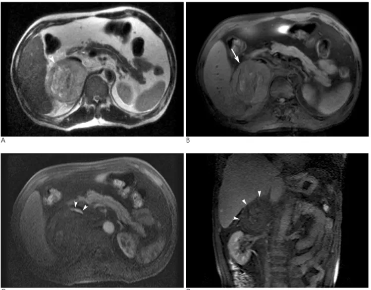
The T2-weighted axial MR images showed a slightly heterogenous mass in the right adrenal gland (Fig. 2A).
There was no detectable fat component within the right adrenal mass on the fat-saturated T1-weighted images (Fig. 2B). The central “whirl” appearing lesion had a hy-
perintense signal on the T2 weighted images and an isointense signal on the T1-weighted images, which are findings consistent with hyperacute hemorrhage (Fig.
2A, B). The gadolinium-enhanced dynamic MR images also depicted only a thin crescent of arterial enhance- ment in the anterior peripheral portion, and this portion might have been a viable tumor component in the hepa- tocellular carcinoma; however, the pathology was not confirmed (Fig. 2C, D).
Palliative excision of the right adrenal gland and he- patic wedge resection at segment V were performed.
Microscopic pathologic examination revealed poorly-dif- ferentiated carcinoma at segment V and in the right
A B
C D
Fig. 1. Abdominal CT scan of a 53-year-old man with right adrenal metastasis from hepatocellular carcinoma.
A. The non-contrast CT scan shows a highly attenuated peripheral portion and hyperdense foci within the central necrotic area in the right adrenal gland (arrow). The adjacent infiltrations and retoperitoneal hemorrhage suggest rupture of the adrenal tumor (ar- rowhead).
B. On the arterial phase, the right adrenal mass is poorly enhanced.
C. This adrenal mass is scarcely enhanced on the delayed phases.
D. An arterial enhancing nodule lies in the subcapsular portion of segment V, and retroperitoneal hemorrhage is also seen (arrow).
adrenal gland, which were both embedded with exten- sive hemorrhage (Fig. 3A, B).
The patient had an uneventful postoperative course and was discharged. Since then, the patient has been do- ing well for 20 months and he has shown no evidence of tumor recurrence or metastasis.
Discussion
After the lung, the adrenal gland is the second most common site of hematogenous metastases from hepato- cellular carcinoma, and this has been found in up to 8.4% of the hepatocellular carcinoma cases at autopsy (1). In another study, the lung, abdominal lymph nodes
and bone were the most common sites of extrahepatic metastasis from hepatocellular carcinoma. Adrenal gland metastasis is the next most common (11%), and other unusual metastatic sites are the brain, gastroin- testinal tract, spleen, seminal vesicle and bladder (3).
The causes of nontraumatic adrenal hemorrhage are categorized into five groups: stress, hemorrhage diasthe- sis or coagulopathy, neonatal stress, underlying adrenal tumors and idiopathic disease (4). Hemorrhagic adrenal metastasis is rare, although adrenal metastases are rather common. On the other hand, primary adrenal cortical carcinoma usually contains areas of hemorrhage and necrosis to variable degrees, as seen on histologic analysis (5). Yet making the accurate differential diagno-
A B
C D
Fig. 2. Abdominal MRI of a 53-year-old man with right adrenal metastasis from hepatocellular carcinoma.
A, B. The T2-weighted axial MR image shows a hyperintense signal portion with a whirl appearance (arrowhead), which is consis- tent with the isointense signal on the T1-weighte images (arrow), suggesting hyperacute hemorrhage.
C, D. Gadolinium-enhanced dynamic axial and coronal T1-weighted images demonstrate a thin crescent of enhancement of only the anterior peripheral portion of the mass on the arterial phase (arrowhead), which may be the viable tumor portion of the hepato- cellar carcinoma.
sis is difficult if the adrenal metastasis displays ruptured hemorrhage, unilateral involvement or necrosis.
Hemorrhage from a benign lesion such as pseudocyst or myelolipoma is discernible due to the water or fat atten- uation on CT scans. Pheochromocytomas characteristi- cally appear markedly hyperintense on T2-weighted MR images and there is marked enhancement, but these tumors often contain areas of hemorrhage and also cys- tic components (4), so pheochoromocytoma can be easi- ly differentiated. The mechanism of spontaneous rup- ture by hepatocellular carcinoma and its metastasis has not been determined. It is believed that a tear in the tu- mor or rupture of a feeding artery causes the intratu- moral pressure to increase expansively and then the he- patocellcular carcinoma ruptures, and especially when the tumor has an encompassing fibrous capsule (6). As a consequence, tearing of the adhesive surfaces or rupture of an adjacent artery occur.
Unfortunately, adrenal metastases from hepatocellu- lar carcinoma are usually not discovered until the pri- mary tumor becomes aggravated due to the underlying advanced liver cirrhosis, and surgery is not indicated at this phase. However, the huge adrenal mass of our pa- tient appeared to be solitary and localized, although it was partially ruptured and the primary single hepatic tumor was small with a noncirrhotic configuration.
Therefore, in such cases, surgical resection should be more strongly considered than the other treatment ap- proaches.
An unusual enhanced pattern of the adrenal metasta- sis was noted during the dynamic CT scan. The CT de- picted only the minimally enhanced peripheral portion and the low attenuated central necrosis of the adrenal mass. The enhanced MR imaging was not useful to dif- ferentiate adrenal metastasis from primary adenocorti- cal carcinoma, except for the detection of the hemor- rhagic foci. The poor enhancement, despite of contrast infusion, on the dynamic CT scan and on the MR im- ages may be the result of scanty arterial vascularity, which was due to the adrenal tumor’s extensive rup- tured hemorrhage.
Although, spontaneous rupture of adrenal metastasis that originates from hepatocellular carcinoma is extrem- ity rare, we should include rupture of adrenal metastasis from hepatocellular carcinoma as part of the differential diagnosis for patients with underlying hepatocellular carcinoma.
References
1. Nakashima T, Okuda K, Kojiro M, Jimi A, Yamaguchi R, Sakamoto K, et al. Pathology of hepatocellular carcinoma in Japan:
232 consecutive cases autopsied in 10 years. Cancer 1983;51:863- 877
2. Fukuoka K, Funatomi T, Ikegami F, Ito M, Shirai T, Tsuchiya H, et al. A case of hepatocellular carcinoma associated with multiple pulmonary tumor thrombi and rupture of its right adrenal metasta- sis. Gan No Rinsho 1987; 33:199-204
3. Katyal S, Oliver JH 3rd, Peterson MS, Ferris JV, Carr BS, Baron RL. Extrahepatic metastases of hepatocellular carcinoma.
A B
Fig. 3. Pathologic photographs of a 53-year-old man with right adrenal metastasis from hepatocellular carcinoma.
A. Light microscopic findings of the adrenal mass. Note the diffuse or trabecular pattern composed of tumor cells with eosinophilic cytoplasm, hyperchromatic nuclei and prominent nucleoli. (H & E stain, ×200)
B. Immunohistochemical staining for alpha-fetoprotein. It reveals shows focal possibility was focused in the cytoplasm of the hepa- tocellular carcinoma. (alpha-fetoprotein, ×200)
Radiology 2000; 216:698-703
4. Kawashima A, Sandler CM, Ernst RD, Takahashi N, Roubidoux MA, Goldman SM, et al. Imaging of nontraumatic hemorrhage of the adrenal gland. Radiographics 1999; 19:949-963
5. Fishman EK, Deutch BM, Hartman DS, Goldman SM, Zerhouni EA, Siegelman SS. Primary adrenocortical carcinoma: CT evalua-
tion with clinical correlation. AJR Am J Roentgenol 1987;148:531- 535
6. Kanematsu M, Imaeda T, Yamawaki Y, Seki M, Goto H, Sone Y, et al. Rupture of hepatocellular carcinoma: predictive value of CT findings. AJR Am J Roentgenol 1992;158:1247-1250
대한영상의학회지 2007;56:273-277
간암에 의한 부신전이의 파열: 1예 보고1
1성빈센트병원 영상의학과
2성빈센트병원 병리과
임채헌・김현진・박수연・황성수・최현주
부신의 종양 파열은 다양한 원인에 따라 드물게 발생한다. 저자들은 처음진단 당시부터 간암에서 전이된 부신 종 양의 자연적인 파열로 나타난 증례를 보고하고자 한다.