Adrenal metastasis in sequentially developed combined hepatocellular carcinoma-cholangiocarcinoma: A case report
Adianto Nugroho1,3, Kwang-Woong Lee1, Kyung-Bun Lee2, Hyo-Shin Kim1, Hyeyoung Kim1, Nam-Joon Yi1, and Kyung-Suk Suh1
Departments of 1Surgery and 2Pathology, Seoul National University College of Medicine, Seoul, Korea,
3Department of Surgery, Fatmawati General Hospital, Jakarta, Indonesia
The incidence of combined hepatocellular carcinoma-cholangiocarcinoma (cHCC-CC) in a single patient accounts for only 0.4 to 14% of all primary liver cancer. However, the prognosis of its intrahepatic cholangiocarcinoma (ICC) compo- nent is poor. We experienced a unique case of a sequentially developed cHCC-CC with adrenal metastasis as the primary presentation and a hidden primary hepatocellular carcinoma. A 65-year-old female with a history of jaundice and abdominal discomfort was diagnosed with S4 ICC measuring 5 cm in diameter, and characterized histologically as papillary adenocarcinoma with intraductal growth, but without any evidence of malignant hepatocyte. S4 segmentec- tomy with hepaticojejunostomy revealed no additional masses. A follow-up CT scan 3 months after surgery showed a right adrenal mass with markedly increased serum AFP (4950 ng/mL), which was treated with right adrenalectomy.
Histopathology revealed a metastatic hepatocellular carcinoma testing positive for AFP, glypican-3, and hepatocytes, but negative for CD-10, inhibin-, EMA, S-100, and cytokeratin-7. Serum AFP level immediately plummeted to 4.1 ng/mL upon adrenal mass removal. A recurrent S7 liver mass was suspected 1 year later with serum AFP value of 7.6 ng/mL, and characteristic CT imaging of HCC. TACE was performed with good response. Adrenal metastasis may manifest as the primary focus of hepatocellular carcinoma in sequentially developed cHCC-CC patients with hidden primary HCC. cHCC-CC should be considered in the differential diagnosis of cholangiocarcinoma with elevated AFP.
(Ann Hepatobiliary Pancreat Surg 2018;22:287-291)
Key Words: Adrenal metastasis; Combined hepatocellular-cholangiocarcinoma; Extrahepatic-recurrence
Received: January 3, 2018; Revised: May 16, 2018; Accepted: May 24, 2018 Corresponding author: Kwang-Woong Lee
Department of Surgery, Seoul National University College of Medicine, 101 Daehak-ro, Jongno-gu, Seoul 03080, Korea Tel: +82-2-2072-2511, Fax: +82-2-766-3975, E-mail: [email protected]
Copyright Ⓒ 2018 by The Korean Association of Hepato-Biliary-Pancreatic Surgery
This is an Open Access article distributed under the terms of the Creative Commons Attribution Non-Commercial License (http://creativecommons.org/
licenses/by-nc/4.0) which permits unrestricted non-commercial use, distribution, and reproduction in any medium, provided the original work is properly cited.
Annals of Hepato-Biliary-Pancreatic Surgery ∙ pISSN: 2508-5778ㆍeISSN: 2508-5859
INTRODUCTION
A combined hepatocellular carcinoma-cholangiocar- cnoma is a relatively rare disease entity, accounting for only 0.4-14% of all primary liver cancer.1-4 The classi- fication of hepatocellular carcinoma-cholangiocarcnoma first developed by Allen Lisa in 1949 consisted of 3 sub- types: 1) type A, characterized by synchronous, separate and autonomous epicenters of hepatocellular carcinoma (HCC) and cholangiocarcnoma (CC) in a single liver; 2) type B, comprising closely admixed and distinguished foci of HCC and CC; and 3) type C, consisting of truly com- bined HCC and CC components originating in the same tumor.1,5
Surgical resection may still lead to intra- and ex-
tra-hepatic recurrence, which is a major concern. Al- though relatively common in hepatocellular carcinoma (HCC), metastasis to adrenal gland rarely occurs in intra- hepatic cholangiocarcinoma (ICC) and combined HCC- CC (cHCC-CC). We experienced a unique case of sequen- tially developed cHCC-CC, classified as Allen Lisa A, with adrenal metastasis as the primary presentation and a hidden primary HCC.
CASE
Sixty-five years old female with a history of jaundice and abdominal discomfort with a high serum alpha-feto- protein (AFP) and low serum chorioembryonic antigen (CEA) (532 ng/ml and 1.9 mg/dl, respectively). No evi-
Fig. 1. Initial preoperative CT scan showing a multiple intra-ductal gradually enhancing lesions within the left intrahepatic duct.
Fig. 2. Liver S4 resection speci- men with intraductal tumor in- filtration suggesting intrahepa- tic cholangiocarcinoma.
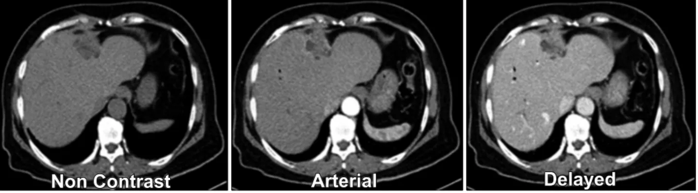
Fig. 3. Post liver resection imaging studies showing a metastatic right adrenal mass.
dence of prior hepatitis infection was found and pre- operative computed tomography (CT) scans showed mul- tiple enhancing lesions intraductally within the left intra- hepatic duct as shown in Fig. 1. She was preoperatively diagnosed with segment 4 mixed type cHCC-CC (Allen Lisa C), and underwent an S4 segmentectomy and hepa- ticojejunostomy. No additional masses were found during surgery. As illustrated in Fig. 2, the pathological result confirmed a 5-cm papillary adenocarcinoma, with intra- ductal growth, without any evidence of malignant hepa-
tocytes.
A follow-up CT scan for disease surveillance revealed a right adrenal mass that gradually enlarged, with a marked increase in serum AFP (4,950 ng/mL). Magnetic resonance imaging (MRI) and positron emission tomog- raphy (PET)-CT also suggested a metastatic lesion in the right adrenal gland (Fig. 3). A percutaneous adrenal gland biopsy confirmed a metastatic HCC (positive for AFP, glypican-3, and hepatocytes, and negative for CD-10, in- hibin-, EMA, S-100, and cytokeratin-7), and right adre-
Fig. 4. Metastatic hepatocellular carcinoma of adrenal gland. Fig. 5. Levels of AFP (ng/mL) and prothrombin in vitamin K absence II (PIVKA-II) PIVKA-II (mAU/mL) in the disease (logarithmic scale).
Fig. 6. CT at 15 months post right adrenalectomy showed intrahepateic recurrence near resected right adrenal, 17 months after transarterial chemoembolization (TACE) showed a small viable HCC and 19 months after radiofrequency ablation (RFA) showed no viable HCC.
nalectomy was performed uneventfully. The specimen is shown in Fig. 4. As shown in Fig. 5, the serum AFP im- mediately declined to 4.1 ng/mL, shortly after adrenalectomy. Unfortunately, a recurrent S7 liver mass was suspected 1 year after adrenalectomy, with serum AFP 7.6 ng/mL and CT imaging characteristic of HCC.
A series of Trans-arterial chemo-embolization (TACE) and RadioFrequency Ablation (RFA) procedures were then performed with favorable responses (Fig. 6).
DISCUSSION
cHCC-CC was first reported by Wells in 1903, and fur- ther classified by Allen and Lisa in 1949 and Goodman in 1985.5,6 It originates in hepatic progenitor cells, the liv- er-specific bipotential adult stem cells, which are activated upon injury to hepatocytes and/or cholangiocytes.7 Thus,
it has the ability to differentiate into hepatocellular carci- noma, cholangiocarcinoma, or both. A few ductal plate malformations, particularly von-Meyenburg complex, show malignant transformation to intrahepatic CC (ICC) suggesting the possibility of combined HCC and ICC originating in ductal plate malformation.8
Histophatologic diagnosis of cHCC-CC, according to World Health Organization (WHO), is characterized by an unequivocally differentiated hepatocellular and biliary component in one tumor, which correspondends to Allen Lisa C (Mixed Type) or Goodman I (Collision tumor).1 Accurate preoperative diagnosis is difficult and often leads to misdiagnosis. cHCC-CC should be highly sus- pected in cases where imaging reveals both HCC and CC features, regardless of tumor marker levels; in the pres- ence of elevated tumor markers (AFP and CA19-9, re- gardless of imaging features) or conflicting imaging and
tumor marker findings.
Initial preoperative diagnosis in our case report was cHCC-CC, based on the contradictory findings: tumor marker level (a very high AFP) and imaging features of cholangiocarcinoma (intraductal gradually enhancing le- sion within the left intrahepatic duct). However, since no additional metastatic lesions were present, liver resection and lymph node dissection was a rational choice,1,3 de- spite the fact that the recurrence rate after surgery was still higher than in hepatocellular carcinoma (disease-free survival of HCC vs. cHCC vs. ICC : 68.2 months vs 23.4 months vs 15.5 months).9
Several prognostic factors of cHCC-CC associated with unfavorable outcome include large tumor size, vascular invasion, lymph node metastasis, and the presence of sat- ellite lesions.6,9 Ariizumi et al.10 reported a median surviv- al of 15.4 months, with a 5-year overall survival rate found in 24% of all patients. Another report by Lee at al.9 showed an overall survival rate of 47.3 months, short- er than HCC (71.7 months), but longer than ICC (21.5 months).
With regards to the nature of disease recurrence and poor survival, close surveillance after surgery is an im- portant aspect of management strategy, as in HCC or ICC.
Early intra- and extra-hepatic recurrence was detected, warranting regular imaging surveillance as well as tumor marker follow-up studies. The incidence of extra-hepatic metastasis is around 13.5 to 42 %,11,12 including metastasis to adrenal glands (12%).11 Presentation of a chol- angiocellular component appeared to be a poor prognostic indicator.13
Since both adrenal glands are almost always included in the scanning area, any changes in size and glandular characteristics may be visualized to detect adrenal in- cidentalinoma,3 an adrenal tumor discovered fortuitously during imaging for other indications.14 Possibility of ma- lignancy increases with large tumor size, heterogeneous lesion and presence of peritoneal lymphadenopathy.
Because of the rich sinusioidal blood flow and its multiple arterial supplies, the adrenal gland is a potential site for metastasis.15 The mechanism of extrahepatic metastasis from primary liver tumor to adrenal glands may involve a direct extension through exophytic growth or systemi- cally through retroperitoneal venous system.12 Poor differ- entiation of malignant lesions from benign adenoma re-
mains a diagnostic challenge in the management of adre- nal incidentalinoma.16
The diagnosis of adrenal metastasis in our case was straightforward—based on the size of the lesion, history of previous malignancy, and low attenuation in pre-con- trast study. However, percutaneous biopsy was indicated for accurate histopathological diagnosis, due to conflicting results based on initial preoperative diagnosis of cHCC-CC and postoperative histopathology of ICC.
The 5-year survival rate of HCC patients with resected adrenal metastasis was reported around 20% to 45%.15 When compare to other modality or no treatment, adrena- lectomy provides the highest survival (11.1 months, 5.7 months, 21.4 months, respectively).17 Disease-free interval of less than 12 months is associated with poor survival.15
Interestingly, we detected a sequentially developed cHCC-CC (Allen Lisa A), with adrenal metastasis as the first presentation of a hidden hepatocellular carcinoma.
Although S4 mass was cholangiocarcinoma, metastasis or recurrent mass showed HCC-like features. Tsalis et al.18 first reported the case of adrenal metastasis as the first presentation of HCC in a 76-year-old male with a marked increase in tumor markers but without initial liver masses.
Pandey et al.19 reported a case of adrenal metastasis from ICC, and the patient is still alive with recurrent disease 2 years after surgery.
The sequentially developed cHCC-CC in this case was unique and invaluable although the S4 mass was chol- angiocarcinoma, whereas metastatic recurrent mass showed HCC-like figures. Our report suggests that a markedly increased level of tumor markers, after resection of a liver malignant lesion, without a visible postoperative liver mass, requires comprehensive evaluation for the pos- sibility of disease recurrence. In conclusion, adrenal meta- stasis may present as the primary focus of HCC in se- quentially developed cHCC-CC patient with hidden pri- mary HCC.
REFERENCES
1. Maximin S, Ganeshan D, Shanbhogue A, Dighe MK, Yeh MM, Kolokythas O, et al. Current update on combined hepatocellular- cholangiocarcinoma. Eur J Radiol Open 2014;1:40-48.
2. Yap AQ, Chen CL, Yong CC, Kuo FY, Wang SH, Lin CC, et al. Clinicopathological factors impact the survival outcome fol- lowing the resection of combined hepatocellular carcinoma and cholangiocarcinoma. Surg Oncol 2013;22:55-60.
3. Park JS, Yoon DS, Kim KS, Choi JS, Lee WJ, Chi HS, et al.
What is the best treatment modality for adrenal metastasis from hepatocellular carcinoma? J Surg Oncol 2007;96:32-36.
4. O'Connor K, Walsh JC, Schaeffer DF. Combined hepatocellular- cholangiocarcinoma (cHCC-CC): a distinct entity. Ann Hepatol 2014;13:317-322.
5. Allen RA, Lisa JR. Combined liver cell and bile duct carcinoma. Am J Pathol 1949;25:647-655.
6. Yap AQ, Chen CL, Yong CC, Kuo FY, Wang SH, Lin CC, et al. Clinicopathological factors impact the survival outcome fol- lowing the resection of combined hepatocellular carcinoma and cholangiocarcinoma. Surg Oncol 2013;22:55-60.
7. Akiba J, Nakashima O, Hattori S, Tanikawa K, Takenaka M, Nakayama M, et al. Clinicopathologic analysis of combined hep- atocellular-cholangiocarcinoma according to the latest WHO classification. Am J Surg Pathol 2013;37:496-505.
8. Terada T. Combined hepatocellular-cholangiocarcinoma ith stem cell features, ductal plate malformation subtype: a case report and proposal of a new subtype. Int J Clin Exp Pathol 2013;6:
737-748.
9. Lee WS, Lee KW, Heo JS, Kim SJ, Choi SH, Kim YI, et al.
Comparison ofcombined hepatocellular and cholangiocarcinoma with hepatocellular carcinoma and intrahepatic cholangiocar- cinoma. Surg Today 2006;36:892-897.
10. Ariizumi S, Kotera Y, KatagiriS, Nakano M, Yamamoto M.
Combined hepatocellular-cholangiocarcinoma had poor outcomes after hepatectomy regardless of Allen and Lisa class or the pre- dominance of intrahepatic cholangiocarcinoma cells within the tumors. Ann Surg Oncol 2012;19:1628-1636.
11. Yasaka K, Gonoi W, Akai H, Katsura M, Akahane M, Kiryu S, et al. Differentiation of adrenal tumors in patients with hep- atocellular carcinoma: adrenal adenoma versus metastasis. Eur J Radiol 2013;82:1213-1218.
12. Chua TC, Morris DL. Exploring the role of resection of extra- hepatic metastasis from hepatocellular carcinoma. Surg Oncol 2012;21:95-101.
13. Park H, Choi KH, Choi SB, Choi JW, Kim DY, Ahn SH, et al. Clinicopathological characteristics in combined hep- atocellular-cholangiocarcinoma: a single center study in Korea.
Yonsei Med J 2011;52:753-760.
14. Germain A, Klein M, Brunaud L. Surgical management of adre- nal tumors. J Visc Surg 2011;148:e250-e261.
15. Sancho JJ, Triponez F, Montet X, Sitges-Serra A. Surgical man- agement of adrenal metastases. Langenbecks Arch Surg 2012;
397:179-194.
16. Birsen O, Akyuz M, Dural C, Aksoy E, Aliyev S, Mitchell J, et al. A new risk stratification algorithm for the management of patients with adrenal incidentalomas. Surgery 2014;156:959-965.
17. Ha TY, Hwang S, Ahn C, Kim KH, Lee YJ, Moon DB, et al.
Resection of metachronous adrenal metastasis after liver re- section and transplantation for hepatocellular carcinoma. Dig Surg 2014;31:428-435.
18. Tsalis K, Zacharakis E, Sapidis N, Lambrou I, Zacharakis E, Betsis D. Adrenal metastasis as first presentation of hep- atocellular carcinoma. World J Surg Oncol 2005;3:50.
19. Pandey D, Lee KH, Wong SY, Tan KC. Adrenal metastasis from intrahepatic cholangiocarcinoma. Liver Int 2007;27:1016.