Mainly Adrenal Gland Involving NK/T-Cell Nasal Type Lymphoma Diagnosed with Delay due to Mimicking Adrenal Hemorrhage
A 29-yr-old man, presented with abdominal pain and fever, had an initial computed tomography (CT) scan revealing low attenuation of both adrenal glands. The initial concern was for tuberculous adrenalitis or autoimmune adrenalitis combined with adrenal
hemorrhage. The patient started empirical anti-tuberculous medication, but there was no improvement. Enlargement of cervical lymph nodes were developed after that and excisional biopsy of cervical lymph nodes was performed. Pathological finding of excised lymph nodes was compatible to NK/T-cell lymphoma. The patient died due to the progression of the disease even after undergoing therapeutic trials including
chemotherapy. Lymphoma mainly involving adrenal gland in the early stage of the disease is rare and the vast majority of cases that have been reported were of B-cell origin. From this case it is suggested that extra-nodal NK/T-cell lymphoma should be considered as a cause of bilateral adrenal masses although it is rare.
Key Words: Bilateral adrenal masses; Extra-nodal lymphoma; Natural killer (NK)/T-cell nasal type lymphoma
Seon Mee Kang1, Woong Ji Kim2, Kyung Ae Lee2, Hong Sun Baek2, Tae Sun Park2 and Heung Yong Jin2
1Division of Endocrinology and Metabolism, Department of Internal Medicine, Seoul National University Bundang Hospital, Seongnam; 2Division of Endocrinology and Metabolism, Department of Internal Medicine, Research Institute of Clinical Medicine, Chonbuk National University Medical School, Jeonju, Korea
Received: 13 January 2011 Accepted: 22 March 2011 Address for Correspondence:
Heung Yong Jin, MD
Division of Endocrinology and Metabolism, Department of Internal Medicine, Chonbuk National University Medical School, 20 Geonji-ro, Deokjin-gu, Jeonju 561-712, Korea
Tel: +82.63-250-2474, Fax: +82.63-254-1609 E-mail: [email protected]
http://dx.doi.org/10.3346/jkms.2011.26.10.1386 • J Korean Med Sci 2011; 26: 1386-1390
INTRODUCTION
Primary extra-nodal lymphoma accounts for one-third of all lymphomas and often involves GI tract, head and neck, bone and skin, lung, and rarely adrenal glands (1). Secondary involve- ment of the adrenal gland with non-Hodgkin’s lymphoma (NHL) has been reported to occur in up to 25% of NHL patients during the course of the disease (2). However, primary adrenal lympho- ma (PAL) or lymphoma involving adrenal gland mainly in the early stage of disease is extremely rare. PAL may present with adrenal insufficiency, fever of unknown origin, or it can also be discovered incidentally on abdominal imaging (3, 4). The vast majority of previously reported cases of PAL or secondary in- volvement of the adrenal gland with NHL were of B-cell origin (1, 3-5).
Here, we report a case of natural killer (NK)/T-cell nasal type lymphoma mainly involving adrenal gland similar to PAL and it was diagnosed with delay due to mimicking adrenal hemorrhage.
CASE DESCRIPTION
A 29-yr-old man was admitted to our hospital after several weeks of lumbar pain and fever up to 38°C on January 19, 2007. He had no significant past medical history. His vital signs were: blood
pressure 130/80 mmHg, heart rate 121/min, and body temper- ature 38.2°C. Upon physical examination, there was no lymph- adenopathy or hepatosplenomegaly. Laboratory tests revealed anemia (hematocrit (Hct) 31.8%, hemoglobin (Hb) 11.8 g/dL), white blood cell (WBC) 3.92 × 10³/μL, elevated aspartate amino- transferase (AST) 78 IU/L and alanine aminotransferase (ALT) 71 IU/L, and markedly elevated lactate dehydrogenase (LDH) 1,591 IU/L. A computed tomography (CT) scan taken prior to admission revealed low attenuation of both adrenal glands, an indication of adrenal hemorrhage or acute adrenalitis (Fig. 1A).
At that time, serum cortisol level was 17.47 μg/dL at 8 AM and ACTH level was 12.1 pg/mL (normal range: 10-90). Chest radi- ography taken at our emergency department showed left sided pleural effusion (Fig. 1B, C). Pleural fluid was aspirated and pleu- ral needle biopsy showed that the fluid was exudate with lym- phocyte predominance and the level of adenosine deaminase (ADA) was 93 IU/L (normal range: 4.3-20.3). Stain for acid-fast bacilli and polymerase chain reaction (PCR) assay on the pleu- ral fluid was negative and pleural biopsy specimen was inade- quate. Magnetic resonance imaging (MRI) of the abdomen sug- gested of adrenal hemorrhage or adrenalitis necessary for differ- ential diagnosis of adrenal tuberculosis (Fig. 2). Therefore, the initial concern was for tuberculous pleurisy and tuberculous adrenalitis, or autoimmune adrenalitis combined with adrenal
A B C Fig. 1. Outside computed tomography (CT) scan of the abdomen and chest radiography taken at admission. (A) CT scan reveals the enlargement surrounding both adrenal glands, especially left side and hyperattenuating fat (arrows) which is suggesting inflammatory condition or hemorrhage. On the chest radiographys, blunting of left costophrenic angle (arrowhead) on the posteroanterior view (B) and fluid shifting (arrowheads) in the left decubitus view (C) demonstrate the presence of pleural fluid in left pleural space.
There is no abnormal consolidative or mass-like lesion in both lung fields.
A B
Fig. 2. Magnetic resonance imaging (MRI) of the abdomen. The enlargement surrounding both adrenal glands which is aggravated compared with the previous CT scan. The isosignal intensity on T1-weighted image (A) and subtle low signal intensity on T2-weighted image (B) around both adrenal glands suggest adrenal hemorrhage or adrenalitis such as adrenal tuberculosis.
A B
Fig. 3. Follow up CT scan of the abdomen. (A) The en- largement surrounding both adrenal glands with low at- tenuation which is more ag- gravated and extended to the retroperitoneal space com- pared with the previous CT scan and MRI. (B) The en- largement of multiple para- aortic lymph nodes which are not evident in the previous CT scan are detected on this study.
hemorrhage. The patient was started on empirical anti-tuber- culous treatment, although there was no direct evidence of My- cobacterium tuberculosis due to the fever and his worsening con-
dition. However, his fever and abdominal pain continued and there was no improvement in his conditin. Ten days after ad- mission, pancytopenia (WBC 2.88 × 10³/μL, Hb 10.7 g/dL, plate-
lets 129 × 103/μL) developed. Bone marrow biopsy was negative for specific hematologic or malignant disorder. Another CT scan of the abdomen revealed an enlargement of the previous adre- nal lesion which extended to the retroperitoneal space (Fig. 3).
Twenty-two days after admission, explorative laparotomy tar- geting the adrenal lesion was performed and some tissue was obtained, which contained only necrotic debris. Despite con- servative management, his condition deteriorated progressive- ly. At that time, with a strong suspicion that he had a malignant disorder, positron emission tomography/CT (PET/CT) scan was taken and it revealed both adrenal gland and multiple cervical, mediastinal and abdominal lymph nodes with intense FDG up- take (Fig. 4). Therefore, a cervical lymph node was excised surgi- cally after thirty eight days from admission. The pathologic find- ing was a monotonous arrangement of small tumor cells with positive nuclear staining with Ki-76 (MIB-1), and immunohis- tochemical staining was positive for CD45, CD30, and CD56 ac-
tivity, but negative for CD20 activity (Fig. 5). These findings con- firmed the diagnosis of non-Hodgkin T-cell lymphoma, natural killer (NK)/T-cell, nasal type. At last, he was referred to the he- mato-oncology department and received combination chemo- therapy of ifosfamide, mesothrexate, etoposide (IMVP-16) and prednisolone forty seven days after admission. Unfortunately, he could not receive further cycles of chemotherapy after first cycle of treatment because of postoperative wound infection and continuous fever. His disease was aggravated despite of the chemotherapy with conservative management, and finally he died on March 20, 2007, 59 days after his first admission.
DISCUSSION
Primary adrenal lymphoma (PAL) or lymphoma involving main- ly adrenal gland without regional lymph node involvement in the early stage of the disease which is difficult to discriminate from PAL is a very rare extra-nodal lymphoma. Patients with ad- renal lymphoma may present with local symptoms such as ab- dominal/lumbar pain or systemic symptoms including fever, fatigue, and weight loss (1, 3-5). Approximately 50% of patients with adrenal lymphoma including PAL experience symptoms of adrenal insufficiency such as vomiting, marked fatigue, skin pigmentation, and hypotension (5). On the other hand, PAL can be discovered incidentally on abdominal imaging (6-8). How- ever, there is no pathognomic appearance on imaging studies to indicate lymphomatous involvement of the adrenal glands (5, 7). When adrenal masses are bilateral, several diagnoses includ- ing cortical adenoma, pheochromocytoma, metastatic disease, lymphoma, infection (e.g., tuberculosis, cryptococcosis etc.), bi- lateral adrenal hemorrhage, and adrenocorticotropic hormone (ACTH)-dependent Cushing’s syndrome are considered (9, 10).
Fig. 4. Positron emission tomography (PET)/CT scan. Variable intensities of FDG up- take is detected in both adrenal glands and surrounding retroperitoneal space, and in the cervical, mediastinal and paraaortic lymph nodes.
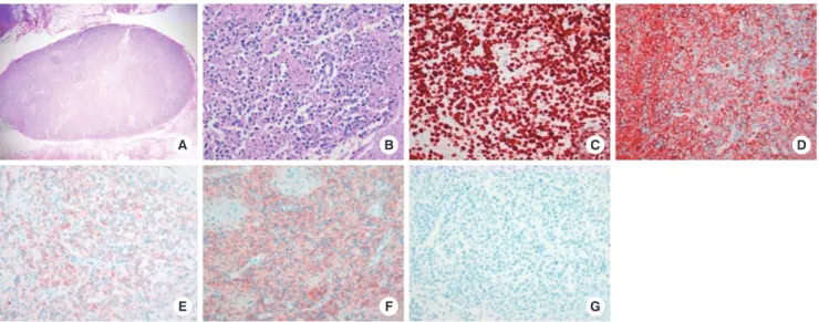
A B C D
E F G
Fig. 5. Pathological finding of surgically excised cervical lymph node. (A) The lymph node is well capsulated with marked degree of necrosis (H&E, × 100), (B) Neoplastic infil- trate of relatively pleomorphic lymphoid cells with scanty cytoplasm, irregular nuclear contour, and prominent nucleoli (H&E, × 400). Immunohistochemical staining demonstrate positive nuclear staining with Ki-67 (MIB-1) (C), and positive for CD45 (D), CD30 (E) and CD56 (F), but negative CD20 activity (G).
Therefore, confirmative diagnosis of PAL is commonly estab- lished by image-guided percutaneous biopsy, surgical explora- tion, or postmortem examination (11-13). As the initial presen- tation of our patient’s tumor in the adrenal gland similar as PAL is extremely rare, our first impression was tuberculous adrenal- itis combined with hemorrhage rather than lymphoma mainly involving adrenal glands.
The definitive diagnosis of tuberculous pleural effusion de- pends on the demonstration of M. tuberculosis in the sputum, pleural fluid, or pleural biopsy specimens (14). The diagnosis can also be established with reasonable certainty by demonstrat- ing granuloma in the parietal pleura or an elevated adenosine deaminase (ADA) level in pleural fluid in the adequate clinical context (15). The most widely accepted cutoff value for pleural fluid ADA is 40 U/L, and higher ADA level is associated with a greater chance of a patient having tuberculosis. Our patient’s pleural fluid was exudate with lymphocyte predominance and ADA level was 93 IU/L. In addition to this pleural fluid finding, the regional characteristic of Korea that tuberculosis is quite pop- ular guided us to make a wrong diagnosis.
Histopathologically, the most common type of PAL or lympho- ma involving adrenal gland is diffuse large B-cell (1, 3-5). Although extranodal NK/T-cell lymphomas are more common in Asia, especially in Korea compared to Western countries (16), prima- ry adrenal NK/T-cell lymphoma or massive adrenal involvement in the early course of diseases is extremely rare. On literature review, we found a few similar cases of NK/T-cell, nasal type lym- phoma involving mainly adrenal gland and the information are listed in Table 1. Unlike upper aerodigestive tract NK/T-cell lym- phoma, extra-upper aerodigestive tract NK/T-cell lymphomas
are often multifocal and pursue an aggressive course (17).
Treatment options for PAL include surgery, combination che- motherapy, surgery followed by chemotherapy and radiothera- py (5). However, the optimal treatment for NK/T-cell lymphoma has not been established yet (18). Glucocorticoid replacement therapy is mandatory when adrenal insufficiency is present. The prognosis of PAL is very poor compared to that of other types of extra-nodal NHL; most follow-up studies showed that patients die of tumor, intercurrent disease or infection within 1 yr of diag- nosis (5, 11, 12, 19). In our case, the patient was treated with com- bination chemotherapy of ifosfamide, mesothrexate, etoposide (IMVP-16). However, he could receive only one cycle of chemo- therapy due to infectious complication and progression of the disease. At admission, there was no evidence of adrenal insuffi- ciency on corticotropin simulation test. As time passes, subse- quent test revealed adrenal insufficiency after admission; we started prednisolone for adrenal hormone supplementation.
Eventually, he expired 59 days after admission, and 22 days after confirmative diagnosis. The rapid deterioration of our patient was similar to patients described in other studies.
NK/T-cell lymphoma is strongly associated with Epstein Barr virus (EBV), suggesting a pathogenic role for the virus (16, 20).
In blood laboratory findings, any of the antibodies to antigen complex, including IgM anti-EBVCA (viral capsid antigen), IgM anti-EBEA (early antigen), and IgM EBNA (nuclear antigen) were not found. However, laboratory tests were not always foolproof.
Unfortunately, we did not perform an analysis of EBV terminal repeat region from excised lymph node specimens. Therefore, we could not definitively conclude whether the patient’s disease was associated with EBV infection or not.
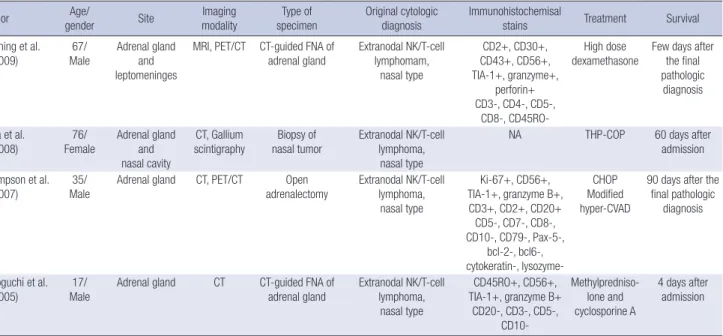
Table 1. Summary of previously reported cases of extranodal NK/T-cell, nasal type lymphoma involving adrenal gland
Author Age/
gender Site Imaging
modality Type of
specimen Original cytologic
diagnosis Immunohistochemisal
stains Treatment Survival
Dunning et al.
(2009)
67/
Male
Adrenal gland and leptomeninges
MRI, PET/CT CT-guided FNA of adrenal gland
Extranodal NK/T-cell lymphomam,
nasal type
CD2+, CD30+, CD43+, CD56+, TIA-1+, granzyme+,
perforin+
CD3-, CD4-, CD5-, CD8-, CD45RO-
High dose dexamethasone
Few days after the final pathologic
diagnosis
Toba et al.
(2008)
76/
Female
Adrenal gland and nasal cavity
CT, Gallium scintigraphy
Biopsy of nasal tumor
Extranodal NK/T-cell lymphoma,
nasal type
NA THP-COP 60 days after
admission Thompson et al.
(2007) 35/
Male Adrenal gland CT, PET/CT Open
adrenalectomy Extranodal NK/T-cell lymphoma,
nasal type
Ki-67+, CD56+, TIA-1+, granzyme B+,
CD3+, CD2+, CD20+
CD5-, CD7-, CD8-, CD10-, CD79-, Pax-5-,
bcl-2-, bcl6-, cytokeratin-, lysozyme-
CHOP Modified hyper-CVAD
90 days after the final pathologic
diagnosis
Mizoguchi et al.
(2005)
17/
Male
Adrenal gland CT CT-guided FNA of adrenal gland
Extranodal NK/T-cell lymphoma,
nasal type
CD45RO+, CD56+, TIA-1+, granzyme B+
CD20-, CD3-, CD5-, CD10-
Methylpredniso- lone and cyclosporine A
4 days after admission
THP-COP: pirarubicin, cyclophosphamide, vincristine, prednisolone; CHOP: cyclophosphamide, doxorubicin, vincristine, prednisolone; CVAD: cyclophosphamide, vincristine, doxorubicin, dexamethasone. MRI, Magnetic resonance imaging; CT, Computed tomography; FNA, Fine needle aspiration.
We described the unusual case of adrenal NK/T-cell nasal type lymphoma diagnosed with delay due to mimicking adrenalitis with hemorrhage which was difficult in the discrimination from adrenal tuberculosis. Adrenal lymphoma, although PAL or dom- inantly adrenal gland involvement is rare, should be considered in the differential diagnosis for an adrenal mass. In addition, ex- tranodal NK/T-cell lymphoma also should be considered in Asian area.
REFERENCES
1. AlShemmari SH, Ameen RM, Sajnani KP. Extranodal lymphoma: a com- parative study. Hematology 2008; 13: 163-9.
2. Rosenberg SA, Diamond HD, Jaslowitz B, Craver LF. Lymphosarcoma:
a review of 1269 cases. Medicine (Baltimore) 1961; 40: 31-84.
3. Mantzios G, Tsirigotis P, Veliou F, Boutsikakis I, Petraki L, Kolovos J, Pa- pageorgiou S, Robos Y. Primary adrenal lymphoma presenting as Addi- son’s disease: case report and review of the literature. Ann Hematol 2004;
83: 460-3.
4. Levy NT, Young WF Jr, Habermann TM, Strickler JG, Carney JA, Stanson AW. Adrenal insufficiency as a manifestation of disseminated non-Hodg- kin’s lymphoma. Mayo Clin Proc 1997; 72: 818-22.
5. Grigg AP, Connors JM. Primary adrenal lymphoma. Clin Lymphoma 2003; 4: 154-60.
6. van den Heiligenberg SM, van Groeningen CJ. Bilateral adrenal enlarge- ment with an unexpected diagnosis. Eur J Intern Med 2007; 18: 249-50.
7. Wang J, Sun NC, Renslo R, Chuang CC, Tabbarah HJ, Barajas L, French SW. Clinically silent primary adrenal lymphoma: a case report and review of the literature. Am J Hematol 1998; 58: 130-6.
8. Wang FF, Su CC, Chang YH, Pan CC, Tang KT, Jap TS, Lin HD, Won J.
Primary adrenal lymphoma manifestating as adrenal incidentaloma. J Chin Med Assoc 2003; 66: 67-71.
9. Arora S, Vargo S, Lupetin AR. Computed tomography appearance of spon-
taneous adrenal hemorrhage in a pheochromocytoma. Clin Imaging 2009; 33: 314-7.
10. Nawar R, Aron D. Adrenal incidentalomas: a continuing management dilemma. Endocr Relat Cancer 2005; 12: 585-98.
11. Dunning KK, Wudhikarn K, Safo AO, Holman CJ, McKenna RW, Pam- buccian SE. Adrenal extranodal NK/T-cell lymphoma diagnosed by fine- needle aspiration and cerebrospinal fluid cytology and immunopheno- typing: a case report. Diagn Cytopathol 2009; 37: 686-95.
12. Thompson MA, Habra MA, Routbort MJ, Holsinger FC, Perrier ND, Waguespack SG, Rodriguez MA. Primary adrenal natural killer/T-cell nasal type lymphoma: first case report in adults. Am J Hematol 2007; 82:
299-303.
13. Tomoyose T, Nagasaki A, Uchihara JN, Kinjo S, Sugaya K, Onaga T, Ohshi- ma K, Masuda M, Takasu N. Primary adrenal adult T-cell leukemia/lym- phoma: a case report and review of the literature. Am J Hematol 2007;
82: 748-52.
14. Gopi A, Madhavan SM, Sharma SK, Sahn SA. Diagnosis and treatment of tuberculous pleural effusion in 2006. Chest 2007; 131: 880-9.
15. Porcel JM. Tuberculous pleural effusion. Lung 2009; 187: 263-70.
16. Ko YH, Ree HJ, Kim WS, Choi WH, Moon WS, Kim SW. Clinicopatholog- ic and genotypic study of extranodal nasal-type natural killer/T-cell lym- phoma and natural killer precursor lymphoma among Koreans. Cancer 2000; 89: 2106-16.
17. Lee J, Kim WS, Park YH, Park SH, Park KW, Kang JH, Lee SS, Lee SI, Lee SH, Kim K, Jung CW, Ahn YC, Ko YH, Park K. Nasal-type NK/T cell lym- phoma: clinical features and treatment outcome. Br J Cancer 2005; 92:
1226-30.
18. Kohrt H, Advani R. Extranodal natural killer/T-cell lymphoma: current concepts in biology and treatment. Leuk Lymphoma 2009; 50: 1773-84.
19. Mizoguchi Y, Nakamura K, Miyagawa S, Nishimura S, Arihiro K, Ko- bayashi M. A case of adolescent primary adrenal natural killer cell lym- phoma. Int J Hematol 2005; 81: 330-4.
20. Kwong YL. Natural killer-cell malignancies: diagnosis and treatment.
Leukemia 2005; 19: 2186-94.