INTRODUCTION
Tumor-infiltrating lymphocytes (TILs) can be found in most cancers with a variable intensity. They are mainly com- posed of CD8+ cytotoxic T-cells, CD4+ helper T-cells and NK cells (1). Although these cells exert antitumor activity, they usually fail to control tumor growth due to other factors such as the presence of regulatory cells and the expression of inhibitory ligands of the tumor cells. The association between TILs and a favorable prognosis has been demonstrated in a few tumors including melanoma and colon cancer (2).
Likewise, in hepatocellular carcinoma, it is known that CD8+ TILs may play a role in the occurrence of tumor cell apoptosis (3), however, CD4+CD25+ regulatory T-cells impair the effector function of them (4, 5). Although there are a few reports that describe hepatocellular carcinoma with lymphoid rich stroma, so called lymphoepithelioma-like hepatocellular carcinoma (6, 7), the phenotypic analysis of TILs in 28 cases of hepatocellular carcinoma revealed that hepatocellular carcinoma tissues had less intensity of lym- phocyte infiltration than the corresponding non-tumor liver
tissues with increased CD4+CD25+ regulatory T-cells.
Here we report an unusual type of TIL composed of imma- ture T-cells in a patient with hepatocellular carcinoma. Because almost TILs in hepatocellular carcinoma are activated type and expressed antigen-experienced phenotypes (8) this find- ing is very exceptional and up to now only one similar case was previously reported. This case further suggests that hep- atocellular carcinoma could occur in association with indo- lent T-lymphoblastic proliferation.
CASE REPORT
A 58-yr-old female patient presented with indigestion and a palpable epigastric mass for two months. The patient was diagnosed as being a hepatitis B s-antigen (HBsAg) carrier 15 yr previously. However, she received no medical treatment.
The patient had no history of excessive alcohol intake. The serum alpha-1-fetoprotein measured 17,400 ng/mL. The white blood cell count was 2,600/mL, and the platelet count was 86,000/mL. The abdominal computed tomography (CT) scan
309
Shin Eun, Youn Kyung Jeon, and Ja June Jang
Department of Pathology, Seoul National University College of Medicine, Seoul, Korea
Address for Correspondence Ja June Jang, M.D.
Department of Pathology, Seoul National University College of Medicine, 28 Daehak-ro, Jongno-gu, Seoul 110-799, Korea
Tel : +82.2-740-8271, Fax : +82.2-3673-5046 E-mail : [email protected]
J Korean Med Sci 2010; 25: 309-12 ISSN 1011-8934
DOI: 10.3346/jkms.2010.25.2.309
Hepatocellular Carcinoma with Immature T-Cell (T-lymphoblastic) Proliferation
Indolent T-lymphoblastic proliferation has been rarely reported in the upper aerodi- gestive tract. The lymphoid cells associated with this condition have the morpho- logical and phenotypical features of immature thymocytes. However, their patho- genesis and biology are unknown. We present an unusual type of tumor infiltrating lymphocytes in a case with hepatocellular carcinoma, presumed to be a T-lym- phoblastic proliferation. A 58-yr-old female patient presented with indigestion and a palpable epigastric mass. The abdominal computed tomography revealed a mass in the S6 region of the liver. A hepatic segmentectomy was performed. Microscop- ic examination showed dense isolated nests of monomorphic lymphoid cells within the tumor. Immunohistochemically, the lymphoid cells were positive for CD3, ter- minal deoxymucleotide transferase (TdT) and CD1a. In addition, they showed dual expression of CD4 and CD8. The polymerase chain reaction used to examine the T-cell antigen receptor gamma gene rearrangement showed polyclonal T-cell pro- liferation. This is the second case of hepatocellular carcinoma combined with indo- lent T-lymphoblastic proliferation identified by an unusual tumor infiltrating lympho- cytes.
Key Words : Lymphocytes, Tumor-Infiltrating; Carcinoma, Hepatocellular; indolent T-lymphoblastic prolifera- tion; Immunohistochemistry
Received : 1 March 2008 Accepted : 27 August 2008
ⓒ 2010 The Korean Academy of Medical Sciences.
This is an Open Access article distributed under the terms of the Creative Commons Attribution Non-Commercial License (http://creativecommons.org/licenses/by-nc/3.0) which permits unrestricted non-commercial use, distribution, and reproduction in any medium, provided the original work is properly cited.
revealed an exophytic mass in the S6 region of the liver. The tumor was confined to the liver; the other organs showed no abnormalities. The patient received hepatic segmentectomy.
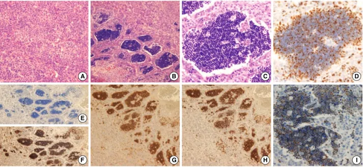
Grossly the tumor measured 5 cm in diameter and had a multinodular confluent pattern with a yellow-tan color. The microscopic examination revealed a poorly differentiated hep- atocellular carcinoma (Edmonson grade 3) with a macrotra- becular and acinar pattern (Fig. 1A). At the periphery, the tumor showed poorer differentiation (Edmonson grade 4) and many giant cells. The surrounding non-neoplastic liver paren- chyma was replaced by cirrhotic nodules, 3-5 mm in diam- eter. Notably, there were foci of extensive infiltrations of lym- phoid cells within the tumor. The lymphoid cells occasion- ally formed dense isolated nests between the cords of tumor cells (Fig. 1B). The lymphoid cells were small with a round nucleus and had scanty cytoplasm. The nucleus was basophilic with indistinct nucleoli. They showed monomorphic features with little cytologic atypism (Fig. 1C). On the immunohis- tochemical staining, the monomorphic lymphoid cells were positive for CD3 (Fig. 1F), terminal deoxynucleotide trans- ferase (TdT) (Fig. 1I) and CD1a. They showed dual expres- sion of CD4 and CD8 (Fig. 1G, H). The above morphologi- cal and immunohistochemical features of the lymphoid cells were consistent with the phenotype of immature T-lympho- blasts. They were all negative for L26 (Fig. 1E), CD79a, gran- zyme, CD68, CD5, and c-kit. They had a relatively high Ki- 67 labeling index (approximately more than 40%) (Fig. 1D).
The polymerase chain reaction to examine the T-cell antigen receptor gamma gene rearrangement, revealed polyclonal T-
cell proliferation.
On the other hand, in the peripheral portion of the tumor, small numbers of polymorphic TILs composed of mature T or B lymphocytes or plasma cells were observed. Some of the polymorphic TILs showed limited infiltration into the por- tal area and lobules of the hepatic parenchyma around the tumor.
Two months later, the patient presented with a chest wall mass. The radiological examination showed recurrence of the tumor in the liver with portal vein involvement, multi- ple lung and rib metastases. Although biopsy was not per- formed, the CT scan favored the possibility of aggravation of hepatocellular carcinoma and the serum alpha-1-fetopro- tein measured 15,000 ng/mL at that time. The operability of the tumor was very low so the patient is receiving conser- vative management.
DISCUSSION
The porcine species is known to have a large percentage of CD4 and CD8 dual expressing peripheral T-cells (9). How- ever, in humans, CD4 and CD8 dual expressing T-cells can be found in thymic cortex in which reside the immature thymic T-lymphocytes, and extrathymic precursor T-cells and T-lym- phoblastic lymphoma/leukemia. Positivity for TdT is observed in cortical thymocytes and immature bone marrow T and B cell precursors undergoing antigen receptor gene rearrange- ment. The immunophenotype found in the lymphocytes of
310 S. Eun, Y.K. Jeon, and J.J. Jang
A B C D
E
F G H I
Fig. 1. Histological and immunohistochemical features. (A) The hepatocellular carcinoma is composed of poorly differentiated cells with trabecular pattern (H&E, ×200). (B) Aggregates of monomorphic lymphoid cells are observed within the hepatocellular carcinoma (H&E,
×100). (C) The lymphocytes show a morphology similar to immature thymocytes (H&E, ×400). (D) Ki67 labeling index is approximately 40% (H&E, ×400). (E) The lymphoid cells are negative for L26 (H&E, ×100). (F) The lymphoid cells are positive for CD3 (H&E, ×100).
(G, H) The lymphoid cells show dual expression of CD4 and CD8 (G, CD4; H, CD8) (H&E, ×100). (I) TdT are expressed in lymphoid cells (H&E, ×400).
the present case was the same as that of precursor (immature) T-cell or T-lymphoblastic lymphoma/leukemia. In addition, they showed a high Ki-67 proliferation index, which made us to considered the possibility of neoplastic precursor T cell proliferation.
T-lymphoblastic lymphoma/leukemia is a high-grade neo- plasm composed of small to medium sized blastic cells. Pati- ents with T-lymphoblastic lymphoma/leukemia frequently present with a mediastinal mass and rapidly progressive dis- ease if untreated. In our case, the proliferating lymphocytes were morphologically and immunohistochemically T lym- phoblasts. However, the presentation was apart from the usual T-lymphoblastic lymphoma. In addition, the polymerase chain reaction study did not show monoclonal T-cell proliferation.
Therefore, the possibility of T-lymphoblastic lymphoma/
leukemia was excluded.
The liver plays an important role in innate and adaptive immunity through the production of acute phase proteins, phagocytosis and removal of activated T-cells (11). Extra- thymic T-cell development and proliferation also could be occur in the mammalian liver, and this function becomes more important with aging (10-12). Therefore, the hepatic T-cell population is quite different from those of other organs.
For example, unlike the peripheral blood, the conventional CD4+ or CD8+ T-cells account only 40% of hepatic CD3+
T-cells. However, CD4+CD8+ dual expressing T-cells and CD4-CD8- dual negative T-cells account for a significant proportion of hepatic T-cells (13). IL-7 induced proliferation of T-cells with intermediate levels of TCR and CD4/CD8 dual negative or CD4 positive phenotype in the murine liver (14).
In the human liver, the hepatic microenvironment including cytokines might influence the distribution of T-cell popula- tions, and increased production of certain cytokines might result in selective expansion of the resident population of T- cells under pathological conditions. In the present case, it would be possible that certain cytokines produced by trans- formed hepatocytes of hepatocellular carcinoma, might have stimulated the expansion of resident CD4+CD8+ dual ex- pressing immature T-cells, or recruited them from the thymic cortex or bone marrow.
Wang et al. previously reported one similar case. They pre- sented a case with hepatocellular carcinoma accompanied by TdT positive T lymphocyte proliferation in the tumor. They suggested that their patient had indolent T-lymphoblastic proliferation combined with hepatocellular carcinoma (15).
Indolent T-lymphoblastic proliferation was first described by Velankar et al. in 1999 in the upper aerodigestive tract (16).
Two years later Strauchen reported a similar phenomenon in the oropharynx in a myasthenia gravis patient (17). In this newly introduced disorder, the infiltrating lymphocytes show- ed lymphoblastic morphology and expressed TdT, and both CD4 and CD8. Although the two patients suffered from mul- tiple recurrences over a long time, they showed no systemic dissemination with or without chemotherapy. Whether this
disorder is a neoplastic proliferation or true hyperplasia of T- lymphoblasts remains unclear. Strauchen reported that the tro- pism of the T-lymphoblasts of the oropharynx due to retained thymic potential of the pharyngeal epithelium might cause the indolent T-lymphoblastic proliferation (17). Wang sug- gested that hepatocellular carcinoma could recruit immature T-lymphocytes into the tumor from the thymic cortex or bone marrow through unknown mechanisms (15).
The clinical course of the present case was more aggressive than that of Wang’s case and showed multiple metastases two months after the initial diagnosis and treatment. If the immature T-cell proliferation accompanying the hepatocel- lular carcinoma was related to a poor patient prognosis should be clarified by further study.
In conclusion, we have reported a second case of hepato- cellular carcinoma with T-cell lymphoblastic proliferation as a type of unusual TIL. The precise mechanism of the T lymphoblastic proliferation in the hepatocellular carcinoma and the clinicopathological significance require further elu- cidation.
REFERENCES
1. Chiou SH, Sheu BC, Chang WC, Huang SC, Hong-Nerng H. Cur- rent concepts of tumor-infiltrating lymphocytes in human malignan- cies. J Reprod Immunol 2005; 67: 35-50.
2. Liakou CI, Narayanan S, Ng Tang D, Logothetis CJ, Sharma P. Focus on TILs: prognostic significance of tumor infiltrating lymphocytes in human bladder cancer. Cancer Immunol 2007; 7: 10-5.
3. Ikeguchi M, Oi K, Hirooka Y, Kaibara N. CD8+ lymphocyte infiltra- tion and apoptosis in hepatocellular carcinoma. Eur J Surg Oncol 2004; 30: 53-7.
4. Fu J, Xu D, Liu Z, Shi M, Zhao P, Fu B, Zhang Z, Yang H, Zhang H, Zhou C, Yao J, Jin L, Wang H, Yang Y, Fu YX, Wang FS. Increased regulatory T cells correlate with CD8 T-cell impairment and poor survival in hepatocellular carcinoma patients. Gastroenterology 2007; 132: 2328-39.
5. Unitt E, Rushbrook SM, Marshall A, Davies S, Gibbs P, Morris LS, Coleman N, Alexander GJ. Compromised lymphocytes infiltrate hep- atocellular carcinoma: the role of T-regulatory cells. Hepatology 2005; 41: 722-30.
6. Chen CJ, Jeng LB, Huang SF. Lymphoepithelioma-like hepatocellu- lar carcinoma. Chang Gung Med J 2007; 30: 172-7.
7. Emile JF, Adam R, Sebagh M, Marchadier E, Falissard B, Dussaix E, Bismuth H, Reyne$s M. Hepatocellular carcinoma with lymphoid stroma: a tumour with good prognosis after liver transplantation.
Histopathology 2000; 37: 523-9.
8. Chen CH, Lee HS, Huang GT, Yang PM, Yu WY, Cheng KC, Lee PH, Jeng YM, Chen DS, Sheu JC. Phenotypic analysis of tumor-infil- trating lymphocytes in hepatocellular carcinoma. Hepatogastroen- terology 2007; 54: 1529-33.
9. Pescovitz MD. Sakopoulos AG, Gaddy JA, Husmann RJ, Zucker- mann FA. Porcine peripheral blood CD4+/CD8+ dual expressing
Hepatocellular Carcinoma with T-lymphoblastic Proliferation 311
T-cells. Vet Immunol Immunopathol 1994; 43: 53-62.
10. Parker GA, Picut CA. Liver immunobiology. Toxicol Pathol 2005;
33: 52-62.
11. Sato K, Ohtsuka K, Hasegawa K, Yamagiwa S, Watanabe H, Asaku- ra H, Abo T. Evidence for extrathymic generation of intermediate T cell receptor cells in the liver revealed in thymectomized, irradiated mice subjected to bone marrow transplantation. J Exp Med 1995;
182: 759-67.
12. Ohteki T, Seki S, Abo T, Kumagai K. Liver is a possible site for the proliferation of abnormal CD3+4-8- double-negative lymphocytes in autoimmune MRL-lpr/lpr mice. J Exp Med 1990; 172: 7-12.
13. Doherty DG, O’Farrelly C. Innate and adaptive lymphoid cells in the human liver. Immunol Rev 2000; 174: 5-20.
14. Miyaji C, Watanabe H, Osman Y, Kuwano Y, Abo T. A comparison
of proliferative response to IL-7 and expression of IL-7 receptors in intermediate TCR cells of the liver, spleen, and thymus. Cell Immunol 1996; 169: 159-65.
15. Wang ZM, Xiao WB, Zheng SS, Sun K, Wang LJ. Hepatocellular carcinoma with Indolent T-lymphoblastic proliferation. Leuk Lym- phoma 2006; 47: 2424-6.
16. Velankar MM, Nathwani BN, Schlutz MJ, Bain LA, Arber DA, Slo- vak ML, Weiss LM. Indolent T-lymphoblastic proliferation: report of a case with a 16-year course without cytotoxic therapy. Am J Surg Pathol 1999; 23: 977-81.
17. Strauchen JA. Indolent T-lymphoblastic proliferation: report of a case with an 11-year history and association with myasthenia gravis.
Am J Surg Pathol 2001; 25: 411-5.
312 S. Eun, Y.K. Jeon, and J.J. Jang