92
서 론
간세포암 환자에서 간문맥의 종양전의 빈도는 높게 보고 되고 있지만 간정맥과 하대정맥을 통하여 우심방으로 연결 되는 종양전의 빈도는 극히 드물다.
하대정맥과 우심방으로 연결되는 종양전을 가지는 간세 포암은 종양전이 하대정맥에서 들어오는 혈류를 완전히 차 단하지 않는 이상 별다른 증상이 없으며 우연히 발견되는 경우가 대부분이다. 본원에서 간정맥을 통하여 하대정맥, 우심방까지 연결된 종양전을 가진 간세포암환자에서 심폐 우회술을 이용하여 간종양과 우심방내의 종양전의 절제가 가능한 예를 경험하였기에 보고하는 바이다.
증 례
63세 남자 환자로 약 10년 전 C형 감염을 진단받고 추적 관리는 하지 않았으며 하지 정맥류를 치료하기 위해 검사 중 우연히 발견된 간좌엽 종괴의 검사를 위해 내원하였다.
내원 당시 이학적 검사상 특이한 소견은 없었으며 하지 부 종은 없었으나 경미한 운동 중 호흡 곤란 증상은 있었다.
혈색소는 14.9 g/dl, 혈소판은 162 K/ul이었으며 알부민 4.1 g/dl, 총빌리루빈 1 mg/dl, AST 106 U/L, ALT 130 U/L, PT 14.7초였으며 차일드스코어는 5점이었다. Anti-HCV가 양성 이었고 태아단백혈청치는 6.45 ng/ml, PIVKA-2는 582 mIU/
ml이었다. 복부CT소견상 3번엽에 4 cm 크기의 종괴소견을 보였고 이 종괴는 좌간정맥과 중간 정맥을 통해 하대정맥
Thrombectomy of Hepatocellular Carcinoma with Tumor Thrombus Extending into Right Atrium Using Cardiopulmonary Bypass and Total Vascular Exclusion
Dong-Shik Lee, M.D., Hong-Jin Kim, M.D., Sung-Su Yun, M.D., Jang-Hoon Lee, M.D.1, Won-Kyu Park, M.D.2 and Jun-Hwan Kim, M.D.3
The extension of HCC with tumor thrombus through the hepatic veins into the right atrium can cause sudden death due to heart failure and pulmonary embolism, even though the primary tumor may be relatively localized. We report a case of hepatic resection and tumor thrombectomy of hepatocellular carcinoma with tumor thrombus extending into right atrium using cardiopulmonary bypass and total hepatic vascular exclusion. A 65-year-old man, with a diagnosis of left liver mass, visited Yeungnam University Hospital. The patient had no noticeable edema in the lower extremities, but had dyspnea on exertion. Laboratory studies revealed a platelet count of 162 K/uL, total bilirubin of 1 mg/dL, albumin of 4.1 g/dL and a prothrombin time of 14.7 second.
In addition, the retention of indocyanine green at 15 min was 13.5 %. The patient was anti-HCV was positive and his levels of alpha-fetoprotein and PIVKA-2 were 6.45 ng/mL and 582 mIU/mL, respectively. An abdominal CT scan, hepatic angiogram and echocardiogram showed a 4 cm sized HCC in the left lobe and a 4 to 5 cm sized tumor thrombus extending into the right atrium through the left and middle hepatic veins. No distant metastasis was detected in other studies. Total operative time, total duration of cardio- pulmonary bypass and cardioplegic time were 13 hours, and 55 and 24 minutes, respectively. The total hepatic vascular exclusion time was 18 minutes. Unfortunately, the patients
from hospital. (J Korean Surg Soc 2005;69:92-96) Key Words: Hepatocellular carcinoma, Tumor thrombus,
Right atrium
중심 단어: 간세포암, 종양전, 우심방
ꠏꠏꠏꠏꠏꠏꠏꠏꠏꠏꠏꠏꠏꠏꠏꠏꠏꠏꠏꠏꠏꠏꠏꠏꠏꠏꠏꠏꠏꠏꠏꠏꠏꠏꠏꠏꠏꠏꠏꠏꠏꠏꠏꠏꠏꠏꠏꠏꠏꠏ Departments of Surgery, 1Chest Surgery, 2Diagnostic radio- logy, 3Internal Medicine, Yeungnam University College of Medicine, Daegu, Korea
책임저자:김홍진, 대구광역시 남구 대명 5동 317-1번지 ꂕ 705-717, 영남대학교 의과대학 부속병원 외과학교실 Tel: 053-620-3580, Fax: 053-624-1213
E-mail: [email protected]
접수일:2005년 1월 28일, 게재승인일:2005년 4월 14일 이 논문은 2004년 6차 세계간담췌외과학회에서 비디오발표하였음.
ꠏꠏꠏꠏꠏꠏꠏꠏꠏꠏꠏꠏꠏꠏꠏꠏꠏꠏꠏꠏꠏꠏꠏꠏꠏꠏꠏꠏꠏꠏꠏꠏꠏꠏꠏꠏꠏꠏꠏꠏꠏꠏꠏꠏꠏꠏꠏꠏꠏꠏꠏꠏꠏꠏꠏꠏꠏꠏꠏꠏꠏꠏꠏꠏꠏꠏꠏꠏꠏꠏꠏꠏꠏꠏꠏꠏꠏꠏꠏꠏꠏꠏꠏꠏꠏꠏꠏꠏꠏꠏꠏꠏꠏꠏꠏꠏꠏꠏꠏꠏꠏꠏꠏꠏꠏꠏꠏꠏꠏꠏꠏꠏꠏꠏꠏ
과 우심방까지 연결되는 양상을 보였으며 우심방내에도 5
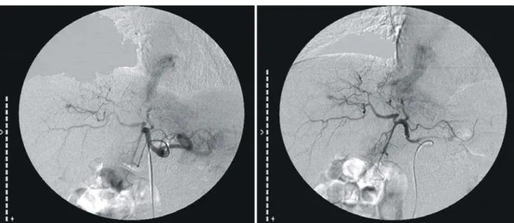
×4 cm 크기의 종괴소견을 보였다(Fig. 1). 혈관촬영상 좌간 동맥에서 혈류공급을 받는 종괴와 좌간정맥과 중간정맥으 로 과혈관성 염색소견이 보이면서 이 염색은 우심방까지 연결되어 나타나는 소견을 보였다(Fig. 2). 심장초음파검사 상 우심방에 5×4 cm의 종괴소견이 보였으며 이는 간상하 대정맥으로부터의 혈류공급을 부분적으로 차단시키는 소 견을 보였다(Fig. 3). 다른 원격부위의 전이 소견은 보이지 않았다.
수술방법: 우심방을 침범한 간세포암 진단하에 수술을 시행하였다. 전신 마취 후 술 중 경식도심초음파를 시행하 면서 종양전의 이탈을 관찰하였다. 먼저 복부 쪽에서 양측 늑골하와 정중절개를 시행하여 간을 노출시켰다. 간경변증 소견이 있었으며 복수는 관찰되지 않았다. 좌간하엽표면부 에 약 5 cm 크기의 원형 종양이 관찰되었다. 술 중 초음파를 시행하여 잔여간의 종양이 없음을 확인한 후 좌측 간동맥 과 간문맥을 결찰하여 좌측간의 혈류 공급을 차단하여 우 엽과 좌엽의 색깔의 변화로 간실질의 절제선을 결정하였으
며 미상엽에서 하대정맥으로 가는 단간정맥을 결찰하여 간 후방 하대정맥의 노출을 용이하게 하였다. 중간정맥의 우 측으로 간절제를 시행하였으며 하대정맥의 노출을 위해 미 상엽의 절제술도 시행하였다. 좌간정맥과 중간정맥의 공통 정맥(common trunk)과 간후방 하대정맥을 완전히 노출시켜 우간과 좌간을 좌간정맥과 중간정맥의 공통정맥만을 하대 정맥에 남겨 놓은 채 분리하였다. 노출된 좌간정맥 및 중간 정맥은 종양전에 의해 확장된 소견을 보였다. 하대정맥에 서 좌간정맥과 중간 정맥을 분리하지 않고 간절제를 모두 마친 후 흉부외과 수술팀이 합류하여 수술을 시행하였다.
정중흉골절개술을 시행하여 심장을 노출시킨 후 심폐혈류 우회술을 시행하기 위해 우측대퇴정맥과 상대정맥으로 정 맥도관을 삽입하였고 대동맥으로 동맥도관을 삽입하였다.
도관이 모두 완성된 후 헤파린을 정맥주사한 후 체외순환 펌프를 가동하여 체외순환을 시행하였다. 심근을 보호하기 위해 심장낭에 얼음슬러쉬를 넣어 저체온을 유발한 다음 심정지액을 투입하여 심정지를 유도하였다. 우심방 절개의 준비가 모두 끝난 후 간하대정맥과 간문맥부의 혈류를 차 Fig. 1. Computed tomography scan showed a filling defect in the right atrium and inferior vein cava and hepatocellular carcinoma in
left lateral section with faint tumor staining in the middle and left hepatic vein.
단하는 total hepatic vascular exclusion (THVE)을 시행한 후 우심방을 절개하여 암종을 확인하였다. 암종은 우심방의 공간을 반 이상 채우고 있는 노란색의 종괴로 우심방벽에 는 심한 유착이 없이 쉽게 박리되었다. 하지만 종양전이 간 실질내에서 간정맥을 거쳐 하대정맥, 우심방까지 하나로 연결이 되어 있어 하나의 종괴로는 제거가 불가능하여 먼 저 복부 쪽에서 하대정맥으로부터 간정맥을 분리시켜 간과 하대정맥 내의 종양전을 제거한 다음 우심방으로부터 우심 방의 종양전을 제거하였다. 종양전이 제거된 하대 정맥과 우심방의 벽은 잔존 종양전이 없이 깨끗하였다.
심폐혈류우회술을 통한 미상엽을 포함한 확대좌엽절제
술과 우심방절개술을 통한 암종제거를 시행하였으며 총 수 술 시간은 720분, 심폐혈류우회 시간은 55분, 저온심장정지 시간은 24분, 간의 총혈류차단 시간은 18분이었다. 총추정 실혈량은 4,500 ml이었다.
육안적 소견으로 약 4 cm 크기의 간세포암이 있었으며 이는 간정맥을 통하여 정맥내 종양전을 형성하였고 간세포 암은 Edmondson-Steiner grade II-III이었으며 부분적으로 피 막형성이 있으며 간세포암의 침범이 있었다(Fig. 4). 환자는 술 후 간부전 소견은 보이지 않았으나 수술 후 1일째부터 심낭에서 출혈 소견이 있어 신선냉동혈장과 적혈구 수혈등 의 대중적 요법으로 치료되었다. 술 후 우측늑막삼출액이 있었으며 1개월 뒤 폐렴소견이 있어 치료하였으나 성인호흡 곤란증후군(ARDS)이 발병되어 술 후 2개월 뒤 사망하였다.
고 찰
간세포암은 간문맥, 간정맥, 하대정맥, 우심방, 폐동맥과 신장정맥 등과 같은 혈관계로 침범하는 경향이 있다. 간문 맥, 간정맥과 하대정맥의 침범은 각각 70.8∼80.8%, 13.3∼
53.3%와 10.8∼13.3%로 보고되고 있으며 간세포암에 의한 종양전이 우심방까지 이어지는 경우는 약 0.67∼1.5%로 극 히 드문 경우로 보고되고 있다.(1,2) 간세포암에 의한 종양 전이 간정맥과 하대정맥을 통하여 우심방까지 연결된 경우 심부전, 부정맥, 심비대, 심낭부종과 Budd-Chiari 증후군을 유발하여 갑작스런 심정지와 폐혈전을 유발할 수 있는 응 급 상황이다. 수술 절제를 고려할 경우 수술 술기의 어려움 과 수술 절제 후에도 기저 간질환을 동반한 암종 자체에 의한 좋지 못한 예후로 인하여 만족스러운 결과는 보이지 Fig. 2. A celiac angiogram showed a hypervascular tumor fed by the left hepatic artery with A-V shunt which extended into the right
atrium.
Fig. 3. A five by four cm sized tumor thrombus showed in the right atrium. The venous return from the inferior vena cava was limited by this tumor thrombus, but not from the superior vena cava.
ꠏꠏꠏꠏꠏꠏꠏꠏꠏꠏꠏꠏꠏꠏꠏꠏꠏꠏꠏꠏꠏꠏꠏꠏꠏꠏꠏꠏꠏꠏꠏꠏꠏꠏꠏꠏꠏꠏꠏꠏꠏꠏꠏꠏꠏꠏꠏꠏꠏꠏꠏꠏꠏꠏꠏꠏꠏꠏꠏꠏꠏꠏꠏꠏꠏꠏꠏꠏꠏꠏꠏꠏꠏꠏꠏꠏꠏꠏꠏꠏꠏꠏꠏꠏꠏꠏꠏꠏꠏꠏꠏꠏꠏꠏꠏꠏꠏꠏꠏꠏꠏꠏꠏꠏꠏꠏꠏꠏꠏꠏꠏꠏꠏꠏꠏ
않고 있다.(3,4) 하지만 이의 유일한 치료 방법은 간절제와 함께 우심방내의 종양전을 제거하는 수술만이 환자의 장기 생존을 기대할 수 있는 유일한 방법이다. 이 질환에 대한 수술의 성공적인 보고는 Fujisaki 등(5)에 의해 1991년에 처 음으로 보고되었으며 문헌고찰에 의하면 간세포암이 간정 맥, 하대정맥을 통하여 우심방까지 이어진 경우 간절제와 종양전을 동시에 함께 제거한 경우는 현재까지 6예만이 보 고되고 있다.(5-8) 최근에 Uemura 등(8)은 우심방으로 연결 된 종양전을 가진 간세포암의 수술에서 심폐우회술을 이용 하지 않고 THVE으로 간절제와 종양전제거를 시행한 증례 보고를 하였는데 이 경우는 하대정맥을 통하여 종양전이 우심방에 걸쳐 있는 소견으로 복부 쪽에서 쉽게 당겨 제거 할 수 있는 경우였다. 하지만 종양전이 우심방 내부를 대부 분 차지하고 있다면 여러 합병증의 위험성이 있더라도 심 폐우회술이 필요하다.
이 수술의 문제점은 간경변증을 동반하는 경우가 대부분 이므로 간절제술의 합병증과 수술시 심폐체외순환에 따르 는 문제점 등이 있을 수 있다. 심폐체외순환을 이용한 간절
제술시 간경변증과 헤파린의 사용으로 심한 출혈을 야기 시킬 수 있으며 술 후 심폐체외순환과 연관되어 파종혈관 내응고(DIC), 성인호흡곤란증후군(ARDS) 등도 나타날 수 있다.(9) 술 중 출혈을 막기 위해 심폐우회술전 간절제를 먼 저 실행한 후 헤파린의 사용이 필요하고 심폐우회술 후 헤 파린의 중화를 위하여 Protamine sulfate의 주사가 필요하다.
또한 술 후 간부전에 대비한 치료와 출혈 방지를 위하여 혈액응고인자의 보충 등 수술 후 세심한 관리가 필요하다.
현재까지 보고된 증례의 예후를 보면 Fujisaki 등(5)은 간 정맥을 통하여 하대정맥, 우심방까지 연결된 종양전을 가 진 간세포암환자에서 심폐우회술을 이용하여 간종양과 우 심방내의 종양전의 제거술을 시행한 후 21개월째 절제부위 에서 재발소견이 있어 방사선 치료와 항암화학약물요법을 시행하여 재발암의 위축소견은 있었으나 장피 누공의 발생 으로 이의 제거 수술 후 성인호흡곤란증후군이 발생하여 술 후 26개월째 사망한 것으로 보고하였으며, Iemura 등(6) 의 보고는 총 3명의 환자 중 두 명의 환자에서 각각 폐전이 와 폐렴으로 4개월째 사망하였고 다른 한 명의 환자는 24개 Fig. 4. Tumor thrombus in the right atrium (A). Tumor in left liver (B). Tumor thrombus in the hepatic vein (C). The hepatocellucar
carcinoma and tumor thrombosis in the hepatic vein (D, H&E stain ×15).
A B
D C
한 치료법이며 수술의 성적을 올리기 위해서는 대량간절제 와 심폐우회술에 따르는 술 후 합병증에 대한 세심한 술 후 관리가 필요할 것으로 생각된다.
REFERENCES
1) Edmondson HA, Steiner PE. Primary carcinoma of the liver.
A study of 100 cases among 48,900 necropsies. Cancer 1954;
7:462-503.
2) Horiike S, Okuno T, Habuchi Y, Kataoka K, Okanoue T, Ta- kino T, et al. Hepatocellular carcinoma presenting as right atrial metastasis: a report of two cases. Jpn J Gastroenterol 1984;81:1094-8.
3) Wu CC, Hseih S, Ho WM, Tang JS, Liu TJ, P'eng FK.
of cardiopulmonary bypass. Surgery 1991;109:214-9.
6) Iemura J, Aoshima M, Ishigami N, Kaneda T, Oba N. Surgery for hepatocellular carcinoma with tumor thrombus in the right atrium. Hepatogastroenterology 1997;44:824-5.
7) Yogita S, Tashiro S, Harada M, Kitagawa T, Kato I. Hepato- cellular carcinoma with extension into the right atrium; report of a successful liver resection by hepatic vascular exclusion using cardiopulmonary bypass. J Med Invest 2000;47:155-60.
8) Uemura M, Sasaki Y, Yanmada T, Eguchi H, Ohigashi H, Doki Y, et al. Surgery for hepatocellular carcinoma with tumor thrombus extending into right atrium: report of a successful resection without the use of cardiopulmonary bypass. Hepato- gastroenterology 2004;51:1259-62.
9) Butler J, Rocker GM, Westaby S. Inflammatory response to cardiopulmonary bypass. Ann Thorac Surg 1993;55:552-9.