INTRODUCTION
Hepatocellular carcinoma (HCC) is the sixth most common cancer worldwide, and it commonly occurs in patients at high risk for this disease, especially those with liver cirrhosis (1). The risk of hepatocarcinogenesis depends on background liver fac- tors, of which fibrosis is a major determinant (2, 3). An impor- tant question is whether hepatocarcinogenesis will accelerate as liver fibrosis progresses. Indeed, a related study reported that a hepatic venous pressure gradient greater than 15 mm Hg is a positive predictor for the development of HCC in patients with decompensated alcoholic cirrhosis (4). However, it is not defi-
nite whether the same relationship is still valid for patients with compensated liver cirrhosis. It is also not definite whether the risk of development of new HCC nodules will increase in HCC patients with portal hypertension who have already received lo- cal curative treatments, compared to those without portal hy- pertension.
Although portal hypertension is conventionally evaluated by measuring the hepatic venous pressure gradient (HVPG) fol- lowing percutaneous catheterization, non-invasive imaging is also widely used in clinical practice (5, 6). In fact, non-invasive diagnosis of clinically relevant portal hypertension (CRPH) is considered useful in the determination of treatment strategies
Tumor Recurrence in Hepatocellular Carcinoma Patients after Radiofrequency Ablation: Portal Hypertension as an Indicator of Recurrence of Hepatocellular Carcinoma
간세포암 환자의 고주파열치료 후 종양 재발: 예후인자로서 문맥고혈압
Seong Won Jang, MD
1, Yun Ku Cho, MD
1*, Ju Won Kim, MD
1, Je Ryung Gil, MD
1, Mi Young Kim, MD
1, Young Lee, MS
2Departments of 1Radiology, 2Research Institute, Veterans Health Service Medical Center, Seoul, Korea
Purpose: To evaluate the effect of portal hypertension on the tumor recurrence in patients with hepatocellular carcinoma (HCC) and without hepatic decompression following radiofrequency ablation (RFA).
Materials and Methods: Treatment-naïve HCC patients within the Milan criteria and with Child-Pugh class A were included in this study, who had performed RFA in our hospital between January 2010 and March 2017. Univariate and multivariate analyses using the Cox proportional hazard model were performed to find the pre- dictors of local or distant tumor recurrence.
Results: Overall, 178 patients were included in this study. Median follow-up period was 40.2 months. The difference in the local tumor progression rates depending on the absence or presence of portal hypertension was not statistically significant (p = 0.195). The 1-, 3-, and 5-year distant intrahepatic tumor spread rates were 6.6%, 29.5%, and 537% in patients without portal hypertension, and 23.4%, 51.9%, and 63.6% in patients with portal hypertension, respectively. The difference was statisti- cally significant (p = 0.011). Univariate and multivariate analysis showed that portal hypertension was an independent predictor for distant intrahepatic tumor spread (p = 0.008).
Conclusion: For HCC patients with Child-Pugh class A, portal hypertension adversely affected distant intrahepatic tumor progression.
Index terms Liver
Portal Hypertension Cirrhosis
Hepatocellular Carcinoma
Received May 21, 2018 Revised June 12, 2018 Accepted June 29, 2018
*Corresponding author: Yun Ku Cho, MD Department of Radiology, Veterans Health Service Medical Center, 53 Jinhwangdo-ro 61-gil, Gangdong-gu, Seoul 05368, Korea.
Tel. 82-2-2225-1442 Fax. 82-2-2224-1433 E-mail: [email protected]
This is an Open Access article distributed under the terms of the Creative Commons Attribution Non-Commercial License (https://creativecommons.org/licenses/by-nc/4.0) which permits unrestricted non-commercial use, distri- bution, and reproduction in any medium, provided the original work is properly cited.
J Korean Soc Radiol 2018;79(5):264-270 https://doi.org/10.3348/jksr.2018.79.5.264
for patients with HCC (6-8). In this study, we evaluated wheth- er the existence of CRPH, which was non-invasively diagnosed, could affect tumor recurrence in HCC patients without hepatic decompensation who underwent radiofrequency ablation (RFA) (9-13).
MaTeRIalS aND MeThODS
Patient Selection
Informed consent was obtained from all the patients after the procedures were fully explained. The Institutional Review Board in our hospital approved the data collection and analysis for this study (IRB No. 2017-02-007). Treatment-naïve patients di- agnosed with HCC within the Milan criteria (a single nodular HCC less than or equal to 5 cm or multiple nodules up to three in number, each less than or equal to 3 cm; no extrahepatic manifestation or vascular invasion) and cirrhotic patients with Child-Pugh class A were recruited for the study. Among these patients, only those with no other coexisting malignancy and who underwent RFA in our hospital between January 2010 and March 2017 were included in the study.
Based on CT or MRI findings, CRPH was determined when at least one of the following surrogate findings was present: 1) varices during upper endoscopy or CT, 2) ascites requiring di- uretic treatment, or 3) splenomegaly (> 12 cm on the largest di- mension) on CT and thrombocytopenia (platelet count below 100000/mm3) (6-8, 14-16). For patients who fulfilled the Milan criteria, but exhibited CRPH, we recommended RFA as an al- ternative primary treatment modality instead of hepatic resec- tion, at our institute (17). In addition, RFA was performed based on the preference of patients against strong recommendations for surgery by the clinicians. The diagnosis of HCC was made using the noninvasive criteria defined by the American Associ- ation for the Study of Liver Disease, consisting of arterial hyper- enhancement with washout on portal-, or delayed-phase imag- es on gadoxetic acid-enhanced liver MRI and/or dynamic CT (17, 18). For patients who did not meet the non-invasive diag- nostic criteria, HCC was diagnosed based on biopsy-proven path- ological confirmation. The primary endpoint was distant intra- hepatic tumor recurrence, and the secondary endpoint was local and overall tumor progression.
Techniques and Equipment for Radiofrequency Ablation
Percutaneous RFA was performed on inpatients under con- scious sedation using a combination of intravenous fentanyl ci- trate (Fentanyl citrate®; Myoungmoon, Hwaseong, Korea) and midazolam (Midazolam; Bukwang, Ansan, Korea) (19). Intra- operative RFA was performed under general anesthesia. We se- lected the RFA device used in each procedure depending on the size and location of the tumor. Internally cooled single or mul- tiple electrodes (clustered or separable) or multitined expand- able electrodes were used when appropriate, according to the tumor size and location. For tumors with diameter greater than 2 cm, the simultaneous use of multiple electrodes was preferred, particularly after November 2011.
Procedures were performed with a 3.5-MHz convex-array transducer by using a free-hand technique. Percutaneous RFA was mostly performed with real-time sonographic guidance.
When sonographic guidance was not technically feasible, CT or fluoroscopy guidance was used. The ablation procedure was terminated when the size of the ablation zone was observed to be large enough to achieve at least 5 mm of safety margin around the index tumors (9). All ablation procedures were performed by a radiologist with 11 years of experience in ablation proce- dures at the start of this study (Y.K.C). Vital signs were moni- tored during the entire procedure.
CT and MRI Imaging Interpretation
CT examinations were performed with 16-, 64-, or 256-slice multidetector CT scanners (SOMATOM Definition Flash, SIE- MENS, Erlangen, Germany; Discovery CT 750HD, GE health- care, ASEAN; SOMATOM SENSATION 64, SIEMENS, Erlan- gen, Germany). The number of tumors was determined using pretreatment CT imaging. MR images were obtained from ei- ther a 1.5-Tesla (T) or 3.0 T superconducting system using an 8-channel or a 32-channel phased-array coil, respectively. The tumor size was determined as the maximal diameter of the nod- ule measured on the pre-ablation CT or MR images taken with- in one month from the ablation procedure.
The presence of any visible portosystemic collateral vessels, including esophageal or gastric varices, was evaluated using mul- tiphasic CT or MR images (20). The spleen size was measured to determine the presence of splenomegaly, and if spleen size
was larger than 12 cm on the axial images, or larger than 13 cm on the coronal images, we considered severe splenomegaly to be present (14). Thrombocytopenia was defined when the plate- let count was below 100000/mm3 (15).
A centrally located tumor was defined when the shortest dis- tance to the right or left main portal vein, or the first branches of the right main portal vein was less than 10 mm (21). A subcap- sular tumor was defined when the tumor was in contact with the liver capsule. The appearance of malignant portal vein tu- mor thrombosis was determined during follow-up based on the neovascularity and early arterial enhancement of portal vein thrombosis, location of thrombosis adjacent to the tumor, direct extension of HCC into the portal vein, or the characteris- tic signal intensity patterns on the heavily T2-weighted or dif- fusion-weighted MR images (22-24). Two radiologists, M.Y.K and J.R.G with 20 and 5 years of experience, respectively, inter- preted the CT and MR images independently. Final decisions were reached by consensus.
Evaluation of Therapeutic Efficacy
An ablative margin was said to be visualized when the outer margin of the treated tumor and that of the surrounding paren- chymal ablated areas were simultaneously visualized on MR images. A minimal ablative margin of 5 mm or greater was con- sidered to be sufficient. A residual viable tumor was judged to be present when a one-month follow-up imaging by CT or MR scanners reveals an enhanced portion within or around the in- dex tumor. If definite evidence of residual unablated foci was not noted during the one-month follow-up imaging, a three- phase or four-phase contrast-enhanced CT was performed three months later. Local tumor progression was defined as the presence, at follow-up, of foci of untreated diseases in tumors that were previously considered to be completely ablated (25).
If local tumor progression was judged to be present, additional RFA or surgical resection was performed when appropriate (26). Transarterial chemoembolization (TACE) or radiation therapy was considered when RFA or surgical resection were neither effective nor feasible.
Statistical Analysis
To investigate the effect of portal hypertension on tumor pro- gression, univariate and multivariate analyses were performed
by using the Cox proportional hazard model. Prognostic fac- tors included in the analysis were age, sex, hepatitis C infection, heavy alcoholics, Child-Pugh score, multinodularity of HCC, presence of HCC nodules > 3 cm, tumor location, operative RFA, and portal hypertension. Parameters that proved to be sig- nificant or marginally significant in the univariate analysis (p <
0.1) were subsequently evaluated using multivariate analysis.
ReSUlTS
Patient Selection
Overall, 178 patients were included in this study. The maxi- mal tumor diameters were 2.0 ± 0.8 cm, mean age was 66.7 ± 5.0 years (ranging from 47 to 87 years old), and median follow- up period was 42.8 months. The follow-up was terminated on June 18th, 2018. All except one patient were followed-up longer than one year, and the early sensor rate within two years was 17.1%. The two groups were similar in terms of baseline charac- teristics except the presence of portal hypertension and the Child- Pugh score (Table 1).
Tumor Progression Rates
After RFA, primary and secondary efficacy in local tumor Table 1. The Baseline Characteristics of Patients. Group 1 and Group 2 Denote thoses Patients without and with Portal Hypertension, and there are 82 and 96 Patients in Each Group, Respectively
Group 1 (n = 82, %)
Group 2
(n = 96, %) p-Value
Age, > 65 years 56 (68.2) 59 (61.4) 0.342
Sex, male 79 (96.3) 92 (95.8) 0.862
Hepatitis C infection 17 (20.7) 21 (21.8) 0.853
Alcoholics 21 (25.6) 24 (25.0) 0.926
Spleen size, >10 cm 36 (43.9) 76 (79.1) 0.000*
Thrombocytopenia 3 (3.6) 45 (46.8) 0.000*
Child-Pugh score 6 3 (3.6) 30 (31.2) 0.000*
Multinodular HCC 10 (12.1) 8 (8.3) 0.394
Presence of tumor, > 3 cm 7 (8.5) 14 (14.5) 0.213 Presence of a dome nodule 19 (23.1) 21 (21.8) 0.836 Presence of subcapsular tumor 43 (52.4) 44 (45.8) 0.380 Tumor adjacent to large vessel 23 (28.0) 28 (29.1) 0.869 Combined TACE and RFA 11 (13.4) 18 (18.7) 0.337
Intraoperative RFA 11 (13.4) 11 (11.4) 0.693
*Statistically significant.
HCC = hepatocellular carcinoma, RFA = radiofrequency ablation, TACE = transarterial chemoembolization
control was 92.1% and 95.5%, respectively. Local tumor pro- gression (LTP) was detected in 39 patients. To treat the local re- current tumors, RFA, TACE, and radiation therapy were per- formed on 22, 16, and 5 patients, respectively. The 1-, 2-, 3-, 4-, and 5-year LTP rates were 9.1%, 16.4%, 21.0%, 29.4%, and
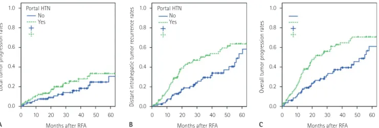
31.9%, respectively. The corresponding rates were 7.5%, 10.6%, 16.0%, 24.7%, and 30.5% in patients without portal hyperten- sion, and 11.9%, 19.8%, 25.4%, 33.3%, and 33.3% in patients with portal hypertension, respectively (Fig. 1A). The difference was not statistically significant (p = 0.195).
Table 2. Univariate Analysis of Prognostic Factors for Recurrence-Free Survival
Prognostic Factors Hazard Ratios Standard Errors p-Value
Age, > 65 years 1.273 (0.798–2.031) 0.238 0.310
Sex, male 1.053 (0.331–3.346) 0.590 0.930
Hepatitis C viral infection 1.064 (0.634–1.786) 0.264 0.814
Alcoholics 1.592 (0.995–2.548) 0.240 0.053
Portal hypertension 1.791 (1.133–2.833) 0.234 0.013*
Child-Pugh score 6 1.774 (1.056–2.981) 0.265 0.030*
Multinodular HCC 2.445 (1.340–4.461) 0.307 0.004*
Presence of tumor, > 3 cm 0.990 (0.475–2.064) 0.375 0.979
Presence of a dome nodule 1.189 (0.714–1.980) 0.260 0.505
Presence of a subcapsular tumor 1.462 (0.934–2.287) 0.228 0.096
Tumor adjacent to large vessel 1.036 (0.637–1.684) 0.248 0.887
Combined TACE and RFA 0.789 (0.425–1.464) 0.316 0.452
Operative RFA 1.172 (0.582–2.359) 0.357 0.657
*Statistically significant.
HCC = hepatocellular carcinoma, RFA = radiofrequency ablation, TACE = transarterial chemoembolization Table 3. Multivariate Analysis of Prognostic Factors for the Recurrence-Free Survival
Prognostic Factors Hazard Ratios Standard Errors p-Value
Portal hypertension 1.870 (1.180–2.961) 0.235 0.008*
Presence of multiple HCC nodules 2.616 (1.431–4.783) 0.308 0.002*
*Statistically significant.
HCC = hepatocellular carcinoma 1.0
0.8
0.6
0.4
0.2
0.0
Local tumor progression rates
0 10 20 30 40 50 60 NoYes
Portal HTN
Months after RFA A
1.0
0.8
0.6
0.4
0.2
Distant intrahepatic tumor recurrence rates 0.0
0 10 20 30 40 50 60 NoYes
Portal HTN
Months after RFA B
NoYes Portal HTN 1.0
0.8
0.6
0.4
0.2
0.0
Overall tumor progression rates
0 10 20 30 40 50 60 Months after RFA
C
Fig. 1. The difference in the recurrence rates depending on the presence of clinically relevant portal hypertension after radiofrequency ablation of hepatocellular carcinoma.
A. Local tumor progression rates were similar between the two curves (p = 0.195).
B, C. Distant intrahepatic tumor recurrence rates (B), and overall tumor progression rates were statistically higher in patients with clinically rele- vant portal hypertension (C) (p = 0.011 and p = 0.003, respectively). Note that the curves for distant intrahepatic recurrence and overall tumor progression were nearly identical.
HTN = hypertension, RFA = radiofrequency ablation
The 1-, 2-, 3-, 4-, and 5-year distant intrahepatic tumor spread rates were 17.2%, 32.5%, 41.5%, 49.6%, and 60.1%, re- spectively. The corresponding rates were 6.6%, 18.9%, 29.5%, 34.0%, and 53.7% in patients without portal hypertension, and 23.4%, 44.3%, 51.9%, 59.6%, and 63.6% in patients with portal hypertension, respectively (Fig. 1B). The difference was statisti- cally significant (p = 0.011). Univariate and multivariate analy- sis showed that portal hypertension and multiplicity of HCC nodules were the two independent adverse predictors of distant intrahepatic tumor recurrence (p = 0.008 and p = 0.002, respec- tively) (Tables 2, 3).
Overall, tumor progression was detected in 94 patients. The 1-, 2-, 3-, 4-, and 5-year overall tumor progression rates were 24.9%, 40.4%, 51.0%, 59.1%, and 65.2%, respectively. The me- dian time to tumor progression was 34.9 months. The 3- and 5-year overall tumor progression rates were 37.3% and 61.0%
in patients without portal hypertension, and 62.7% and 70.6%
in patients with portal hypertension, respectively (Fig. 1C). The difference was statistically significant (p = 0.003). To treat the recurrent tumors, RFA, TACE, and radiation therapy were per- formed on 67, 42, and 29 patients, respectively.
DISCUSSION
Most HCCs develop as a result of liver cirrhosis caused by chronic liver inflammation. Chronic viral infections, alcoholic hepatitis, or non-alcoholic steatohepatitis are well known un- derlying causes of liver cirrhosis and hepatocarcinogenesis (3).
The development and progression of liver fibrosis are known to be associated with hepatocyte death and a subsequent inflam- matory response, both of which involve reactive oxygen species accumulation in injured hepatocytes (27). Considering that the risk of human HCC recurrence after hepatectomy seems to be correlated with protein oxidation in the liver, increased oxidative stress in liver parenchymal cells may explain the close relation- ship between liver fibrosis and hepatocarcinogenesis (3).
It is well known that liver fibrosis can act as an adverse prog- nostic factor for survival because of hepatic decompensation as well as the development of HCC (4). Therefore, reversal of liver fibrosis before the development of portal hypertension, if possi- ble, will be clinically important in reducing the development of HCC as well as hepatic decompensation (28, 29). When we con-
sider that portal hypertension signifies advanced liver fibrosis, it would be logical to conclude that the risk of HCC development increases as liver cirrhosis progresses (4). A previous study re- ported that in patients with decompensated alcoholic cirrhosis, a baseline hepatic vein-portal vein gradient greater than 15 mm Hg is a weak, but independent predictive factor for the develop- ment of HCC (relative risk = 1.128) (4).
This study showed that CRPH is an indicator for tumor pro- gression in patients without hepatic decompensation. This was because distant intrahepatic tumor recurrence rate, but not the LTP rate, was much higher in HCC patients with portal hyper- tension. In contrast, CRPH had little effect on overall survival according to our unpublished data. The reason may be that many other factors related to the treatment of recurrent HCC could affect survival outcomes.
It seems that the potential for the development of HCC or in- trahepatic metastasis increases as liver fibrosis progresses, even for patients without hepatic decompression. For the other prog- nostic factor (recurrence-free survival) which was identified in this study using multivariate analysis, it could be inferred that multinodularity was also an independent significant factor for tumor progression (1, 30).
This study has several limitations. First of all, non-invasive imaging tools were used to determine portal hypertension (31).
Although portal hypertension is conventionally evaluated by measuring the HVPG, non-invasive imaging tools are also widely used in clinical practice (5, 6). Secondly, the significance of the results may be limited because of its retrospective nature. A fu- ture prospective multi-center study may reinforce the signifi- cance of this study. Finally, this study was conducted for patients who were already diagnosed with HCC. Therefore, the results of this study may not be applicable to high risk patients who have not been diagnosed with HCC yet.
In conclusion, even among the HCC patients without hepatic decompensation, a higher distant intrahepatic tumor progres- sion rate was noted in patients with portal hypertension. Early treatment of liver fibrosis in the reversible stage will be clinically important to prevent the development of HCC, as well as hepat- ic decompensation.
RefeReNCeS
1. El-Serag HB, Mason AC. Rising incidence of hepatocellular carcinoma in the United States. N Engl J Med 1999;340:745- 750
2. Lok AS. Hepatitis B: liver fibrosis and hepatocellular carci- noma. Gastroenterol Clin Biol 2009;33:911-915
3. Sakurai T, Kudo M, Umemura A, He G, Elsharkawy AM, Seki E, et al. p38α inhibits liver fibrogenesis and consequent he- patocarcinogenesis by curtailing accumulation of reactive oxygen species. Cancer Res 2013;73:215-224
4. Kim MY, Baik SK, Yea CJ, Lee IY, Kim HJ, Park KW, et al. He- patic venous pressure gradient can predict the development of hepatocellular carcinoma and hyponatremia in decom- pensated alcoholic cirrhosis. Eur J Gastroenterol Hepatol 2009;21:1241-1246
5. Thabut D, Moreau R, Lebrec D. Noninvasive assessment of portal hypertension in patients with cirrhosis. Hepatology 2011;53:683-694
6. Choi JW, Chung JW, Lee DH, Kim HC, Hur S, Lee M, et al. Por- tal hypertension is associated with poor outcome of trans- arterial chemoembolization in patients with hepatocellular carcinoma. Eur Radiol 2018;28:2184-2193
7. Bruix J, Sherman M; American Association for the Study of Liver Diseases. Management of hepatocellular carcino- ma: an update. Hepatology 2011;53:1020-1022
8. European Association for the Study of the Liver; European Organisation for Research and Treatment of Cancer. EASL- EORTC clinical practice guidelines: management of hepato- cellular carcinoma. J Hepatol 2012;56:908-943
9. Shiina S, Tateishi R, Arano T, Uchino K, Enooku K, Nakagawa H, et al. Radiofrequency ablation for hepatocellular carci- noma: 10-year outcome and prognostic factors. Am J Gas- troenterol 2012;107:569-577; quiz 578
10. Peng ZW, Lin XJ, Zhang YJ, Liang HH, Guo RP, Shi M, et al.
Radiofrequency ablation versus hepatic resection for the treatment of hepatocellular carcinomas 2 cm or smaller: a retrospective comparative study. Radiology 2012;262:1022- 1033
11. Nakazawa T, Kokubu S, Shibuya A, Ono K, Watanabe M, Hi- daka H, et al. Radiofrequency ablation of hepatocellular carcinoma: correlation between local tumor progression
after ablation and ablative margin. AJR Am J Roentgenol 2007;188:480-488
12. Lee DH, Lee JM, Lee JY, Kim SH, Yoon JH, Kim YJ, et al. Ra- diofrequency ablation of hepatocellular carcinoma as first- line treatment: long-term results and prognostic factors in 162 patients with cirrhosis. Radiology 2014;270:900-909 13. Kei SK, Rhim H, Choi D, Lee WJ, Lim HK, Kim YS. Local tumor
progression after radiofrequency ablation of liver tumors:
analysis of morphologic pattern and site of recurrence. AJR Am J Roentgenol 2008;190:1544-1551
14. Bezerra AS, D’Ippolito G, Faintuch S, Szejnfeld J, Ahmed M.
Determination of splenomegaly by CT: is there a place for a single measurement? AJR Am J Roentgenol 2005;184:1510- 1513
15. Perri RE, Chiorean MV, Fidler JL, Fletcher JG, Talwalkar JA, Stadheim L, et al. A prospective evaluation of computer- ized tomographic (CT) scanning as a screening modality for esophageal varices. Hepatology 2008;47:1587-1594 16. Lee CM, Jeong WK, Lim S, Kim Y, Kim J, Kim TY, et al. Diag-
nosis of clinically significant portal hypertension in patients with cirrhosis: splenic arterial resistive index versus liver stiffness measurement. Ultrasound Med Biol 2016;42:1312- 1320
17. Forner A, Llovet JM, Bruix J. Hepatocellular carcinoma. Lan- cet 2012;379:1245-1255
18. Bruix J, Sherman M; Practice Guidelines Committee, Amer- ican Association for the Study of Liver Diseases. Manage- ment of hepatocellular carcinoma. Hepatology 2005;42:
1208-1236
19. Lang EV, Chen F, Fick LJ, Berbaum KS. Determinants of in- travenous conscious sedation for arteriography. J Vasc In- terv Radiol 1998;9:407-412
20. Lupescu I, Masala N, Capsa R, Câmpeanu N, Georgescu SA.
CT and MRI of acquired portal venous system anomalies. J Gastrointestin Liver Dis 2006;15:393-398
21. Curley SA, Marra P, Beaty K, Ellis LM, Vauthey JN, Abdalla EK, et al. Early and late complications after radiofrequency ab- lation of malignant liver tumors in 608 patients. Ann Surg 2004;239:450-458
22. Tublin ME, Dodd GD 3rd, Baron RL. Benign and malignant portal vein thrombosis: differentiation by CT characteristics.
AJR Am J Roentgenol 1997;168:719-723
23. Li C, Hu J, Zhou D, Zhao J, Ma K, Yin X, et al. Differentiation of bland from neoplastic thrombus of the portal vein in pa- tients with hepatocellular carcinoma: application of sus- ceptibility-weighted MR imaging. BMC Cancer 2014;14:
590
24. Catalano OA, Choy G, Zhu A, Hahn PF, Sahani DV. Differen- tiation of malignant thrombus from bland thrombus of the portal vein in patients with hepatocellular carcinoma: ap- plication of diffusion-weighted MR imaging. Radiology 2010;254:154-162
25. Kang TW, Rhim H, Lee MW, Kim YS, Choi D, Lim HK. Termi- nology and reporting criteria for radiofrequency ablation of tumors in the scientific literature: systematic review of compliance with reporting standards. Korean J Radiol 2014;
15:95-107
26. Ahmed M, Solbiati L, Brace CL, Breen DJ, Callstrom MR, Char- boneau JW, et al. Image-guided tumor ablation: standard- ization of terminology and reporting criteria--a 10-year
update. J Vasc Interv Radiol 2014;25:1691-1705.e4 27. Ghany MG, Kleiner DE, Alter H, Doo E, Khokar F, Promrat K,
et al. Progression of fibrosis in chronic hepatitis C. Gastro- enterology 2003;124:97-104
28. Ismail MH, Pinzani M. Reversal of hepatic fibrosis: patho- physiological basis of antifibrotic therapies. Hepat Med 2011;3:69-80
29. Gieling RG, Burt AD, Mann DA. Fibrosis and cirrhosis revers- ibility - molecular mechanisms. Clin Liver Dis 2008;12:915- 937
30. Okuda K, Ohtsuki T, Obata H, Tomimatsu M, Okazaki N, Hasegawa H, et al. Natural history of hepatocellular carci- noma and prognosis in relation to treatment. Study of 850 patients. Cancer 1985;56:918-928
31. Cho KC, Patel YD, Wachsberg RH, Seeff J. Varices in portal hypertension: evaluation with CT. Radiographics 1995;15:
609-622
간세포암 환자의 고주파열치료 후 종양 재발:
예후인자로서 문맥고혈압
장성원
1· 조윤구
1* · 김주원
1· 길제령
1· 김미영
1· 이 영
2목적: 간기능이 보존된 간세포암 환자에서 간문맥 고혈압이 고주파열치료 후 종양 재발에 미치는 영향을 평가한다.
대상과 방법: 2010년 1월에서 2017년 3월 사이에 Milan criteria 및 Child-Pugh class A를 가진 신규 간세포암 환자 중 본 원에서 고주파열치료를 시행한 환자가 본 연구에 포함되었다. 종양 재발에 대한 예측인자를 찾기 위해 Cox proportional hazard model을 이용한 단변량 및 다변량 분석을 수행하였다.
결과: 모두 178명의 환자가 본 연구에 포함되었다. 추적 관찰 기간의 중앙값은 42.8개월이었다. 국소 재발률은 문맥고혈 압 여부에 따라 뚜렷한 차이를 유발하지 않았다(p = 0.195). 3년 및 5년 원위부 간내 종양 재발률은 문맥고혈압이 없는 환자의 경우 각각 29.5%와 53.7%, 그리고 문맥고혈압이 있는 환자의 경우 51.9%와 63.6%였으며 두 군 사이의 차이는
통계적으로 유의하였다(p = 0.011). 단변량 및 다변량 분석에서 문맥압항진은 원위부 간내 종양 재발에 대한 독립적인 예
측 인자이었다(p = 0.008).
결론: Child-Pugh class A를 가진 간세포암 환자의 경우, 문맥고혈압은 종양 재발에 불량 예후인자로 작용하였다.
중앙보훈병원 1영상의학과, 2의학연구소