서 론
어류의 후각(olfaction)은 먹이탐색, 포식자 회피, 동종 인식, 서식처 인지, 이주와 회유와 같은 다양한 생태적 행동들을 유 발시키는 생체 내 감지능력으로써, 물속에 녹아있는 화학물 질을 감지하는 것으로부터 촉발된다(Yamamoto, 1982). 특히, 경골어류의 후각기관은 비강(olfactory chamber) 또는 비낭 (accessory nasal sac)이 해부적으로 구강(oral cavity) 및 새강 (branchial cavity)쪽으로 개방되지 않고 서로 분리되어 있어 고 등한 척추동물들과는 다르게 호흡의 기능을 전혀 수행하지 않 는 폐쇄적인 구조를 나타낸다(Zeiske et al., 1992). 이러한 구조 적 특징으로 인하여, 전비공(anterior nostril)을 통해서 유입된 물은 어류의 소화관(digestive tract)으로 들어가지 않고 일반적
으로 후내강(olfactory lumen)을 거쳐서 다시 후비공(posterior nostril)을 통해서만 밖으로 배출되게 된다(Zeiske et al., 1992).
따라서 물이 통과되는 경로인 전비공, 후비공, 비강, 비낭 조직 및 형태는 종의 후각민감성, 유영방향, 생태적 니치(niche) 등 에 따라 다양한 특성을 나타내게 되며, 유속 및 수량, 부유물 특성과 같은 유입되는 서식처 환경의 물리적인 요소와 깊이 관 련된다(Hara, 1986; Atta, 2013).
국내에서 보고된 납자루아과 Acheilognathinae 어류는 모두 3속 15종으로써 절멸된 것으로 추정되는 서호납줄갱이 Rho
deus hondae를 제외한 14종이 현재 분포하고 있다(Kim et al., 2014). 본 속 대부분의 종들은 한국 고유종으로써 서식처 바닥 의 조개류 내에 산란관을 삽입하여 알을 낳는 독특한 산란습성 과 화려한 체색을 보유하고 있어 관상 및 학술적 가치가 매우 높은 종들로 평가되고 있다(Smith and Hartel, 1999; Kim and Park, 2002). 그중 각시붕어 R. uyekii는 유속이 완만하고 수초 가 비교적 많은 우리나라 서해와 남해로 흐르는 하천과 저수
—123 — http://www.fishkorea.or.kr
†김현태, 윤승운은 논문 작성에 동등하게 기여하였음.
* Corresponding author: Jong Young Park Tel: 82632703344, Fax: 82632703362, Email: [email protected]
ISSN: 1225-8598(Print), 2288-3371(Online). DOI: https://doi.org/10.35399/ISK.31.3.1
Accepted: August 14, 2019
한국고유종 각시붕어 Rhodeus uyekii 후각기관의 형태 및 조직, 세포화학적 특성 연구
김현태†·윤승운†·이용주1·박종영
*
전북대학교 자연과학대학 생명과학과, 전북대학교 부설 생물다양성연구소, 1전주교육대학교 과학교육과
Morphology, Histology, and Histochemistry of the Olfactory Organ in Korean Endemic Fish, Rho deus uyekii(Pices, Cyprinidae) by Hyun Tae Kim†, Seung Woon Yun†, Yong Joo Lee1 and Jong Young Park* (Department of Biological Science and Institute for Biodiversity Research, College of Natural Sciences, Chonbuk National University, Jeonju 54896, Republic of Korea; 1Jeonju National University of Education, Jeonju 55101, Republic of Korea)
ABSTRACT The olfactory organ of the Korean endemic fish, Rhodeus uyekii, a mussel-spawning species, was researched anatomically, histologically and histochemically, for looking into a relation to the physical and chemical condition of its habitat and ecological habit, using stereo microscopy, light microscopy and scanning electron microscopy. The external structure of the olfactory organ consists of the semicircular-shaped anterior nostril with arched shape at its forward position, posterior nostrils and the nasal flap. Within the olfactory chamber, it has the rosette structure with 14 to 15 lamellae which is largely divided into the sensory and non-sensory regions. The sensory region has the olfactory recep- tor neurons, the supporting cells, the basal cells, the lymphatic cells, and the plasma cells, while the non-sensory region has the stratified epithelial cells, the mucous cells with sulfomucin and 1 type of unidentified cell. In particular, the arched feature in the anterior nostril and the mucous cell of sulfomu- cin were unique.
Key words: Rhodeus uyekii, olfactory organ, structure, histology, histochemistry
지 등에 서식하는 전장 4~5cm(성어)의 소형종으로서, 그동안 인공교잡실험(Suzuki and Jeon, 1988), 생식주기(An, 1995), 골 격학적 연구(Kim, 1997), 산란관 신장(Chae, 2001), 자연잡종 (Kim et al., 2010), 떡납줄갱이 R. notatus 간 정교배체의 세포 유전학적 분석(Kim et al., 2012), 수컷의 혼인색과 암컷의 배 우자 선택(Jeon and Suk, 2012), 개체군 생태(Kim et al., 2015), 시각세포(Kim and Park, 2016) 등 다양한 연구들이 진행되어 왔다. 그러나 감각기관을 포함한 종의 외부형태 및 조직학적 특성들은 종의 서식처나 생태적 습성에 적응한 많은 증거들을 제시해 오고 있지만(Hara, 1986; Kasumyan, 2004), 아직까지 각시붕어에 관한 연구는 이루어진 바 없다. 일반적으로 잉어 목 어류의 후각기관은 많은 수의 후판과 뚜렷한 눈을 보유하 는 것으로 알려진 “eye-nose fishes”로 구분되고 있어(Ojha and Kapoor, 1973) 그동안 먹이 감지나 환경특성 인지 측면에 있어 서 시각, 미각, 후각에 초점되는 많은 연구들이 진행되어져 왔 다(Bhute and Baile, 2007; Ghosh and Chakrabarti, 2013, 2014;
Samajdar and Mandal, 2016). 그중 납자루아과 어류들은 일부 종들이 비슷한 형태를 나타내지만 서식장소의 유속, 하상구조, 수변부의 식생의 선호도가 각각 다른 것으로 알려져 있다(Kim and Park, 2002). 각시붕어는 그중 비교적 가장 느린 유속의 수 환경을 선호하는 것으로 알려져 있다. 따라서 본 연구에서는 이러한 서식처의 차이가 각시붕어 후각기관에 어떻게 반영되 어 있는지 확인하기 위해서, 본 기관의 형태, 조직 및 세포화학 적 특성을 묘사하고 서식처 환경 및 생태적 습성과의 연관성을 분석해 보고자 하였다.
재료 및 방법
1. 실험재료
2019년 3월부터 4월까지 전라남도 담양군 무정면 평지리의 오례천(35°16ʹ59ʺN, 127°03ʹ41ʺE)에서 족대(망목 4×4mm)를 이용하여 각시붕어(체장 36.3~45.1mm) 15마리를 채집하였 다. 그 후 현장에서 즉시 0.1% maminobenzoic acid ethylester methanesulfonate 수용액(MS222, Sigma, USA)을 이용하여 개 체들을 마취시킨 후 10% 중성 포르말린 용액과 2.5% 중성 글 루타르알데히드 용액(pH 7.4 2.5% G.A.; EMS, USA)에 고정 하였다. 본 연구종의 실험은 전북대학교의 “실험동물의 관리와 사용에 관한 지침”(Institutional Animal Care and Use Commit
tee)의 절차에 따라 수행되었다.
2. 실험방법
각시붕어 후각기관의 해부학적 특징을 관찰하기 위해 실 체현미경(Carl Zeiss, Germany) 하에서 커터칼(Surgical blade
stainless No. 10, 11, Japan)을 이용하여 두부로부터 절개한 후 디지털 카메라(TG3, Olympus, Japan)로 촬영하였다.
조직 및 세포의 일반적인 특징을 알아보기 위해 10% 포르 말린 용액에 고정된 표본의 두부로부터 후각기관을 절개하여 탈수와 투명화처리 후 일반적인 파라핀 포매법으로 24시간 처 리하여 block(Parablast, Oxford)을 제작하였다. 포매된 block 은 microtome(Leica 820, Germany)을 이용하여 5μm로 각각 절편 되었으며 Harris’s hematoxylineosin(HE)로 대조 염색 (Gurr, 1956) 후 광학현미경(LM; LE REL 4.4, Carl Zeiss, Ger
many) 하에서 관찰 및 촬영하였다. 점액세포(mucous cell)의 조직화학적 특성(pH, 다당류의 종류)을 판별하기 위해 아래와 같은 특수염색법을 추가로 실시하였다: 1) Masson trichrome (Masson, 1929), 2) periodic acid Schiff(PAS) and diastase/PAS, 3) hematoxylin, Weigert iron and toluidine blue, 4) alcian blue (AB pH 1.0, pH 2.5), 5) mild methylation, methylationsaponi
fication and Aldehyde fuchsin-AB.
비강 내 로제트구조(rosette structure)의 특징과 후각세포들 의 분포 및 미세구조를 알아보기 위해 2.5% G.A. 용액에 고정 된 후각기관을 두부로부터 절개하여 같은 용액에 재고정하였 다. 그 후 중성 1% OsO4 용액(pH 7.4)에 2차 고정, 알코올의 상승된 농도 순으로 탈수, t부탄올에 처리하여 임계점 건조 (VFD21S; Vaccum Device Co., Ltd., Japan)하였으며, 오스뮴 코팅(HPC1SW; Vacuum Device Co., Ltd.)한 후 주사전자현미 경(SEM; SUPRA40VP, Carl Zeiss, Germany)으로 촬영하였다.
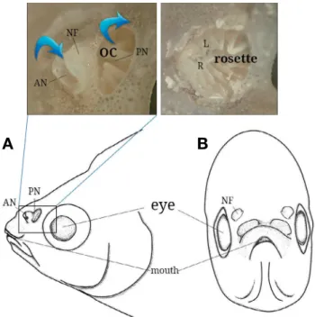
Fig. 1. Anatomical diagram of the front(left) and side(right) views in the head of Rhodeus uyekii. Above photographs indicate external(left) and internal(right) structure. AN, anterior nostril; L, lamellae; NF, nasal flap; OC, olfactory chamber; PN, posterior nostril; R, raphe.
A B
결 과
1. 해부학적 구조
한국 고유종 각시붕어의 후각기관(olfctory organ)은 주둥이 등 쪽에 좌우 한 쌍으로 위치해 있으며, 각각 전비공(anterior nostril), 후비공(posterior nostril), 비경계판(nasal flap)으로 구
성되었다(Fig. 1A and B). 전비공(직경 0.57~0.86mm, 길이 0.4~0.6mm)은 앞쪽에 피부로부터 돌출된 아치형(archshape) 의 조직이 발달한다. 후비공(직경 0.94~1.17mm)은 피부로부 터 돌출되지 않는 반원형의 유출구로써 전비공과 차이를 보였 다. 비경계판은 피부 위로 돌출된 얇은 조직판으로 전비공과 후비공을 구분하는 격벽의 형태이다. 후각기관의 내부는 비강 내 14~15개의 후판(lamella)들과 중심부의 후선(olfactory
A B
C D
E F
G
Fig. 2. Histology of the olfactory epithelium of Rhodeus uyekii, stained with hematoxylin and eosin(A, C, E), Masson’s trichrome(B, D), and alcian blue(pH 2.5)periodic acid Schiff(G). A, B), its rosette structure, and a number of lamellae consisting the sensory and the nonsensory epi
theliums; C, D), the sensory epithelium having the ORNs, the SCs, the BCs, the LCs, the PCs, the CNC, and the connective tissue with the FBCs, the PMCs, and blood capillaries; E~G), the nonsensory epithelium containing the SECs, the LCs, the MCs and the UCs. Arrowhead, mucous cell; BC, basal cell; CNC, ciliated non-sensory cell; FBC, fibroblast cell; LC, lymphatic cell; ORN, olfactory receptor neuron; PC, plasma cell;
PMC, pigment cell; SC, supporting cell; SEC, stratified epithelial cell; UC, unidentified cell. Bars indicate 200μm in A, B, and 50μm in C~H, respectively.
raphe)으로 구성된 로제트 구조가 발달했다(Figs. 1 and 3A).
후판들은 내강으로부터 돌출된 혀 모양으로 비강의 측면과 바 닥면과 연결되고 후선을 중심으로 방사형으로 배열되었다.
2. 조직학적 특징 및 세포화학적 특성
비강 내 후판은 감각(sensory epithelium)과 비감각(nonsen
sory epithelium)영역으로 구성되었다. 감각상피는 후판의 양면 에 발달하는 반면에 비감각상피는 후판의 가장자리 즉, 비강 내벽과 후선의 연접부에 발달했다(Fig. 2A and B). 감각상피는 거짓중층원주상피층(pseudostratified columnar layer)으로써 표 면에 많은 섬모들이 관찰되며(Fig. 3C), 후감각뉴런(olfactory receptor neuron), 지지세포(supporting cell), 기저세포(basal cell), 림프구(lymphatic cell), 형질세포(plasma cell)와 같은 다 양한 후각세포들로 이루어져 있었다(Fig. 2C and D). 후감각뉴 런은 세포질이 상피층 표면과 기부 쪽으로 뻗어있는 양극성 세 포로서(Fig. 2B), HE와 Masson’s trichrome 염색에서 핵은 짙 은 보라색을 보이고 세포질은 다홍색으로 염색되었다. 지지세 포는 감각뉴런과는 다르게 돌출된 세포질을 나타내지 않는 원 주세포로서 세포표면에 많은 섬모들을 보유하며, 이것의 핵은 원형 또는 타원형이고 Masson’s trichrome 염색에서 짙은 보라 색을 나타내었다(Fig. 2C). 기저세포는 상피층 하부에 위치한 타원형의 세포로서 세로와 가로로 기저막 바로 위에 배열되고, HE 염색에서는 짙은 다홍색을 나타내었다. 림프구는 작은 원 형의 세포로서 핵에 비해 극히 적은 세포질 공간을 보여주며 핵은 HE와 Masson’s trichrome 염색에서 각각 짙은 자주색과 보라색으로 염색되었다. 형질세포는 림프구보다 큰 원형의 세 포로써 핵과 세포질의 염색성이 뚜렷하게 구분되었다. 핵은 세 포질측면에 위치하며 Masson’s trichrome 염색에서 짙은 보라 색으로 염색되고 세포질은 진한 적색을 나타내었다(Fig. 2D).
감각상피층 아래의 결합조직에서는 3~4개의 적혈구가 관찰 되는 모세혈관, 색소세포, 섬유아세포가 확인되었다(Fig. 2C and D). 색소세포는 기저막 아래에 불규칙하게 배열되어 있으
며 HE와 Masson’s trichrome 염색에서는 검정색으로 나타났 다. 섬유아세포는 평평한 긴 타원형의 세포로써 HE와 Mas
son’s trichrome 염색에서는 주변기질로 뻗어나는 세포질이 관 찰되지 않았다. 핵은 Masson’s trichrome 염색에서 보라색으로 염색되며 작은 핵소체들이 소수 관찰되었다(Fig. 2C and D).
비감각상피는 중층편평상피층(stratified squamous layer)으 로써 층상상피세포(stratified epithelial cell), 림프구, 점액세포, 비감각섬모세포(ciliated nonsensory cell), 미확인세포(uniden
tified cell)들로 구성되었다(Fig. 2E~G). 층상상피세포는 비감 각상피층을 구성하는 주된 세포들로 원 또는 타원형의 핵과 상대적으로 넓은 세포질공간을 나타냈다. 핵은 세포질의 다양 한 공간에 위치하는데 HE염색에서는 진한 자주색, Masson’s trichrome 염색에서는 진한 남색, ABPAS 염색에서는 진한 청 색을 나타냈다(Fig. 2E and F). 비감각상피의 림프구는 감각상 피에서 보여지는 형태와 세포화학적 특징과 같았다. 점액세포 는 상피층 표면에 위치하는 단세포샘(unicellular gland)으로써 핵은 세포 바닥에 평평한 형태로 나타났다. 또한 점액세포는 다음과 같이 고용된 특수염색에서 다양한 색(적색, PAS, dia
Fig. 3. Scanning electron micrographs of the rosette structure of Rhodeus uyekii. A), the rosette structure with a number of lamellae; B), the sen
sory epithelium with olfactory receptor neurons; C), the surface of the sensory epithelium. Bars indicate 200μm in A, 10μm in B and C, respec
tively.
A B C
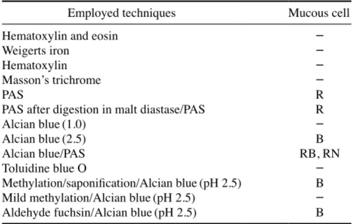
Table 1. The mucous cell stainability of the olfactory organ in Rho
deus uyekii, identified by each technique
Employed techniques Mucous cell
Hematoxylin and eosin -
Weigerts iron -
Hematoxylin -
Masson’s trichrome -
PAS R
PAS after digestion in malt diastase/PAS R
Alcian blue(1.0) -
Alcian blue(2.5) B
Alcian blue/PAS RB, RN
Toluidine blue O -
Methylation/saponification/Alcian blue(pH 2.5) B Mild methylation/Alcian blue(pH 2.5) - Aldehyde fuchsin/Alcian blue(pH 2.5) B B, blue; R, red; RB, reddish blue; RN, reddish black; -, no reaction
stase/PAS; 청색, AB pH 2.5, Methylation/saponification/AB, Aldehyde fuchsin/AB; 청적색, 적흑색, ABPAS pH 2.5; 음성, HE, Weigerts iron, Hematoxylin, Masson’s trichrome, AB pH 1.0, Toluidine blue O, Mild methylation/AB)을 나타내었다 (Table 1). 비감각섬모세포는 상피층의 가장 위쪽에 위치했다.
또한 5~10개의 세포들이 서로 연접해있어 하나의 세포 그룹 을 형성하였으며 HE와 Masson’s trichrome염색에서 각각 연 하고 진한 보라색을 나타냈으며 핵소체들이 뚜렷하게 관찰되 었다. 섬모와 핵 사이의 세포질은 HE와 Masson’s trichrome 염색에서 각각 선홍색과 진한 적청색으로 염색되었다(Fig. 2E and F). 미확인세포는 비감각상피층 가장 아래에서 관찰되며 기저막 바로 위에 위치했다. 세포질공간보다 핵의 크기가 두 드러지게 컸으며 원통과 타원형을 나타내며 HE와 Masson’s trichrome염색에서 각각 선홍색과 진한 흑적색으로 염색되었 다(Fig. 2E~G).
고 찰
한국 고유종 각시붕어 후각기관의 외부형태는 전비공, 후비 공, 그리고 이들 사이에 위치한 비경계판으로 구성되며, 14~15 개의 후판들로 이루어진 로제트 구조를 비강 내에 보유한다. 경골어류 비공의 모양과 발달 위치는 종에 따라 서식처의 물 리적 특성, 유영행동, 먹이습성 등을 반영한 독특한 특징을 나 타내지만 비공 수는 생태적 습성보다는 분류군의 진화적 위치 에 관련되는 경향성이 더 크다(Yamamoto, 1982; Zeiske et al., 1992). 상어류, 가오리류와 같은 연골어류와 현존하는 경골어 류들은 모두 물의 유입과 출구가 다른 2개의 구멍을 주둥이 위 쪽 또는 아래쪽에 보유하지만, 칠성장어류(lamprey)나 먹장어 류(hagfish)와 같은 하등한 어류에서는 두부의 등 쪽에 물의 유 입구와 유출구가 같은 경로인 1개의 구멍을 나타낸다(Thorn
hill, 1967). 따라서, 각시붕어의 주둥이 위에 나타난 2개(전비 공, 후비공)의 비공 수는 무악류(agnatha)와 비교되는 경골어 류의 진화적 위치를 반영하는 것으로 생각된다.
각시붕어의 비공은 반원형으로써 다른 잉어목 어류들과 전 체적으로 비슷한 특징을 공유하지만 그중 전비공은 앞쪽에 아 치형의 조직 돌출을 보이고 있어 아직까지 한국산 어류에서 알 려지지 않은 매우 독특한 구조적 특징을 나타낸다. Kim(2018) 은 한국산 어류 17종 후각기관의 형태적 분류에서 i) 피부와 평 행한 반원형, ii) 앞쪽의 돌출된 조직을 갖는 반원형, iii) 피부와 평행한 원형, iv) 돌출된 튜브형 등의 4 type을 제시한 바 있다.
각시붕어의 전비공은 이 중에서 ii) type인 앞쪽에 돌출된 조직 을 갖는 반원형에 해당하는 형태로써 같은 잉어목 어류의 쉬 리, 돌마자(각각 i type)와 구조적 차이를 나타낸다. 경골어류에 서 후각기관을 포함한 주둥이 형태의 다양성은 주로 섭식행동
에 관련된 생태적 니치(ecological niche)와 서식처 선호도에 매 우 밀접하게 연관된 것으로 알려져 있다(Lauder, 1982; White
ley, 2007). 튜브형의 돌출된 조직을 갖는 전비공은 대부분 유 속이 거의 없는 수환경에 서식하거나 활동성이 약한 어류들에 게서 발견되는데(Kasumyan, 2004), 다른 잉어목 어류들과 비 교해 아치형 조직을 갖는 각시붕어의 전비공은 뒤쪽의 비경계 판과 함께 위의 iv) type과 비슷한 형태를 나타낸다. 이러한 구 조는 유속이 거의 없는 서식처의 물리적 특성과 활동성이 다소 약한 각시붕어의 유영습성, 그리고 섭식행동에 적응된 결과로 판단된다.
각시붕어의 비강 내에는 14~15개의 후판으로 구성되는 로 제트 구조가 발달한다. 본 연구어종이 속하는 잉어목 어류에서 후판 수는 Tribolodon hakonensis 황어 30개, Pseudogobio eso
cinus 모래무지 10개, Zacco platypus 피라미 21개, Cyprinus carpio 잉어 31개, Microphysogobio yaluensis 돌마자 22~24개 로 다양하게 알려져 있어 종마다 많은 수적 차이를 나타내고 있다. 일반적으로 후판의 수적 증가는 비강 내 후상피(olfactory epithelium)의 발달 표면적을 증가시켜 후각기관의 효율성과 감각의 민감도를 높이는 것으로 알려져 있는데(Zeiske et al., 1976), 위의 선행연구들과 비교해 볼 때 각시붕어의 후판은 다 른 잉어목 어류들보다 적은 수를 나타내고 있어 상대적 후각 민감도는 낮을 것으로 추측된다.
일반적으로 경골어류의 후각상피는 감각영역과 비감각영역 으로 나누어지며, 이와 같은 조직분화는 각각의 상피층을 구성 하는 세포들의 기능적 차이를 야기한다(Hara, 1994). 각시붕어 의 후각상피 역시 감각과 비감각영역으로 나누어지는데, 감각 상피에서는 후감각뉴런, 지지세포, 기저세포 등이 있어 외부의 화학적 자극의 인지, 전달, 상피층의 기계적지지, 세포학적 재 생을 수행할 수 있을 것으로 생각되며 이러한 기본 세포 구성 은 경골어류에서 흔하게 보고되고 있다(Hara, 1986). 그 외 감 각상피에서는 림프구, 형질세포가 확인된다. 림프구는 Bcell과 Tcell, 그리고 자연살상세포(natural killer cell)로 크게 분류되 는데(Bone and Moore, 2008), 이 들은 주로 감염저항(infective resistance), 과민증(hypersensitiveness), 이식편 거부반응(graft rejection) 등과 같은 세포성 면역(cellmediated immunity)을 일으킨다(Nakanishi et al., 2002). 형질세포는 많은 양의 항체 를 분비하는 혈청항체생성세포이며 림프구로부터 변형된다 (Wang et al., 2019). 따라서 각시붕어의 다음과 같은 면역세포 특성들은 서식처에서의 세균, 박테리아, 그 밖의 면역반응과 관 련된 생물학적 요인들로부터 후각기관의 세포들을 보호하는 환경에 대한 세포학적 적응으로 생각된다.
각시붕어의 비감각상피는 층상상피세포, 점액세포, 비감각섬 모세포, 미확인세포 1종류가 확인되는데, 층상상피세포와 점액 세포는 다른 경골어류에서도 확인되는 일반적인 세포 구성이 다(Hara, 1986; Kasumyan, 2004; Kim et al., 2016). 그중 점액
세포의 세포화학적 조성은 PAS, AB(pH 2.5)PAS, Aldehyde fuchsin/AB에서 양성반응을 보여 중성과 산성의 황점액소(sul
fomucin)로 확인되는데, 이와 같은 탄수화물 성질은 정관(sper
matic duct)(Lahnsteiner and Patzner, 1990), 구강(Varute and Jirge, 1971), 아가미(Reid, 1995), 소화관(Fanta and Ribeiro, 2000) 등 다양한 생체 내 기관들에 광범위하게 존재하며, 한국 산 어류의 후각기관에서는 개소갱(Kim et al., 2018a)과 돌마자 (Kim et al., 2018b)에서 보고된 바 있다. 그러나 아직까지 어류 에서 황점액소의 정확한 기능은 보고된 바 없으나 척추동물에 서 뮤신(mucin)은 일반적으로 외부로부터 물리 및 화학적 영 향이 잦은 생체 내 조직에서 항균 작용을 하는 것으로 알려져 있다(Frenkel and Ribbeck, 2015).
각시붕어 감각상피 내의 비감각섬모세포들은 많은 경골어 류에서 비강 내에서 확인되지만 발생위치 및 분포양상은 매우 다양하다고 알려져 있다(Hansen and Zeiske, 2005). 일반적으 로 비감각섬모세포들은 많은 섬모를 세포표면에 가지고 있어 감각상피의 섬모성 지지세포와 비슷한 기능과 형태로 비감각 상피 내에서 발생하게 된다(Zeiske et al., 1992; Hansen and Zeiske, 2005). 감각상피 내에서의 비감각섬모세포의 발생은 붕어에서 유일하며(Hansen et al., 1999), 잉어목 어류를 포함한 다른 경골어류에서 알려진 바 없다. 각시붕어의 비감각상피층 에서 발생하는 비감각섬모세포의 형태와 분포는 다른 어류들 에서 선행된 연구결과와 일치하였다(Hara, 1986). 비감각섬모 세포의 운동성 섬모들은 상피층 위로 흐르는 물이나 고여 있 는 점액의 이동에 도움을 주는데(Doving et al., 1977; Zeiske et al., 1992), 이러한 특징들은 각시붕어의 서식처와 같은 거의 정체되어 있거나 느리게 흐르는 수환경에 물리적 특성에 적응 된 양상으로 사료된다.
본 실험에서 이용된 조직화학염색법은 염료의 특이성과 민 감성, 그리고 염료가 착색된 조직이나 세포를 통과하는 빛의 파장, 효소활성을 통한 반응물의 변화양상을 현미경을 이용 해 특정 결과(색이나 대조의 차이)를 확인함에 목적을 둔다 (Lavis, 2011). 또한, 이용된 염료의 pH는 산성, 염기성, 중성, 양성염료로 나눌 수 있어 조직이나 세포의 pH 성향을 확인할 수 있기도 하다(Lavis, 2011). 이러한 염색법의 특징들로 인해 각시붕어 후각조직 또는 세포의 서로 간의 경계와 차이를 잘 보여주었으며, 특히 특정 후각 세포들의 염색 특이성을 잘 확 인할 수 있었다.
각시붕어 후각기관의 해부 및 조직, 세포화학적 결과들은 유 속이 느리거나 정체된 서식처의 물리적 특성을 잘 반영하며, 특히 고유한 일부 후각 특성(전비공의 아치형 조직, 황점액소) 들은 추후 다른 종들과 비교되는 분류학적 특징으로 이용될 수 있을 것으로 판단된다.
요 약
한국 고유종 각시붕어 Rhodeus uyekii의 후각기관과 서식처 의 물리 및 화학적 특성 그리고 생태적 습성과의 연관성을 알 아보기 위해 실체현미경, 광학현미경, 주사전자현미경을 이용 하여 해부 및 조직, 세포화학적 특성을 조사하였다. 후각기관의 외부형태는 반원의 전비공과 후비공, 비경계판으로 구성되는 데, 특히 전비공의 앞쪽에는 아치형의 조직이 돌출되어 있다.
비강 내에는 14~15개의 후판으로 구성되는 로제트 구조를 보 유하며 후판은 감각상피와 비감각상피로 구분된다. 감각상피 는 후감각뉴런, 지지세포, 기저세포, 림프구, 형질세포로 구성되 는 반면에 비감각세포는 층상상피세포, 점액세포, 비감각섬모 세포, 미확인세포가 1종류 확인된다. 점액세포의 세포화학적 특성은 산성 및 중성의 황점액소(sulfomucine)로 나타났다.
사 사
본 논문은 2017년도 정부(교육부)의 재원으로 한국연구재단 의 지원을 받아 수행된 기초연구사업임(NRF2017R1D1A1B 03028268).
REFERENCES
An, C.M. 1995. Effects of photoperiod and water temperature on the reproductive cycle of the springspawning bitterling, Rho
deus uyekii. Korean J. Ichthyol., 7: 4355.(in Korean) Atta, K.I. 2013. Morphological, anatomical and histological studies
on the olfactory organs and eyes of teleost fish: Anguilla an
guilla in relation to its feeding habits. J. Basic Appl. Zool., 66: 101108.
Bhute, Y.V. and V.V. Baile. 2007. Organization of the olfactory sys
tem of the Indian Major Carp Labeo rohita(Hamilton): a scanning and transmission electron microscopic study. J.
Evol. Biochem. Physiol., 43: 342349.
Bone, Q. and R. Moore. 2008. Biology of fishes. (3rd ed.) Taylor &
Francis Group Pub., Milton Park, Abingdon, New York.
478p.
Chae, B.S. 2001. Elongation of the ovipositor in Korean rose bitter
ling, Rhodeus uyekii(Pisces: Cyprinidae). Korean J. Ichthy
ol., 13: 111116.(In Korean)
Døving, K.B., M. DuboisDauphin, A. Holley and F. Jourdan. 1977.
Functional anatomy of the olfactory organ of fish and the ciliary mechanism of water transport. Acta Zool., 58: 245
255.
Fanta, E. and C.A.O. Ribeiro. 2000. Microscopic morphology and histochemistry of the digestive system of a tropical fresh
water fish Trichomycterus brasiliensis(Lütken)(Siluroidei, Trichomycteridae). Revista Brasil. Zool., 17: 953971.
Frenkel, E.S. and K. Ribbeck. 2015. Salivary mucins protect surfac
es from colonization by cariogenic bacteria. Appl. Environ.
Microbiol., 81: 332338.
Ghosh, S.K. and P. Chakrabarti. 2013. Studies on the morphology of the olfactory organ in the freshwater teleost, Labeo bata (Hamilton). Mesopot. J. Mar. Sci., 28: 163174.
Ghosh, S.K. and P. Chakrabarti. 2014. Structural characterization of the olfactory epithelium of freshwater olive barb, Puntius sarana(Hamilton, 1822). Int. J. Aquat. Biol., 2: 147-154.
Gurr, G.T. 1956. A practical manual of medical and biological stain
ing techniques. Interscience, New York, pp. 1-99.
Hansen, A., H.P. Zippel, P.W. Sorensen and J. Caprio. 1999. Ultra
structure of the olfactory epithelium in intact, axotomized, and bulbectomized goldfish, Carassius auratus. Micros. Res.
Tech., 45: 325338.
Hansen, A. and B.S. Zielinski. 2005. Diversity in the olfactory epi
thelium of bony fishes: development, lamellar arrangement, sensory neuron cell types and transduction components. J.
Neurocytol., 34: 183208.
Hara, T.J. 1986. Role of olfaction in fish behaviour. In: Pitcher, T.J.
(ed.), The Behaviour of Teleost Fishes. Springer, US, pp.
152176.
Hara, T.J. 1994. Olfaction and gustation in fish: an overview. Acta Physiol. Scand., 152: 207217.
Jeon, H.B. and H.Y. Suk. 2012. Male colors and female mate prefer
ence in Korean rosy bitterling, Rhodeus uyekii(Cyprinidae:
Acheilognathinae). Korean J. Ichthyol., 24: 263271.(in Korean)
Kasumyan, A.O. 2004. The olfactory system in fish: structure, func
tion, and role in behavior. J. Ichthyol., 44: S180.
Kim, B.S., E.J. Kang, H. Jang and I.S. Park. 2012. Morphometric traits and cytogenetic analysis in induced cross and recipro
cal hybrid between Rhodeus uyekii and R. notatus. Korean J.
Ichthyol., 24: 151159.(in Korean)
Kim, C.H., W.O. Lee, Y.J. Kang and J.M. Baek. 2010. Occurrence of a natural intergeneric hybrid, Rhodeus uyekii×Acheilogna
thus signifer(Pisces; Cyprinidae) from Jojongcheon Bukhan River, Korea. Korean J. Ichthyol., 22: 225229.(In Korean) Kim, D.M., H.B. Jeon and H.Y. Suk. 2014. Tanakia latimarginata, a
new species of bitterling from the Nakdong River, South Korea(Teleostei: Cyprinidae). Ichthyol. Explor. Freshwaters, 25: 5968.
Kim, H.S., J.G. Ko, W.S. Choi and J.Y. Park. 2015. Population ecol
ogy of Korean rose bitterling, Rhodeus uyekii(Pisces: Achei
lognathinae) in the Bongseocheon, Mankyeonggang(river), Korea. Korean J. Ichthyol., 27: 7885.(in Korean)
Kim, H.T. 2018. A comparative anatomy and histology of the olfac
tory organ of 17 fishes in South Korea. Ph. D. dissertation, Chonbuk national university, South Korea, 175pp.
Kim, H.T., Y.J. Lee and J.Y. Park. 2016. An anatomical and histo
chemical study of the olfactory organ in rice-fish Oryzias
sinensis(Pisces: Adrianichthyidae) in South Korea. Korean J. Ichthyol., 28: 223228.
Kim, H.T., Y.J. Lee and J.Y. Park. 2018a. Functional anatomy and histology of the olfactory organ in Korean Eel Goby, Odon
tamblyopus lacepedii(Pisces: Gobiidae). Appl. Microsc., 48:
1116.
Kim, H.T., Y.J. Lee, H.S. Kim and J.Y. Park. 2018b. Structure and histological characters of the olfactory organ in Korean en
demic fish, Microphysogobio yaluensis(Cypriniformes, Cy
prinidae). Korean J. Ichthyol., 30: 161166.(in Korean) Kim, I.J. 1997. A osteological study of Rhodeus uyekii. Korean J.
Ichthyol., 9: 130140.(in Korean)
Kim, I.S. and J.Y. Park. 2002. Freshwater fishes of Korea. Kyo-Hak Publishing Co. Ltd., Korea, pp. 6667.(in Korean)
Kim, J.G. and J.Y. Park. 2016. A comparative study on the visual cells in two Korean bittering fishes. Korean J. Ichthyol., 28:
6771.(in Korean)
Lahnsteiner, F. and R.A. Patzner. 1990. The spermatic duct of blen
niid fish(Teleostei, Blenniidae): fine structure, histochemis
try and function. Zoomorphology, 110: 6373.
Lauder, G.V. 1982. Patterns of evolution in the feeding mechanism of actinopterygian fishes. American Zool., 22: 275-285.
Lavis, L.D. 2011. Histochemistry: live and in color. J. Histochem.
Cytochem., 59: 139145.
Masson, P. 1929. Some histological methods. Trichrome stainings and their preliminary technique. J. Tech. Methods, 12: 75- 90.
Nakanishi, T., U. Fischer, J.M. Dijkstra, S. Hasegawa, T. Somamoto, N. Okamoto and M. Ototake. 2002. Cytotoxic T cell func
tion in fish. Dev. Comp. Immunol., 26: 131-139.
Ojha, P.P. and A.S. Kapoor. 1973. Structure and function of the olfac
tory apparatus in the freshwater carp, Labeo rohita(Ham.
Buch.). J. Morphol., 140: 7785.
Reid, S.D. 1995. Adaptation to and effects of acid water on the fish gill. In: Biochemistry and Molecular Biology of Fishes. El
sevier, Amsterdam, pp. 213227.
Samajdar, I. and D. Mandal. 2016. Histological organization and ul
trastructures of the apical surface of the olfactory epithelium of a carp, Labeo bata(Hamilton). Int. J. Pure Appl. Zool., 4:
134141.
Smith, D.G. and K.E. Hartel. 1999. Margaritiferidae mollusca: Uni
onoida: possible hosts for Rhodeus(Pisces: Cyprinidae). Pol.
Arch. Hydrobiol., 46: 277281.
Suzuki, N. and S.R. Jeon. 1988. Hybridization experiments in Achei
lognathine fishes(Cyprinidae), an intrageneric hybrid be
tween Rhodeus uyekii and R. ocellatus smithi. Korean J. Lim., 21: 129142.(in Korean)
Thornhill, R.A. 1967. The ultrastructure of the olfactory epithelium of the lamprey Lampetra fluviatilis. J. Cell Sci., 2: 591602.
Varute, A.T. and S.K. Jirge. 1971. Histochemical analysis of mucos
ubstances in oral mucosa of mouthbreeding cichlid fish and seasonal variations in them. Histochem., 25: 91102.
Wang, X., G.L. Hao, B.Y. Wang, C.C. Gao, Y.X. Wang, L.S. Li and
J.D. Xu. 2019. Function and dysfunction of plasma cells in intestine. Cell Biosci., 9: 26.
Whiteley, A.R. 2007. Trophic polymorphism in a riverine fish: mor
phological, dietary, and genetic analysis of mountain white
fish. Biol. J. Linn. Soc., 92: 253-267.
Yamamoto, M. 1982. Comparative morphology of the peripheral ol
factory organs in teleosts. In: Hara, T.J.(ed.), Chemorecep
tion in Fishes. Elsevier, Amsterdam, pp. 3959.
Zeiske, E., J. Kux and R. Melinkat. 1976. Development of the olfac
tory organ of oviparous and viviparous cyprinodonts(Tele
ostei). Zeit. Zool. Syst. Evolut., 14: 3440.
Zeiske, E., B. Theisen and H. Breucher. 1992. Structure, develop
ment, and evolutionary aspects of the peripheral olfactory system. In: Hara, T.J.(ed.), Fish Chemoreception. Springer, Netherlands, pp. 1339.