—281—
INTRODUCTION
In fish, the olfaction exerts a functional role in an aspect of homing migration, feeding, reproduction, and fright reaction (Hara, 1986). In teleost, the olfactory organ responsible for olfaction has mainly rosette-like structure within a pair of nasal chamber or sac; this struc- ture is comprised of lamella from only one to dozens which is covered by sensory epithelium (Yamamoto, 1982). The receptor cells which develop in sensory epi- thelium perceive the odorant released by exterior envir- onment; the chemical characteristic of this receptor cells is closely related to habitat and food of fish (Yamamoto, 1982; Singh and Singh, 1986; Singh, 1994). Accordingly, the structural variation of the olfactory organ, including the shape, arrangement and distributional pattern of receptor cells and epithelium, probably vary consider- ably from species to species, and has been used in taxo- nomic character (Hara, 1975; Yamamoto, 1982; Singh and Singh, 1986).
Scartelaos gigas, known as the mudskipper, is distrib-
uted in intertidal zone of Korea, China and Taiwan (Mur- dy, 1989; Lin et al., 1994; Randall and Lim, 2000; Park et al., 2008), and has been known to inhabit in mudflats with more than 99% mud (Kim et al., 2011). This species which has much commercial value may is being endan- gered by restricted habitat compared with Boleophthal- mus pectinirostris (Park et al., 2008). Additionally, the conservation of S. gigas is getting more and more impor- tant, because of decrease in number caused not only by environmental pollution by human activities but also overfishing (Kim et al., 2005; Park et al., 2008; Kim et al., 2011). Unfortunately, a few of previous studies on this species has yet been reported in Korea, comparison with B. pectinirostris to be abundantly studied in exten- sive fields (Chung et al., 1989; Chung et al., 1991; Ryu et al., 1995; Jeong et al., 2004; Kim and Jeong, 2007).
The purpose of this study was to investigate the charac- teristics of olfactory organs focusing on the relation bet- ween its ecological habits linked with food and habitat.
MATERIALS AND METHODS
Scartelaos gigas, ranging from 160.1 to 175.5 mm standard length were collected by fishermen between
A Study on the Structure of Peripheral Olfactory Organ in the Korean Mudskipper, Scartelaos gigas (Pisces, Gobiidae)
By Hyun Tae Kim, Yong Joo Lee
1, Jong Sung Park and Jong Young Park*
Faculty of Biological Science and Institute for Biodiversity, College of Natural Science, Chonbuk National University, Jeonju, Korea
1Jeonju National University of Education, Jeonju 560-757, Korea
ABSTRACT An olfactory organ in Scartelaos gigas, so-called mudskipper known as adaptation to an amphibious lifestyle, was investigated anatomically and histologically. S. gigas possessed the pair- ed olfactory organ comprising respectively the one elongated canal and two nasal sacs, lacrimal and ethmoidal nasal sac. The sensory epithelium developed partly in the canal contained four distinct types of cells: (1) receptor cell with 3 to 4 cilia in number, (2) supporting, (3) basal, (4) mucus cell. The sensory epithelium was also of transitional layer as multi cellularity structure. The non-sensory epithe- lium had no sensory elements. The two nasal sacs possessed typically a lot of mucin droplets. These results might be considered that anatomical structure and histological characters of the olfactory organ showing in S. gigas is adapted to semi-aquatic life associated with its ecological habit and habitat.
Key words : Scartelaos gigas, olfactory organ, ecological habit, mudskipper
*Corresponding author: Jong Young Park Tel: 82-63-270-3344 Fax: 82-63-270-3362, E-mail: [email protected] ISSN: 1225-8598 (Print), 2288-3371 (Online)
Accepted: December 20, 2014
http://www.fishkorea.or.kr
August and October 2013 in Dangchon-ri, Shinan-gun, Jeollanam-do, Korea, 35�01′56′′N, 126�09′02′′E (Fig. 1).
Across twenty specimens collected by casting a net, ten were fixed in 10% neutral buffered formalin solution; the living remainders were taken to the laboratory. To exam- ine the anatomical structure of the olfactory organ by stereoscopic microscope (SM), the organs were dyed using a stock solution of hematoxylin, devised by Jaku- bowski (1967, 1975). A light microscope (LM, Carl Zeiss, Germany) and a scanning electron microscope (SEM, Carl Zeiss, SUPRA40VP, Germany) were used for the histological and morphological characteristics of the olfactory epithelium. For histological examination, the olfactory organ resected from the head, was dehydrated through a standard ethanol series to 100%, cleared in xylene, and then embedded in wax (Paraplast, Oxford).
Five-micrometer sections were deparaffinized and stain- ed with hematoxylin and eosin (Gurr, 1956) for general
histology. The sections were also combined alcian blue- periodic acid Schiff (AB-PAS, pH 2.5) to ascertain a mu- cus cell. For study of observation through SEM, living specimens were anaesthetized with MS-222, and olfac- tory organ of specimens were dyed using a stock solution of hematoxylin for 1 hour. After dyeing, olfactory tissues were resected from head of specimen and prefixed in 2.5% glutaraldehyde solution with 0.1 M phosphate buf- fer (pH 7.4) for 24 hours, and postfixed in 1% osmium tetroxide solution with 0.1 M phosphate buffer, and de- hydrated gradually by ethanol series, and dried by criti- cal point drier with liquid CO2and coated with osmium tetroxide by ion sputtering, and then were examined un- der a SEM. A cell type of determination was referred to Yamamoto (1982).
RESULTS
1. External morphology
The paired olfactory organs set anteriorly in the head are made up of the canal and two nasal sacs. The elon- gated canals which are connected from anterior nostril (0.62~1.07 mm diameter) at upper lip to lacrimal sac are located in each left and right side of snout, and have the sensory epithelium partly developed inside the wall.
The end part of the anterior nostril situated at upper lip forms the short tentacle, and the posterior that is a slit form. The nasal sacs fall into ethmoidal and lacrimal nasal sac. The ethmoidal nasal sac is located just below the eye and has the posterior nostril (1.25~1.76 mm dia- meter) where water is drained outside, whereas lacrimal nasal sac is ventrally arranged and is linked with the end of canal (Fig. 2).
2. Light microscopy
Histologically, in the canal which forms the closely
Fig. 2.The anatomical structure of olfactory organ in Scartelaos gigas. The red broken line within the canal indicates distributional pattern of the sensory epithelium. AN, anterior nostril; C, canal; E, ethmoidal nasal sac; L, lacrimal nasal sac; PN, posterior nostril.
Eye
PN
C
C
AN Mouth Mouth
Eye Fig. 1.Habitats of Scartelaos gigas (Dangchon-ri, Shinan-gun, Jeol-
lanam-do, Korea).
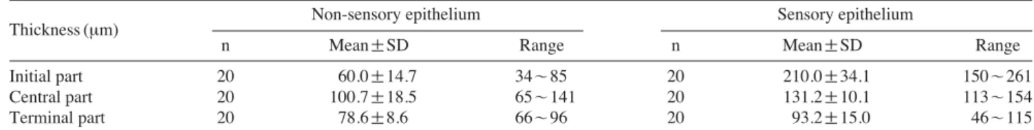
circle and the elongated-oval in the cross-sectional mor- phology of each part, the olfactory epithelium falls into two types, sensory at the dorsal and non-sensory epithe- lium at the ventral and is getting increase of the sensory and decrease of the non-sensory from initial to terminal in ratio covering a total area (Fig. 3A-C). The thickness of sensory epithelium in the canal is 210 μm (vs 60 μm in non-sensory), 131 μm (vs 100.7 μm), 93.2 μm (vs 78.6 μm), respectively (Table 1). Accordingly, the sensory epi- thelium is the thickest at the initial part while the non- sensory is at the central. The sensory epithelium is of transitional layer and multi-cellularity structure which is made of the receptor, supporting, basal and mucus cells.
The receptor cells ruddily stain with eosin is situated in
between supporting cells and is being elongated vertical- ly. The supporting cells which are a major component of the epithelium show irregularly and relatively small cell body in the sensory epithelium but the vertically long cell body in the non-sensory epithelium. The basal cells as cuboidal form being arranged side by side at the basement membrane exhibit a big cell body relatively than other type of cells (Fig. 3D).
The apical part in sensory epithelium has mucus cells that possess the nucleus situated at the bottom. Through AB (pH. 2.5)-PAS staining method, the mucus cells showed a strong positive reaction as a reddish-purple tint (Fig. 3E). The non-sensory epithelium comprises only supporting and basal cells (Fig. 3F).
Fig. 3.Histological characteristics of each part of the canal, sensory and non-sensory epithelium stained with AB-PAS (pH 2.5) (E, F) and hematoxylin-eosin (A-D). (A), the terminal part is the more elongated form and each part are typically the sensory at dorsal and the non-sensory epithelium at ventral. (B), the central part is the elongated form; (C), the initial part is the closely circle form. (D), the sensory epithelium with a unequable surface structure is multi cell structure consisting of the receptor cell, the supporting cell and basal cell. (E), the apical part in sensory epithelium has the neutral mucus cell. (F), the non-sensory epithelium comprises supporting and basal cell. Star, sensory epithelium; n, non- sensory epithelium; solid arrow, receptor cell; arrowhead, mucus cell; broken arrow, basal cell; LP, lamina propria; SC, supporting cells. Bars indicate 100 μm in (A), (B), (C) and 20 μm in (D), (E), (F), respectively.
3. Scanning electron microscopy
In study of SEM observation, the sensory epithelium developed in the canal is not only the elongated corni- form entirely in distributional type (Fig. 2), but is also characterized by presence of receptor cell as only cilia- ted. In receptor cell, the cilia arising in apical part of the cell are 3 or 4 in number and the distance between this cells is 2~2.5 μm, roughly. A few long kinocilias are also distributed on the surface (Fig. 4A). On the other hand, on non-sensory epithelium the surface is furrowed and sensory elements were not observed (Fig. 4B). On the epithelial surface of two nasal sacs, the mucin drop- lets occur commonly in both of the sacs (Fig. 4C).
DISCUSSION
Scartelaos gigas possesses the paired olfactory organ comprising respectively the one elongated canal and two nasal sacs, lacrimal and ethmoidal nasal sac. The feature in the anatomical structure of the olfactory organ in S.
gigas resemble that of Boleophthalmus boddarti and S.
histophorus as a congener with the S. gigas, but is dif- ferent from two species above, having lacrimal and eth- moidal nasal sac in the position of nasal sac. In addition, until now, most of species in Periophthalmus known as fish more terrestrial than the S. gigas have only one nasal sac (Horn et al., 1999). Thus, the degree of adaption from sea to land environment may be causative of extensive variation on place of nasal sac. But, the hypothesis in
the point of this view is needed further comparative stu- dies with olfactory organ of each other species in Gobi- idae. In teleost, the nasal sac expanded from olfactory cavity not only provides the environment of interaction between odorant and receptor cell, but also may be evol- utionally correlated with structural variation of morpho- logy (Hara and Zielinski, 2007). Most of species in Per- ciforms have only two nasal sacs, except for a few spe- cies belonging to Oxudercinae, amphibious fish (Kuciel et al., 2013).
Yamamoto (1982) suggested the category of olfactory organ classified by arrangement of the sensory epithe- lium, number of lamella, their shape and organization and type of receptor cells, and opined that this organ in fish is relation with their ecological habit or olfactory ability. S. gigas is characterized by absence of rosette- like structure and the occurrence of sensory area in olfac- tory canal, similarly to previous studies in Oxudercinae, but there are evident differences in view from arrange- ment of sensory epithelium and type of receptor cells (Kuciel et al., 2013). S. gigas is entirely similar to B.
boddarti and S. histophorus as a cuneiform in distribu- tional pattern of sensory epithelium, but is different in aspect of that arrangement of sensory field shows no con- tinuous in detail. Continuously, Periophthalmus barba- rus, P. variabilis and P. chrysospilos is of form of islet.
In morphology of receptor cell, B. boddarti has giant cell whereas S. gigas has only receptor cell with cilia (Kuciel et al., 2013). It may be expected that ability of olfaction in fish is closely related on the size of olfac- tory organ with distribution of sensory epithelium and
Table 1.Measurements of the olfactory epithelial thickness in the canal. N==number of determinations
Non-sensory epithelium Sensory epithelium
Thickness (μm)
n Mean±SD Range n Mean±SD Range
Initial part 20 60.0±14.7 34~85 20 210.0±34.1 150~261
Central part 20 100.7±18.5 65~141 20 131.2±10.1 113~154
Terminal part 20 78.6±8.6 66~96 20 93.2±15.0 46~115
Fig. 4.Scanning electron micrographs of the surface of epithelium in olfactory organ of Scartelaos gigas. (A), the long kinocilias (arrowhead) and receptor cells with cilia (solidarrow) showing on the surface of sensory epithelium in the canal. (B), the surface of non-sensory epithelium in the canal is furrowed and have no sensory elements. (C), there are a lot of mucin droplets over the epithelium in nasal sacs. MD, mucin droplet.
Bars indicate 2 μm, 30 μm, 50 μm, respectively.
receptor cell (Yamamoto, 1982). In fish, lamella folded on inner wall in olfactory cavity or rosette built of one or several lamella increase surface of olfactory epithe- lium; this structure is related to odorant sensitivity (Mer- edith and Kajiura, 2010). Moreover, fish having high olfactory sensitivity, Oncorhynchus mykiss and Salmo gairdneri possess 4 or more cilia radiating a receptor cell (Hara, 1986; Zielinski and Hara, 1988), but P. bar- barus, congener with S. gigas, known as low sensitivity fish does not exceed 4 in number of cilia (Kuciel et al., 2011).
The mucus secreted over olfactory epithelium provi- des function of intermediate in between receptor cells and environments to help in binding chemical odorants, and protect the cell against hazard materials in habitat (Shephard, 1994). As the mucus droplets in S. gigas is unique, and were observed extensively only in two nasal sacs that does not have sensory elements, but not in canal. Presence of substances like a mucin mass in olfac- tory organ was reported in Sperrata aor and Labeo bata (Chakrabarti and Ghosh, 2011; Ghosh and Chakrabarti, 2011). It is considered that the presence of the mucin droplet and mucus cell in our result may be a distinctive form to adapt to its dry and environment extremely and changeably with hazardous materials, as well as mudflat in intertidal zone.
In epithelium of canal in S. gigas, the layers of two distinctive types, sensory and non-sensory epithelium, occur dorsally and ventrally, respectively, and receptor cell is only in epithelium at dorsal. Especially, among the structures above, it is considered that the transitional layer as multi-cell comprising sensory epithelium in the canal may be a role in keeping the cells of olfactory epi- thelium through changing the size of space in canal, affected by force letting in water (Kuciel, 2013). Mud- skippers in Gobiidae have been known to do ventilation by sniffing; this performance improves circulation of water or gas exchange in nasal sac (Nevitt, 1991; Murphy and Stacey, 2002; Belanger et al., 2006; Meunier et al., 2013).
Mudskippers belonging to Gobiidae have a special and prominent morphology in eyes for surviving life, inclu- ding avoiding predator and feeding out of water (Sayer, 2005). So, Peripheral olfactory characters of S. gigas is concluded to rely less on detecting environmental che- mistry by olfaction than vision, and to protect epithelial tissue against dry in olfactory organ during ebb tide; the mudskipper is included as eye fish in category of Teich- mann (1954), and to support this result further study on vision of S. gigas is needed. From this study, we confir- med that the characteristic of olfactory organ in S. gigas is adapted to semi-aquatic life associated with its ecol- ogical habits.
REFERENCES
Belanger, R.M., L.D. Corkum, W. Li and B.S. Zielinski.
2006. Olfactory sensory input increases gill ventila- tion in male round gobies (Neogobius melanostomus) during exposure to steroids. Comparative Bioche- mistry and Physiology Part A: Mol. Integr. Physiol., 144: 196-202.
Chakrabarti, P. and S.K. Ghosh. 2011. The structural organ- ization and functional aspects of the olfactory epithe- lium of tigerperch, Terapon jarbua (Forsskal, 1775) (Perciformes: Terapontidae). Turk. J. Zool., 35: 793- 799.
Chung, E.Y., B.S. Ryu and J.R. Kim. 1989. A study on the process of the ovarian maturation of the blue-spotted mud hopper, Boleophthalmus pectinirostris (Linna- eus). Mar. Dept. Res., Kunsan National University, 1 (1989): 19-36. (in Korean)
Chung, E.Y., C.M. Ah and T.Y. Lee. 1991. Sexual matura- tion of the blue-spotted mud hopper, Boleophthalmus pectinirostris (Linnaeus). Bull Korean Fish Soc., 24:
167-176. (in Korean)
Ghosh, S.K. and P. Chakrabarti. 2013. Architectural pattern of different cells lining the olfactory epithelium of long-whiskered catfish, Sperata aor (Hamilton). Mes- opot. J. Mar. Sci., 28: 69-80.
Gurr, G.T. 1956. A practical manual of medical and biolo- gical staining techniques. Interscience, New York, pp. 1-99.
Hara, T.J. 1975. Olfaction in fish. Progress in neurobiology, Oxford, 5: 271-335.
Hara, T.J. 1986. Role of olfaction in fish behaviour. In: The behaviour of teleost fishes. Springer US, pp. 152-176.
Hara, T.J. and B. Zielinski. 2007. Olfaction. Sensory Sys- tems Neuroscience, Academic press, San Diego/Lon- don, pp. 1-43.
Horn, M.H., K.L.M. Martin and M.A. Chotkowski. 1999. Int- ertidal Fishes: Life in Two Worlds. Academic Press, San Diego, 399 pp.
Jakubowski, M. 1967. A method for the manifestation of lat- eral-line canals and their neuromasts in fishes. Cop- eia, 1: 234-235.
Jakubowski, M. 1975. Anatomical structure of olfactory organs provided with internal nares in the Antarctic fish Gymnodraco acuticeps Boul. (Bathydraconidae).
Bull. Acad. Pol. Sci., Ser. Sci. Biol., 23: 115-120.
Jeong, S.J., K.H. Han, J.K. Kim and D.S. Sim. 2004. Age and growth of the blue spot mudskipper (Boleoph- thalmus pectinirostris) in the mud flat of southwest- ern Korea. J. Korean Fish Soc., 37: 44-50. (in Kor- ean)
Kim, J.K. and S.J. Jeong. 2007. Growth estimation of 0-aged blue spot mudskipper Boleophthalmus pectinirostris using length frequency data. J. Korean Fish Soc., 40:
50-52. (in Korean)
Kim, J.K., H.J. Baek, J.W. Kim, D.S. Chang and J.I. Kim.
2011. Sexual maturity and early life history of the mudskipper Scartelaos gigas (Pisces, Gobiidae): imp- lications for conservation. Fish Aquat. Sci., 14: 403- 410.
Kim, J.K., J.Y. Kim, J.I. Kim, S.T. Kim, S.D. Hwang, Y.I.
Seo, D.S. Sim, D.S. Chang, J.I. Choi, J.H. Park, S.Y.
Kim, S.J. Jeong, E.K. Chu, K.H. Han and C.W. Oh.
2005. Population dynamics of mudskippers in the southwestern Korea. In: NFRDI (ed) NFRDI Techni- cal Report in 2004. NFRDI, Busan, pp. 225-272. (in Korean)
Kuciel, M. 2013. The mechanism of olfactory organ ventila- tion in Periophthalmus barbarus (Gobiidae Oxuder- cinae). Zoomorphology, 132: 82-85.
Kuciel, M., K. ˙Zuwala and M. Jakubowski. 2011. A new type of fish olfactory organ structure in Periophtha- lmus barbarus (Oxudercinae). Acta Zool., 92, 276- 280.
Kuciel, M., K. Zuwala and U. Satapoomin. 2013. Compara- tive morphology (SEM) of the peripheral olfactory organ in the Oxudercinae subfamily (Gobiidae, Per- ciformes). Zool. Anz. A J. Comp. Zool., 252: 424- 430.
Lin, P.L., K.T. Shao and J.P. Chen. 1994. Five new records of coastal fishes from western Taiwan. Zool. Stud., 33: 174-176.
Meredith, T.L. and S.M. Kajiura. 2010. Olfactory morpho- logy and physiology of elasmobranchs. The Journal of experimental biology, 213: 3449-3456.
Meunier, B., B. White and L.D. Corkum. 2013. The role of fanning behavior in water exchange by a nest-guard- ing benthic fish before spawning. Limnology &
Oceanography, 3: 198-209.
Murdy, E.O. 1989. A taxonomic revision and cladistic ana- lysis of the Oxudercine Gobies (Gobiidae: Oxuder- cinae). Records of the Australian Museum Suppl., 11:
1-93.
Murphy, C.A. and N.E. Stacey. 2002. Methyl-Testosterone Induces Male-Typical Ventilatory Behavior in Res- ponse to Putative Steroidal Pheromones in Female Round Gobies (Neogobius melanostomus). Hormo- nes and Behavior, 42: 109-115.
Nevitt, G.A. 1991. Do fish sniff? A new mechanism of olf- actory sampling in pleuronectid flounders. J. Exp.
Biol., 157: 1-18
Park, K.D., J.K. Kim, D.S. Chang, J.I. Kim and C.W. Oh.
2008. Age and growth of the mudskipper, Scartelaos gigas (Perciformes, Gobiidae) from Korea. Animal Cells and Systems, 12: 305-311.
Randall, J.E. and K.K.P. Lim. 2000. A checklist of the fishes of the South China Sea. Raffles Bull. Zool. Suppl., 8: 569-667.
Ryu, B.S., I.S. Kim and Y. Choi. 1995. Ecology and life his- tory of Boleophthalmus pectinirostris in Korea. J.
Korean Fish. Soc., 28, 316-324. (in Korean)
Sayer, M.D. 2005. Adaptations of amphibious fish for sur- viving life out of water. Fish and Fisheries, 6: 186- 211.
Shephard, K.L. 1994. Functions for fish mucus. Reviews in Fish Biology and Fisheries, 4: 401-429.
Singh, N. 1994. Scanning electron microscopic study of the olfactory epithelium of four coldwater hillstream tel- eosts from Garhwal hills (India). Journal of Biosci., 19: 91-102.
Singh, W. and H.R. Singh. 1986. A SEM study of the olfac- tory rosettes of some hillstream fishes. Folia Morph- ologica, 34: 331-335.
Teichmann, H. 1954. Vergleichende Untersuchungen an der Nase der Fische. Zeitschrift fur Morphologie und Okologie der Tiere, 43: 171-212. (in German) Yamamoto, M. 1982. Comparative morphology of the peri-
pheral olfactory organs in teleosts. In: Hara TJ (ed.), Chemoreception in fishes. Elsevier Scientific Pub.
Co., pp. 39-59.
Zielinski, B. and T.J. Hara. 1988. Morphological and physi- ological development of olfactory receptor cells in rainbow trout (Salmo gairdneri) embryos. J. Comp.
Neur., 271: 300-311.
한국산 남방짱뚱어 Scartelaos gigas의 후각기관 구조에 관한 연구
김현태∙이용주1∙박종성∙박종영
전북대학교 자연과학대학, 1전주교육대학교 과학교육과
요 약 :수중과 육상의 이중적인 생활을 하는 남방짱뚱어 Scartelaos gigas (amphibious mudskipper)의 후각기 관을 해부학과 조직학적으로 연구하였다. 그 결과 후각기관은 좌우 한 쌍이 존재하며 각 기관은 한 개의 가느다 란 긴 관 (an elongated canal)과 사비강과 누비강의 두 개의 비강 (ethmoidal and lacrimal nasal sacs)으로 구성되 어 있었다. 각 관의 내벽에 발달한 감각 상피는 3~4개의 섬모를 가지는 감각 수용기 세포 (receptor cell), 지지 세포 (supporting cell), 기저세포 (basal cell) 그리고 점액세포 (mucus cell)들로 구성되는 다세포성 구조로 이루어 져 있었다. 반면에 비감각 상피는 감각기를 갖지 않는 주름진 표면만을 가지고 있었다. 또한 두 개의 비강은 상 피 표면에 점액 방울 (mucin droplet)을 가지고 있었다. 따라서 남방짱뚱어의 이러한 후각기관의 특징은 생태적 습성과 서식처에 관련된 반수중 생활 (semi-aquatic life)에 적응한 특징으로 사료된다.
찾아보기 낱말 :Scartelaos gigas, 후각기관, 생태적 습성, 서식처