—179— ISSN: 1225-8598 (Print), 2288-3371 (Online)
서 론
일반적으로 어류의 난모세포는 여포세포층(follicular layer) 과 방사대 (zona radiata) 그리고 난세포질 (oocyte plasma membrane) 등 3층으로 구성되어 있다. 특히 여포세포층은 theca cell과 follicular cell (==granulosa cell)로 구성되는데 theca cell은 방어기능을 수행하는 반면에 여포세포는 호흡, 난모세포와 배 발생에 영양공급, 효소분비, 난황 및 steroid 형성 등 많은 기능을 수행하고 있다 (Kjesbu and Kryvi, 1989; Hirai, 1993; Riehl and Greven, 1993). 이러한 다양한 기능 이외에도 일부 어류에서는 여포세포층에서의 난막 구 조변화 (egg membrane, adhesive material, adhesive mem-
brane, egg envelope, vitelline envelope)가 나타나기도 한다.
이러한 난막구조는 종의 산란습성과 관련된 부착기능 (ad- hesive function), 수압 조절, 배아 보호 기능뿐 아니라 분류 학적 목적으로도 널리 이용되어 왔다(Blaxter, 1969; Lagler et al., 1977; Laale, 1980; Groot and Alderdice, 1985; Erickson and Pikitch, 1993; Thiaw and Mattei, 1996; Park and Kim, 2001a, b; Kim and Park, 2013). 또한 체외수정을 선택한 대 부분의 담수어류의 정자 머리에는 첨체를 보유하지 않기 때문에 난자의 동물극 쪽에 위치한 난문(micropyle)을 통 해 수정이 이루어지는데 이들의 난문은 종마다 모양과 크 기가 다양하게 나타남으로써 종특이성을 가지고 있다. 일반 적으로 어류에서 난문은 다른 종의 정자가 침입하는 것을 방해하고 다수정을 방지하는 역할을 수행하는 형태적 특이 성을 가지고 있어 종을 분류하는 중요한 특징으로 이용되 고 있다(Park, 1996; Yoon, 1999; Chen et al., 1999; Mori-
멸종위기 어류 돌상어 Gobiobotia brevibarba의 난자형성과정과 난막의 형태
최웅선∙김재구∙박종영*
전북대학교 자연과학대학 생명과학과, 전북대학교 생물다양성연구소
Structure of Egg Envelope and Oogenesis of the Korean Endangered Fish Gobiobotia brevibarba (Pisces:
Cyprinidae)
by Wung Sun Choi, Jae Goo Kim and Jong Young Park* (Department of Biological Science and Institute for Biodiversity Research, College of Natural Sciences, Chonbuk National University, Jeonju 561-756, Korea)ABSTRACT A histological study on the egg envelope and oogenesis of Gobiobotia brevibarba (Pisces, Cyprinidae) was carried out by a light microscope and a scanning electron microscope. Various developmental cells appeared in the ovary caught during May 2014, spawning season. For the relative area of oocyte, the ovary consisted of mature stage (74.5%), a vitellogenic stage (yolk granule stage, 16.6% and yolk vesicle stage, 6.6%) and previtellogenic stage (perinucleolus stage 2.2%), which means its spawning season. The cytoplasm of the perinucleolus oocyte is acidic and many nucleoli are locat- ed at the inner side of the nuclear membrane. The yolk vesicles, an early vitellogenic stage, has a follic- ular layer and a zona radiata clearly. Numerous villi, called an egg envelope, begin to form on the zona radiata. The yolk granules, an another vitellogenic stage, proceeds and they show a strong eosinophilic nature. Such yolk granules appeared between the yolk vesicles occupying most cytoplasm, and as the stage proceeds, there are some yolk masses fused with each other. Egg envelope is covered with plenty of villi (2~
~3 μμm in the length) over the entire egg surface.Key words : Egg envelope, villi, oogenesis, Gobiobotia brevibarba, endangered fish
*Corresponding author: Jong Young Park 82-63-270-3344 Fax: 82-63-270-3362, E-mail: [email protected]
Accepted: September 25, 2014
http://www.fishkorea.or.kr
sawa, 1999; Debus et al., 2002).
돌상어는 잉어목 잉어과 모래무지아과에 속하는 담수어 류로 물이 깨끗하고 유속이 빠르며, 바닥에 자갈이 깔린 곳 에 서식하고 주로 수서곤충을 먹고 산다. 우리나라의 한강, 임진강, 금강에만 서식하는 한국고유종 현재 멸종위기 야생 생물 II급으로 지정되어 보호받고 있다 (Kim and Park, 2002). 돌상어에 관한 연구는 난발생 및 초기생활사(Ko et
al., 2011), 식성 (Choi et al., 2001b), 산란생태 (Choi et al.,2001a) 등은 있었지만 난자형성과정과 난막의 형태에 관한 연구는 아직까지 보고된 바 없고, 현재 서식처 교란으로 인 해 개체수가 급격히 감소하고 있는 현 실정에서 생식세포 형성과정에 대한 연구의 필요성이 절실하다. 따라서 돌상어 의 난자형성과정과 산란특성에 따른 부착구조를 조사하여 국내에 출현하는 3종의 꾸구리속(Gobiobotia) 어류 연구에 대한 기초자료를 얻고자 한다.
재료 및 방법
1. 실험재료
돌상어 포획은 금강유역환경청의 포획허가(허가번호 제 2014-7)를 받은 후 2014년 5월에 충남 금산군 제원면 용화 리 일대에서 족대(망목 4×4 mm)를 이용하여 5개체(전장 8.5~10.3 cm)를 채집하였다. 이후 살아있는 상태의 돌상어 를 실험실로 운반하여 실험을 진행하였다.
2. 실험방법
1) 광학현미경 관찰
채집된 5개체 중 3개체의 난소를 적출하여 4�C의 10%
formalin에 12시간 고정한 후 12시간 수세하였다. 일반적인 ethanol 탈수법으로 탈수한 다음 xylene으로 치환하여 paraf- fin으로 포매하였고 block을 제작하였다. Block은 microtome (Leica-820, Germany)으로 5 μm 간격으로 절단하여 hema- toxylin과 eosin으로 이중 염색하여 표본을 제작하였다. 광학 현미경(Carl zeiss, AX10, Germany)을 이용하여 난소를 관찰 하였고 난소 내 분화단계별 세포의 면적은 Axio vision (Carl zeiss, Germany)으로 0.01 μm2까지 측정하여 백분율로 환산 하였다.
2) 주사전자현미경 관찰
2014년 5월에 채집한 성체로부터 직접압박법을 통해 얻 은 난을 재료로 0.1 M phosphate buffer (pH 7.4)로 조정된 2.5% glutaraldehyde에 4�C에서 4시간 전고정 후 1% osmi- um tetroxide (OSO4)로 90분간 후고정하여 ethanol series를 통해 탈수시켰다. Tert-butyl alcohol (TBA)을 이용하여 동결
건조시킨 뒤 OSO4를 증착시켜 주사전자현미경(JEOL JSM- 6400, Japan)으로 관찰하였다.
결 과
산란기의 돌상어 난소는 체강의 등 쪽에 낭상형으로 배치 되어 있었으며, 길이는 체강과 비슷하였다. 제작된 슬라이드 를 광학현미경으로 관찰한 결과, 난소 내에는 염색인기 (chromatin-nucleolus stage), 주변인기(peri-nucleolus stage), 난황포기(yolk vesicle stage), 난황구기(yolk granule stage), 성숙기(mature stage) 등 다양한 분화단계의 생식세포들이 존재하였다(Fig. 2A). 이러한 난세포들의 상대면적은 성숙 란(74.5%)이 가장 많은 부분을 차지하고 있었으며, 난황물 질이 형성되는 난황구기(16.6 %), 난황포기(6.6%), 그리고 주변인기(2.3%) 순으로 구성되었다(Figs. 1, 2).
염색인기의 난세포는 주변인기로 진행되었고 핵막의 안 쪽에는 여러 개의 인(nucleolus)들이 배열하고 있었으며, 세 포질은 hematoxylin에 강하게 염색되었다. 난세포의 외측에 는 단층의 편평상피세포(squamous epithelial cell)로 구성된 여포세포층이 뚜렷하게 발달하기 시작하였다. 주변인기의 난세포는 직경 98.9~215.4 μm (평균 160.1±39.5 μm)로 다 양한 크기를 보였다(Fig. 2B). 난황포기의 난세포에서는 핵 내에 존재하는 인들의 크기가 작아지면서 핵막 인근으로 이 동하였고, 세포질에는 외측에서부터 난황포(yolk vesicle)가 형성되기 시작하였다 (Fig. 2C). 난황구기의 난세포에서는 호산성의 난황 과립들이 핵막 주변의 내질층으로부터 피질
180 최웅선∙김재구∙박종영
Fig. 1.Cell-type distribution represented by relative area of four dif- ferent types of oocytes in ovaries of Gobiobotia brevibarba in May 2014. MS, mature stage; PNS peri-nucleolus stage; YGS, yolk granule stage; YVS, yolk vesicle stage.
90
80
70
60
50
40
30
20
10
0 PNS YVS YGS MS
Relative area of oocyte in ovary
%
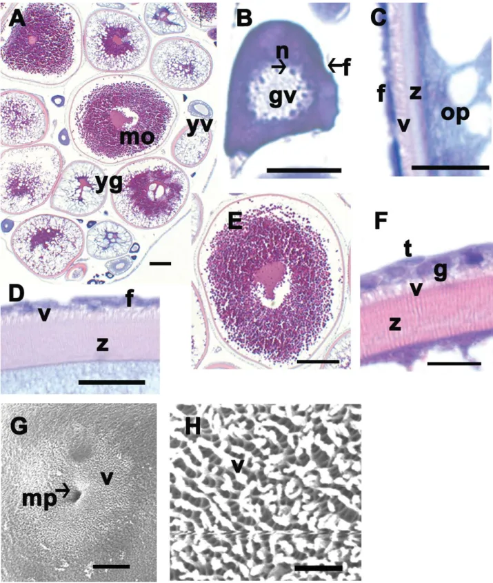
Fig. 2.Photos of a light microscopy (A-F) and a scanning electron microscopy (G and H). A-F, Transverse section with hematoxylin and eosin staining. A, There are various development stages in the full-grown ovary. B, Early perinucleolus stage with many nucleolus arranged along inner nuclear membrane. C, Late yolk vesicle stage during vitellogenesis. The formation of follicular layer (f), villus (v) on the zona radiata (z) begins rapidly. D, Early yolk granule stage. E, Mature stage. F, Follicular layer has theca cell (t) and granulosa cell (g). H, Lots of villi (v) cover the whole full grown oocyte (z). f, follicular layer; gv, germinal vesicle; mo, mature oocyte; mp, micropyle; n, nucleolus; op, ooplasm; v, villi; yg, yolk granule; yv, yolk vesicle; z, zona radiata. Scales indicate 200 μm (A), 50 μm (B), 10 μm (C), 20 μm (D), 200 μm (E), 10 μm (F), 67 μm (G), 7 μm (H), respectively.
층으로 확산되었다(Fig. 2D). 발달과정이 진행됨에 따라 성 숙기의 난세포는 평균 1151.3±64.7 (1052.2~1251.3) μm까 지 커졌으며 이미 형성된 난황 과립들은 서로 융합하면서 세포질의 대부분을 차지하고 있었다(Fig. 2E). 난황구기에 서 성숙기로 진행되면서 여포세포와 theca cell로 구성된 2 층의 여포세포층이 증가하였으며, 세포질과 여포세포층 사 이에 줄무늬모양(striated appearance)의 방사대는 두꺼워졌 다(Fig. 2D). 성숙란 시기의 방사대는 약 10~15 μm 두께를 가지며 호산성을 보였다(Fig. 2D, F). 방사대와 여포세포층 사이에 존재하는 융모(villi)는 난황포기의 난세포에서 출현 하기 시작하여 성숙란에서는 그 길이가 2~3 μm까지 길어 졌다(Fig. 2C, D, F, G, H).
주사전자현미경을 이용하여 관찰한 결과, 성숙란의 표면 에는 깔때기 모양의 난문이 관찰되었으며, 그 외경은 약 13 μm였으나 내경이 점차 작아지는 특징을 보였다(Fig. 2G).
또한 난문을 제외한 대부분의 난표면에는 2~3 μm 길이의 미세융모가 일정하게 분포하고 있었다(Fig. 2H).
고 찰
경골어류 성어의 난소는 나출형과 낭상형으로 구분되는데 (Park,1996; Parenti and Grier, 2004; Koya and Munoz, 2007), 돌상어의 난소는 체강의 앞쪽에서 좌우 한 쌍으로 나타났 으며 항문 부근에서 융합된 낭상형으로 체강의 길이와 비슷 한 크기로 존재하고 있었다. 이러한 특징은 모래무지아과 어류인 Coreoleuciscus splendidus (Kim et al., 2010), Pseu-
dorasbora parva (Kim et al., 2007)의 난소와 유사한 형태를보이고 있었다. 이는 여울부에 서식하는 저서성 어류 모래 무지아과에서 나타나는 특징으로 보인다. 부레를 갖는 Rhyn-
chocypris oxycephalus (Kim et al., 2009)와 해산어류인 양볼락과 어류 Scorpaena notata (Munoz et al., 2002)의 난소들 은 쌍으로 구성되어 있어 돌상어와는 차이를 보였다.
성숙한 돌상어의 난소 내에는 핵 안에 인이 산재한 주변 인기와 세포질 외측에 난황포가 형성되는 난황포기, 난황포 에 난황구를 축적하여 난황물질이 핵막 주변의 내질층으로 부터 피질로 확산 중인 난황구기 등 다양한 분화단계의 생 식세포들이 관찰되었고 성숙기 난세포의 평균 크기는 1151.3 (1052.2~1251.3) μm로 이전의 난발생 연구(Ko et al., 2011) 와 거의 일치하였다. 또한 성숙기 난세포의 최외각에는 편 평한 모양의 여포세포가 층을 이루고 있었으며 약 10~15 μm 두께의 방사대가 관찰되었다. 일반적으로 난막의 두께는 부성란이 아닌 침성란, 난태생어류보다 난생어류, 친어가 알 을 보호하지 않는 경우, 유속이 더 빠른 곳에 서식하는 어 류에서 더 두꺼운 것으로 알려져 있다(Guraya, 1986). 따라 서 두꺼운 난막을 가지는 돌상어는 난생의 침성란으로 유속
이 빠른 곳에서 산란하는 생태학적 특성을 잘 반영한다고 사료된다.
돌상어의 난막 표면을 광학현미경과 주사전자현미경으로 관찰한 결과, 많은 수의 융모가 관찰되었는데 이러한 미세 융모는 난황포가 형성되는 시기에 발달하기 사작하여 난황 물질들이 형성됨에 따라 더욱 뚜렷해졌다. 이와 같이 난막 에 존재하는 다양한 구조들은 부착기능, 수압 조절, 배아 보 호 기능 등을 수행하는 것으로 알려져 있다(Laale, 1980;
Hiromi, 1984; Rizzo et al., 2002). 특히 저서성 어류인 한국 산 미꾸리과 어류는 난막표면 구조물을 과립형 (granular form), 융모형(villous form), 사상형(filamental form), 거치형 (saw-shaped form), 소구형 (hillock-shaped form), 울타리형 (fence-shaped form) 그리고 무구조형(non-structural form) 등 7가지로 분류하였고, 이러한 구조는 산란 장소와 깊은 관계 가 있는 것으로 보고하였다(Park 1996). 또한 이러한 난막 의 부착구조는 서식환경과 밀접한 생태적 연관성을 보이고 있으며 일부 분류군에서는 분류학적 특징으로 이용되기도 한다 (Groot and Alderdice, 1985; Hirai, 1993; Yoon, 1999;
Park and Kim, 2001a, 2001b; Rizzo et al., 2002; Kim et al., 2011; Kim and Park, 2013). 돌상어의 난막에서는 외경이 크 고 내경이 작아지는 깔때기 모양의 한 개의 난문이 관찰되 었는데 이는 종의 특이성을 보일 뿐 아니라 다른 종의 정자 가 침입하는 것을 방어하고 다수정 방지 기능을 수행하는 것으로 알려져 있다(Grierson and Neville, 1981; Cameron and Hunter, 1984). 또한 일부 어류에서는 난문의 수가 2개 이상 나타나고 있어 중요한 분류학적 특징으로 이용되고 있 다(Chen et al., 1999; Morisawa, 1999; Debus et al., 2002).
이상과 같이 돌상어의 난자형성과정과 난막의 미세구조 에 대한 연구는 국내의 멸종위기 야생생물로 지정된 꾸구리 속 어류 3종의 생태학적 특성을 파악할 수 있는 기초자료와 분류학적 특성을 제공할 수 있을 것으로 생각된다.
요 약
잉어과(Pisces, Cyprinidae)에 속하는 멸종위기 어류인 돌 상어 Gobiobotia brevibarba의 난막미세구조 및 난자형성과 정을 광학현미경과 주사전자현미경을 이용하여 관찰하였다.
2014년 5월의 돌상어 난소 내에는 다양한 발생단계의 생식 세포들이 관찰되었다. 난모세포들의 상대면적은 성숙란(74.5
%), 난황물질이 형성되는 난황구기(16.6%), 난황포기(6.6%), 그리고 미숙한 발달단계인 주변인기(2.3%) 순으로 구성되 어 있었다. 주변인기 단계의 세포질은 hematoxylin에 강하 게 염색되며, 핵막의 안쪽에는 다수의 인들이 존재하였다.
난황형성물질인 난황포기와 초기 난황구기 단계에서는 여 포층과 방사대가 뚜렷하게 형성되기 시작되었으며, 방사대
182 최웅선∙김재구∙박종영
위에는 미세융모의 난막구조가 더욱 뚜렷해졌다. 난황구기 및 성숙란의 단계에서는 난황구 일부가 융합되면서 강한 호산성을 보였다. 또한 성숙란의 표면에는 깔때기 모양의 난문이 관찰되었으며, 2~3 μm 길이의 미세융모가 일정하 게 분포하였다.
인 용 문 헌
Blaxter, J.H.S. 1969. Development: eggs and larvae. In: Hoar, W.S., D.J. Randall and E.M. Donalson (eds.), Fish physiology.
Academic Press, New York, pp. 177-252.
Cameron, I.L. and K.E. Hunter. 1984. Regulation of the permeabil- ity of the medaka fish embryo chorion by exogeneous sodi- um and calciumions. J. Exp. Zool., 231: 447-454.
Chen, K.C., K.T. Shao and J.S. Yang. 1999. Using micropylar ultra- structure for species identification and phylogenetic infer- ence among four species of Sparidae. J. Fish Biol., 55: 288- 300.
Choi, J.S., H.K. Byeon and O.K. Kwon. 2001a. Reproductive Ecology of Gobiobotia brevibarba (Cyprinidae). Korean J. Ichthyol., 13: 123-128. (in Korean)
Choi, J.S., O.K. Kwon, J.H. Park and H.K. Byeon. 2001b. Feeding Habit of Gobiobotia brevibarba (Cyprinidae) from the Hong- cheon River, Korea. Korean J. Ichthyol., 13: 230-236. (in Korean)
Debus, L., M. Winkler and R. Billard. 2002. Structure of micropyle surface on oocytes and caviar grains in Sturgeons. Internat.
Rev. Hydrobiol., 87: 585-603.
Erickson, D.L. and E.K. Pikitch. 1993. A histological description of shortspine thorny head, Sebastolobus alascanus, ovaries:
structures associated with the production of gelatinous egg masses. Env. Biol. Fish., 36: 273-282.
Grierson, J.P. and A.C. Neville. 1981. Helicoidal architecture of fish eggshell. Tiss. Cell, 13: 818-830.
Groot, E.P. and D.F. Alderdice. 1985. Fine structure of the external egg membranes of five species of Pacific salmon and steel- head trout. Can. J. Zool., 63: 552-566.
Guraya, S.S. 1986. Monographs in developmental biology, The cell and molecular biology of fish oogenesis. Karger, 18: 111- 147.
Hirai, A. 1993. Fine structure of the egg membrane in four species of Pleuronectinae. Japanese J. Ichthyol., 40: 227-235.
Hiromi, O. 1984. Electron microscopic study on adhesive material of pacific herring (Clupea pallasi) eggs. Japanese J. Ichthyol., 30: 404-411.
Kim, C.H., J.G. Kim and J.Y. Park. 2011. Structure of egg envelope and oogenesis of Kichulchoia multifasciata (Pisces, Cobiti- dae). Korean J. Electron Microscopy, 41: 189-196. (in Korean) Kim, D.H., B.S. Chang, H.S. Jung, Y.C. Teng, S. Kim and K.J. Lee.
2009. The oogenesis of Chinese minnow, Leuciscinae,
Teleostei. Korean J. Electron Microscopy, 39: 237-243. (in Korean)
Kim, D.H., K.J. Lee, S. Kim and Y.K. Deung. 2007. A study on the oogenesis of false dace (Pseudorasbora parva). Korean J.
Electron Microscopy, 37: 65-72. (in Korean)
Kim, D.H., K.J. Lee, Y.C. Teng, W.J. Kim and S. Kim. 2010. The oogenesis of Coreoleuciscus splendidus, Cyprinidae, Teleo- stei. Korean J. Electron Microscopy, 40: 9-14. (in Korean) Kim, I.S. and J.Y. Park. 2002. Freshwater fishes of Korea. Kyo-hak
Publishing Co., Ltd., pp. 140-141. (in Korean)
Kim, J.G. and J.Y. Park. 2013. Reproductive Cycle of the Korean loach, Kichulchoia multifasciata (Pisces, Cobitidae). Korean J. Ichthyol., 25: 216-221. (in Korean)
Kjesbu, O.S. and H. Kryvi. 1989. Oogenesis in cod, Gradus morhua L., studied by light and electron microscopy. J. Fish Biol., 34: 735-746.
Ko, M.H., S.Y. Park, I.R. Lee and I.C. Bang. 2011. Egg development and early life history of the endanger species Gobiobotia brevibarba (Pisces, Cyprinidae). Korean J. Limnol., 44: 136- 143. (in Korean)
Koya, Y. and M. Munoz. 2007. Comparative study on ovarian struc- tures in scorpaenids: possible evolutional process of repro- ductive mode. Ichthyol. Res., 54: 221-230.
Laale, H.W. 1980. The perivitelline space and egg envelopes of bony fishes, a reviews. Copeia, 210-226.
Lagler, K.F., J.E. Bardach, R.R. Miller and D.R.M. Passono. 1977.
2nd ed. Ichthyology. John Wiley & Sons. pp. 268-310.
Morisawa, S. 1999. Fine structure of micropylar region during late oogenesis in eggs of the hagfish Eptatretus burgeri (Agnatha).
Develop. Growth Differ., 41: 611-618.
Munoz, M., M. Casadevall and S. Bonet. 2002. The ovarian mor- phology of Scorpaena notata shows a specialized mode of oviparity. J. Fish Biol., 61: 877-887.
Parenti, L.R. and H.J. Grier. 2004. Evolution and phylogeny of gonad morphology in bony fishes. Integr. Comp. Biol., 44:
333-348.
Park, J.Y. 1996. A morphological study on the gonad of the species in the family Cobitidae (Pisces, Cypriniformes) from Korea.
Dissertation, Chonbuk National Univ., pp. 6-158. (in Korean) Park, J.Y. and I.S. Kim, 2001a. Fine structure of oocyte envelopes
of the related cobitid species in the genus Iksookimia (Cobiti- dae), Ichthyological Res., 48: 71-75.
Park, J.Y. and I.S. Kim, 2001b. Ultrastructure of the external egg en- velopes in two Cobitid fishes (Cobitidae). Korean J. Ichthyol., 13: 19-23.
Riehl, R. and H. Greven. 1993. Fine structure of egg envelopes in some viviparous goodeid fishes, with comments on the rela- tion of envelope thinness to viviparity. Can. J. Zool., 71: 91- 97.
Rizzo, E., Y. Sato, B.P. Barreto and H.P. Godinho. 2002. Adhesive- ness and surface patterns of eggs in neotropical freshwater teleosts. J. Fish Biol., 61: 615-632.
Thiaw, O.T. and X. Mattei. 1996. Ultrastructure of the secondary egg envelope of Cyprinodontidae of the genus Epiplatys Gill, 1862 (Pisces, Teleostei). Acta. Zool., 77: 161-166.
Yoon, J.M. 1999. Ultrastructure of egg micropyles and zona radiata in three aquacultural teleosts. Korean J. Ichthyol., 11: 126- 133.
184 최웅선∙김재구∙박종영