ISSN 2234-3806 • eISSN 2234-3814
Ann Lab Med 2015;35:423-428
http://dx.doi.org/10.3343/alm.2015.35.4.423
Comparison of FcR γ-Deficient and CD57+ Natural Killer Cells Between Cord Blood and Adult Blood in the Cytomegalovirus-Endemic Korean Population
Hee Jo Baek, M.D.1, Da-Woon Kim, M.D.2,3, Minh-Trang Thi Phan, M.S.2,4,5, Ju-Sun Kim, M.S.5, Ji-Hoon Yang, M.S.6, Jeong Il Choi, M.D.6, Je-Jung Lee, M.D.5, Myung-Geun Shin, M.D.2, Dong-Wook Ryang, M.D.2, Sang-Ki Kim, D.V.M.7, Seung-Hwan Lee, Ph.D.8, Hoon Kook, M.D.1, and Duck Cho, M.D.2,4,5,*
Department of Pediatrics1, Chonnam National University Hwasun Hospital, Chonnam National University Medical School, Gwangju; Department of Laboratory Medicine2, Chonnam National University Medical School, Gwangju; Department of Laboratory Medicine3, KS Hospital, Gwangju; Center for Creative Biomedical Scientists4, Chonnam National University, Gwangju; Research Center for Cancer Immunotherapy5, Chonnam National University Hwasun Hospital, Hwasun; Department of Anesthesiology and Pain Medicine6, Chonnan National University Medical School and Hospital, Gwangju;
Department of Companion & Laboratory Animal Science7, Kongju National University, Yesan, Korea; Department of Biochemistry, Microbiology and Immunology8, University of Ottawa, Ottawa, Canada
Background: FcRγ-deficient natural killer (NK) cells (g¯NK cells) have been associated with cytomegalovirus (CMV) infection. However, the frequency of g¯NK cells in a CMV-en- demic area (i.e., Korea) has not yet been studied. We examined the frequency of g¯NK cells and expression of CD57 on NK cells in cord blood (CB) and adult blood (AB).
Methods: Of the 24 AB samples collected, 95.8% (23/24) were CMV IgG+/IgM-, while 100% of the 13 healthy CB samples were CMV IgG+/IgM-. We performed whole-blood flow cytometry assays to analyze intracellular FcRγ and CD3ζ expression of CD3-/CD56dim NK cells from 13 CB and 24 AB samples, and surface CD57 expression on CD3-/CD56dim/ CD16+ NK cells from 13 CB and 19 AB samples.
Results: All CMV seropositive AB samples contained g¯NK cells (23/23), and the median proportion of g¯NK cells in the CD3-/CD56dim NK cell pool was 35.0% (range: 11-77%).
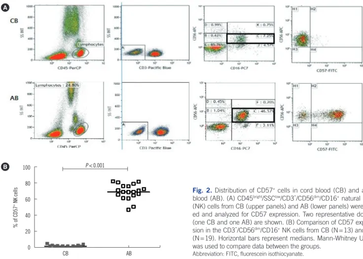
CD57+ NK cells in the CD3-/CD56dim/CD16+ NK cell population were detected in all 19 AB samples tested, but not in any CB samples.
Conclusions: Our data suggest that g¯NK cells and CD57+ NK cells are present at a very high frequency in CMV-seropositive AB, but rare in CMV-naïve CB.
Key Words: Adult, Blood, Cord, Cytomegalovirus, Infection, FcRγ, Natural killer cell
Received: August 24, 2014
Revision received: December 3, 2014 Accepted: April 2, 2015
Corresponding author: Duck Cho Department of Laboratory Medicine, Chonnam National University Hwasun Hospital, 322 Seoyang-ro, Hwasun-eup, Hwasun-gun, Jeollanam-do 519-809, Korea
Tel: +82-61-379-7951 Fax: +82-61-379-7984 E-mail: [email protected]
*Current address: Deparment of Laboratory Medicine and Genetics, Samsung Medical Center, Sungkyunkwan University School of Medicine, 81 Irwon-ro, Gangnam-gu, Seoul 135-710, Korea
Tel: +82-2-3410-2403 Fax: +82-2-3410-2719 E-mail: [email protected] Co-corresponding author: Hoon Kook Department of Pediatrics, Chonnam National University Hwasun Hospital, 322 Seoyang-ro, Hwasun-eup, Hwasun-gun, Jeollanam-do 519-809, Korea
Tel: +82-61-379-7696 Fax: +82-61-379-7697
E-mail: [email protected]
© The Korean Society for Laboratory Medicine This is an Open Access article distributed under the terms of the Creative Commons Attribution Non-Commercial License (http://creativecom- mons.org/licenses/by-nc/3.0) which permits unrestricted non-commercial use, distribution, and reproduction in any medium, provided the original work is properly cited.
INTRODUCTION
Natural killer (NK) cells are innate immune cells that are an in- tegral part of the immune response to certain microbial infec- tions and tumors. In particular, NK cells play a crucial role in the control of herpes virus infection, such as cytomegalovirus (CMV) [1]. NK cell function is regulated by the balance of signals from activating receptors (i.e., NKG2C, NKG2D, NKp30, NKp44, NKp46, CD16), which recognize ligands on tumors and virus- infected cells, and inhibitory receptors (i.e., killer cell immuno- globulin like receptor, NKG2A) that are specific for major histo- compatibility complex class I molecules [1-3].
For the stimulatory signals from NK cell-activating receptors (i.e., CD16, NKp46, NKp30), transmembrane signaling adaptors for signal transduction are required. NK cells express immunore- ceptor tyrosine-based activation motif (ITAM)-bearing adaptor proteins (i.e., FcRγ and CD3ζ), which transmit biochemical sig- nals through ITAMs [2-4]. Recently, Hwang et al. [4] identified a distinct subset of human NK cells that are deficient in FcRγ, but express normal levels of CD3ζ, called FcRγ-deficient NK cells (g¯NK cells). g¯NK cells were readily detectable in 32.0%
(39/122) of healthy blood donors in the United States and were confined to the CD56dim population. A subsequent study by the same group also reported that the presence of g¯NK cells is strongly associated with previous exposure to CMV and that g¯NK cells express significantly higher CD57 and NKG2C levels, but lower NKG2A levels, than conventional NK cells [5].
NK cells expand in response to chronic infections, particularly human CMV. CMV drives expansion of NKG2C+ NK cells, which preferentially acquire CD57 [6]. Therefore, CD57 can be con- sidered a CMV infection-associated marker of NK cells and can be used to determine the association between g¯NK cells and prior CMV infections [5].
CMV infection in the general population is asymptomatic; how- ever, the infection mostly establishes life-long latent infection and can be reactivated when hosts become immunocompromised.
CMV is found in all geographic locations, socioeconomic groups, and ages. Between 36.3% and 90.8% of adults in the United States are CMV-seropositive [7]. Interestingly, some countries show extremely high CMV-seroprevalence, with that in Korea es- timated to be 96% (552/575 individuals) [8-10].
The frequency of g¯NK cells has been studied only in the US population. However, the frequency of g¯NK cells in a CMV-en- demic area (i.e., Korea) has not yet been investigated. We hy- pothesized that the CMV-endemic Korean population has a high frequency of g¯NK cells, and attempted to determine the fre-
quency of g¯NK cells in both CMV-seropositive and CMV-sero- negative individuals. Since CMV IgG-negative samples from healthy adult donors in CMV-endemic Korea are rare, we de- cided to analyze g¯NK cells in umbilical cord blood (CB), which is considered to be CMV-naïve [6].
Here, we examined the frequency of g¯NK cells in CB and adult blood (AB) samples from the Korean population. We also investigated the expression of CD57 on NK cells in AB and CB.
METHODS 1. Ethics statement
All study samples were obtained following acquisition of written informed consent from the study participants or the mothers of infants in the case of CB samples, in accordance with the Decla- ration of Helsinki. This research protocol was reviewed and ap- proved by the institutional review board of Chonnam National University Hwasun Hospital (CNUHH) (Permit Number: 2012- 126).
2. Blood samples
All adults enrolled in this study were healthy and free of HIV, hepatitis B virus, and hepatitis C virus infections. EDTA blood samples from 37 subjects (24 AB and 13 CB [collected at birth from full-term neonates]) left over from ABO/RhD blood typing or complete blood count analysis were used for the study of g¯NK cells, and leftover serum or plasma were used for mea- surement of anti-CMV IgG and IgM. Among these samples, 19 AB and 13 CB samples were used for the assessment of CD57+ NK cells. All samples were obtained from CNUHH and Chon- nam National University Hospital.
3. Measurement of anti-CMV IgG and IgM
Samples were processed within 24 hr of collection. The pres- ence of anti-CMV IgG and IgM antibodies in the sera of 24 Ko- rean healthy adult donors was determined by chemiluminescent microparticle immunoassay (CMIA) (Architect; Abbott Laborato- ries, Abbott Park, IL, USA). Of the AB samples, 95.8% (23/24) were CMV IgG+/IgM- and 4.2% (1/24) were CMV IgG-/IgM-, whereas 100.0% (13/13) of the CB samples were CMV IgG+/ IgM- (Table 1). Although all CB samples were CMV IgG+, these CB samples were regarded as CMV-negative, because anti-CMV IgG antibodies can cross the placenta. Naitou et al. [11] re- ported that qualitative nested PCR for CMV DNA was negative in 40 CB plasma samples. This report supports the assumption that CB is a CMV-naïve sample. In addition, Foley et al. [6] also
used CB in their study to avoid confounding effects from adult donors who may have encountered CMV previously. On the ba- sis of their findings, the CB samples from 13 healthy neonates used in this study were regarded as CMV-negative controls.
4. g¯NK cells analysis
Signaling adaptors (FcRγ and CD3ζ) were detected by using a modification of the previously described method [4]. Instead of using peripheral blood mononuclear cells (PBMCs), whole blood samples (50 µL) were stained for flow cytometry analysis by using phycoerythrin (PE)-conjugated anti-CD3 (clone UCHT1; BD Biosciences, San Jose, CA, USA) and PE-conju- gated Cy5-anti-CD56 antibodies (clone B159, BD Biosciences).
After surface labeling, the cells were washed, fixed, and perme- abilized (IntraPrep kit, Beckman Coulter; Fullerton, CA, USA), according to the manufacturer’s instructions. For detection of intracellular FcRγ and CD3ζ expression, fixed and permeabi- lized cells were stained with fluorescein isothiocyanate (FITC)- anti-FcεRIγ (FcRγ) (Millipore, Temecula, CA, USA) or FITC-anti- CD247 (CD3ζ) (Biolegend, San Diego, CA, USA) antibodies.
Samples were acquired with FACSCalibur system (Becton Dick- inson, San Jose, CA, USA), and the resulting data were ana- lyzed by using CellQuest software (Becton Dickinson). On the basis of these two signal adaptors’ intracellular expression of CD3-/CD56dim NK cells, a distinct subset of human NK cells were identified as g¯NK cells; these cells are deficient for FcRγ expression, but express normal levels of CD3ζ. We used 10%
as an arbitrary cut-off value to define the presence or absence of g¯NK cells for this study.
5. CD57 analysis
Whole blood from AB and CB samples was stained with the fol- lowing antibodies: peridinin chlorophyll protein complex (PerCP)-anti-CD45 (clone 2D1; BD Biosciences), Pacific blue- anti-CD3 (UCHT1, Beckman Coulter), allophycocyanin (APC)- anti-CD56 (clone N901, Beckman Coulter), CD16-PC7 (clone 3G8, Beckman Coulter), and FITC-anti-CD57 (clone HNK-1;
BD Biosciences). After incubation, the red blood cells in whole blood samples were lysed as described above. Samples were acquired with NAVIOS flow cytometer (Beckman Coulter). Dur- ing sample acquisition, low side-scatter (SS) and bright CD45 staining were used to set an electronic gate around the lympho- cyte population. The expression of CD57 on NK cells from the CD3-/CD56dim/CD16+gates was analyzed by using Kaluza soft- ware (Beckman Coulter).
6. Statistical analysis
Median values along with ranges were reported, and a nonpara- metric Mann–Whitney U test was used to compare data be- tween groups. P value of less than 0.05 was considered statisti- cally significant.
RESULTS
1. Distribution of g¯NK cells in CB and AB
We determined the frequency of g¯NK cells in the CD3-/CD56dim NK cell population. Only one AB sample showed 9.8% g¯NK cells, and was thus designated as g¯NK cell-negative, according to our arbitrarily chosen cut-off value of 10%. In the remaining AB samples, the proportion of g¯NK cells ranged from 11% to as high as 77% (median 35%) (Fig. 1A, B). The one AB donor who had 9.8% g¯NK cells was CMV IgG-/IgM-.
We then analyzed the frequency of g¯NK cells in the 13 CB samples. Among the 13 CB samples (all samples were anti- CMV IgG+/IgM-, with no clinical evidence of congenital CMV in- fection), only one sample was designated as g¯NK cell-positive, as it showed 33% of g¯NK cells in the CD3-/CD56dim NK cell pool. The proportion of g¯NK cells in CB samples was signifi- cantly lower than that in AB samples (P <0.001; Fig. 1C).
2. Distribution of CD57
+NK cells in CB and AB
We gated CD45bright/SSClow/CD3-/CD56dim/CD16+ NK cells from 19 AB and 13 CB samples and analyzed the expression of CD57 (Fig. 2A). When CD57 positivity was defined as at least 10% of the CD3-/CD56dim/CD16+ NK cell pool, we could detect CD57+ NK cells in all 19 AB samples tested, with positivity vary- Table 1. Cytomegalovirus antibody status and frequency of g¯NK
cells in healthy adult and cord blood in the Korean population Adult blood Cord blood Anti-CMV IgG, N (%)
Positive 23 (95.8) 13 (100.0)
Negative 1 (4.2) 0 (0.0)
Anti-CMV IgM, N (%)
Positive 0 (0.0) 0 (0.0)
Negative 24 (100.0) 13 (100.0)
g¯NK cells, median % (range)
Prior CMV infection* 35 (11-77); N=23 NA Presumptive CMV naïve† 10; N=1 2 (0-33); N=13
*indicates anti-CMV IgG+ adult blood; †indicates IgG- adult blood or anti- CMV IgG+ cord blood.
Abbreviations: g¯NK cells, FcRγ-deficient natural killer cells; CMV, cytomega- lovirus; NA, not applicable.
ing from 50.5% to 82.0%. In contrast, less than 10% of these NK cells were detected in all 13 CB samples tested (Fig. 2B).
DISCUSSION
In the present study, among the 24 AB samples, 95.8% (23/24)
were CMV IgG+/IgM-, while 100% of the 13 healthy CB samples were CMV IgG+/IgM-. Studies from other CMV-endemic areas, such as Africa and Asia, also demonstrated a high maternal CMV-seroprevalence (90-100%) [12], consistent with our re- sults. In this study, whole blood was used rather than PBMCs for analysis of g¯NK cells and CD57+ NK cells. Single platform Fig. 1. Identification of FcRγ-deficient human NK cells (g¯NK cells) and distribution of g¯NK cells in cord blood (CB) and adult blood (AB).
(A) Representative flow cytometry plots from one CB and one AB samples. CD3-/CD56dim NK cells in CB express both CD3ζ and FcRγ, whereas NK cells in AB express CD3ζ with low levels of FcRγ. (B) Diagram showing the proportion according to the percentage of g¯NK cells among the CD3-/CD56dim NK cells in CB and AB. (C) Comparison of g¯NK cells between CB (N=13) and AB (N=24). Horizontal bars represent medians. Mann-Whitney U test was used to compare data between the groups.
100 80 60 40 20
0 CB AB
P <0.001
% of g-NK cells
A
C B
g-NK cells
CB AB
≤10% 11-40% >40%
92.3%
n=12 62.5%
n=15 33.3%
n=8 8.0%n=1
4.2%n=1
Fig. 2. Distribution of CD57+ cells in cord blood (CB) and adult blood (AB). (A) CD45bright/SSClow/CD3-/CD56dim/CD16+ natural killer (NK) cells from CB (upper panels) and AB (lower panels) were gat- ed and analyzed for CD57 expression. Two representative donors (one CB and one AB) are shown. (B) Comparison of CD57 expres- sion in the CD3-/CD56dim/CD16+ NK cells from CB (N=13) and AB (N=19). Horizontal bars represent medians. Mann-Whitney U test was used to compare data between the groups.
Abbreviation: FITC, fluorescein isothiocyanate.
100 80 60 40 20
0 CB AB
P <0.001
% of CD57+ NK cells A
B
flow cytometry with a lyse-no-wash procedure was used to ana- lyze AB and CB samples to overcome the technical difficulties associated with limited CB volumes. Compared with the density gradient separation method for PBMCs isolation, this method reduces loss of any particular lymphocyte subclass because sample manipulation is minimized [13]. For a more clear-cut discrimination between g¯NK cells and conventional NK cells, an arbitrary cut-off of 10% was chosen, rather than the 3% cut- off used by Hwang et al. [4].
The frequency of g¯NK cells in AB from individuals with prior CMV infection and that in CMV-naïve CB were determined. All CMV-seropositive AB samples contained g¯NK cells (23/23), and the proportion of g¯NK cells in the CD3-/CD56dim NK cell pool was 35.0% (range, 11-77%). Our results are consistent with a previous report that prior CMV infection is associated with a high frequency of g¯NK cells [5]. In addition to the high fre- quency of g¯NK cells, we also found that the proportions of g¯NK cells among CD3-/CD56dim NK cells were relatively high com- pared with those found in healthy US adults [4]. In contrast with
CMV, it has been reported that infection with two common her- pes viruses (HSV-1 and HSV-2) was not associated with a high frequency of g¯NK cells [5].
Recently, CMV has been reported to induce the expansion of CD94/NKG2C+ NK cells in healthy donors as well as in HIV- or hantavirus-infected patients and leukemia patients. The per- centage of CD94/NKG2C+ NK cells remains elevated even after therapeutic intervention and in asymptomatic CMV+ donors who likely contracted the virus during childhood [14-18]. Another study found that NKG2C+ NK cells proliferated and acquired CD57 during acute human CMV infection in solid-organ trans- plant recipients [19]. Furthermore, CD57+NKG2C+ NK cells can be detected in CMV+ healthy adults several years after the pri- mary infection.
In the present study, we examined the expression of CD57 on NK cells in CMV-endemic Korean AB and CB samples. The pro- portion of CD57+ NK cells in the CD3-/CD56dim/CD16+ cell popu- lation in AB was significantly higher than that in CB (P <0.001;
Fig. 2B). This finding is consistent with a previous study showing
that CD56dim NK cells derived from CB almost completely lacked surface expression of CD57 [20]. However, a significant propor- tion of CD57+ NK cells in the CD56dim/CD16+ cell subset was also reported in CMV-seronegative young donors [21]. These findings suggest that the different proportions of CD57+ NK cells in AB and CB are associated not only with CMV exposure but also with other factors, such as the immaturity of CB.
We present for the first time a comparative analysis of g¯NK cells in AB and CB in the CMV-endemic Korean population.
Compared with AB from the US population, AB from the CMV- endemic Korean population had a high frequency of g¯NK cells and CD57+ NK cells, whereas CB samples had a very low fre- quency of g¯NK cells and CD57+ NK cells.
Authors’ Disclosures of Potential Conflicts of Interest
No potential conflicts of interest relevant to this article were re- ported.
Acknowledgments
The authors thank Mr. Yoohyun Kim for his excellent technical assistance. This research was supported by Basic Science Re- search Program through the National Research Foundation of Korea (NRF) funded by the Ministry of Education, Science, and Technology (2010-0023757) and by a grant (CRI13047-21) from Chonnam National University Hospital Research Institute of Clinical Medicine.
REFERENCES
1. Muntasell A, Vilches C, Angulo A, López-Botet M. Adaptive reconfigura- tion of the human NK-cell compartment in response to cytomegalovi- rus: a different perspective of the host pathogen interaction. Eur J Im- munol 2013;43:1133-41.
2. Lanier LL. Natural killer cell receptor signaling. Curr Opin Immunol 2003;15:308-14.
3. Lanier LL. Up on the tightrope: natural killer cell activation and inhibi- tion. Nat Immunol 2008;9:495-502.
4. Hwang I, Zhang T, Scott JM, Kim AR, Lee T, Kakarla T, et al. Identifica- tion of human NK cells that are deficient for signaling adaptor FcRɣ and specialized for antibody-dependent immune functions. Int Immunol 2012;24:793-802.
5. Zhang T, Scott JM, Hwang I, Kim S. Cutting edge: antibody-dependent memory-like NK cells distinguished by FcRɣ deficiency. J Immunol
2013;190:1402-6.
6. Foley B, Cooley S, Verneris MR, Pitt M, Curtsinger J, Luo X, et al. Cyto- megalovirus reactivation after allogeneic transplantation promotes a lasting increase in educated NKG2C+ natural killer cells with potent function. Blood 2012;119:2665-74.
7. Staras SA, Dollard SC, Radford KW, Flanders WD, Pass RF, Cannon MJ.
Seroprevalence of cytomegalovirus infection in the United States, 1988- 1994. Clin Infect Dis 2006;43:1143-51.
8. Lopo S, Vinagre E, Palminha P, Paixao MT, Nogueira P, Freitas MG. Se- roprevalence to cytomegalovirus in the Portuguese population, 2002- 2003. Euro Surveill 2011;16. pii:19896.
9. Cannon MJ, Schmid DS, Hyde TB. Review of cytomegalovirus serop- revalence and demographic characteristics associated with infection.
Rev Med Virol 2010;20:202-13.
10. Sohn YM, Park KI, Lee C, Han DG, Lee WY. Congenital cytomegalovirus infection in Korean population with very high prevalence of maternal immunity. J Korean Med Sci 1992;7:47-51.
11. Naitou H, Mimaya J, Horikoshi Y, Takashima Y, Amano K, Morita T.
Qualitative and quantitative detection of cytomegalovirus DNA in sera by PCR as a clinical marker. Biol Pharm Bull 1998;21:1371-5.
12. Gaytant MA, Steegers EA, Semmekrot BA, Merkus HM, Galama JM.
Congenital cytomegalovirus infection: review of the epidemiology and outcome. Obstet Gynecol Surv 2002;57:245-56.
13. Tamul KR, Schmitz JL, Kane K, Folds JD. Comparison of the effects of Ficoll-Hypaque separation and whole blood lysis on results of immuno- phenotypic analysis of blood and bone marrow samples from patients with hematologic malignancies. Clin Diagn Lab Immunol 1995;2:337- 42.
14. Lopez-Vergès S, Milush JM, Schwartz BS, Pando MJ, Jarjoura J, York VA, et al. Expansion of a unique CD57+NKG2Chi natural killer cell sub- set during acute human cytomegalovirus infection. Proc Natl Acad Sci U S A 2011;108:14725-32.
15. Björkström NK, Lindgren T, Stoltz M, Fauriat C, Braun M, Evander M, et al. Rapid expansion and long-term persistence of elevated NK cell num- bers in humans infected with hantavirus. J Exp Med 2011;208:13-21.
16. Marcenaro E, Carlomagno S, Pesce S, Della Chiesa M, Parolini S, Moretta A, et al. NK cells and their receptors during viral infections. Im- munotherapy 2011;3:1075-86.
17. Gumá M, Budt M, Sáez A, Brckalo T, Hengel H, Angulo A, et al. Expan- sion of CD94/NKG2C+ NK cells in response to human cytomegalovirus- infected fibroblasts. Blood 2006;107:3624-31.
18. Gumá M, Cabrera C, Erkizia I, Bofill M, Clotet B, Ruiz L, et al. Human cytomegalovirus infection is associated with increased proportions of NK cells that express the CD94/NKG2C receptor in aviremic HIV-1-pos- itive patients. J Infect Dis 2006;194:38-41.
19. Della Chiesa M, Marcenaro E, Sivori S, Carlomagno S, Pesce S, Moretta A. Human NK cell response to pathogens. Semin Immunol 2014;26:
152-60.
20. Björkström NK, Riese P, Heuts F, Andersson S, Fauriat C, Ivarsson MA, et al. Expression patterns of NKG2A, KIR, and CD57 define a process of CD56dim NK-cell differentiation uncoupled from NK-cell education.
Blood 2010;116:3853-64.
21. Campos C, Pera A, Sanchez-Correa B, Alonso C, Lopez-Fernandez I, Morgado S, et al. Effect of age and CMV on NK cell subpopulations. Exp Gerontol 2014;54:130-7.