서 론
잉어과(Cyprinidae)에 속하는 모래주사속(Microphyso
gobio)은 동아시아에 28종이 보고되어 있으며(Huang et al., 2017), 그 중 됭경모치 Microphysogobio jeoni, 모래주사 M.
koreensis, 배가사리 M. longidorsalis, 여울마자 M. rapidus, 돌 마자 M. yaluensis의 5종은 모두 국내에만 분포하는 고유종 이다(National Institute of Biological Resources, 2013). 이 가 운데 돌마자는 다른 종과 비교하여 한강, 금강, 영산강, 낙동 강 및 대부분의 독립하천 수역에 가장 광범위하게 분포하고 있다. 또한 활동성이 비교적 느리고 유속이 완만한 자갈이나 모래바닥에 서식하면서, 부착조류 및 수서 곤충을 주로 섭식 하는 것으로 알려져 있다(Kim and Park, 2002). 어류의 감각
기관과 이를 포함한 외부 형태는 독특한 생태적 습성과 서식 처 환경에 적응한 진화적 결과로 알려져 있는데(Motta and Kotrschal, 1991), 특히 후각기관의 해부 및 조직학적 특성들 은 서식처의 물리 및 화학적 요소들과 매우 밀접한 연관성을 나타내는 것으로 보고하고 있다(Ghosh and Chakrabarti, 2009, 2011; Kim et al., 2016). 잉어목 어류에서 후각기관에 대한 연 구가 일부 보고된 바 있으나(Hansen and Eckart, 1998; Ghosh and Chakrabarti, 2011; Waryani et al., 2013), 돌마자와 같은 저 서 표층성(benthopelagic) 또는 표층성(pelagic) 어류에 관한 후각기관 연구는 매우 미비한 실정이다. 따라서 본 연구는 한 국 고유종 돌마자 후각기관의 구조와 조직학적 특성을 분석하 여, 서식처 및 생태적 습성과의 연관성을 규명하고 종 보전 및 분류학적 연구의 기초자료를 제공하고자 하였다.
—161 — http://www.fishkorea.or.kr
* Corresponding author: Jong Young Park Tel: 82632703344, Fax: 82632703362, Email: [email protected]
ISSN: 1225-8598(Print), 2288-3371(Online)
Accepted: September 29, 2018
돌마자 Microphysogobio yaluensis (Cypriniformes, Cyprinidae) 후각기관의 구조 및 조직학적 특성 연구
김현태·이용주1·김형수2·박종영*
전북대학교 생명과학과, 1전주교육대학교 과학교육과, 2국립수산과학원 중앙내수면연구소 내수면양식연구센터
Structure and Histological Characters of the Olfactory Organ in Korean Endemic Fish, Microphysogobio yaluensis(Cypriniformes, Cyprinidae) by Hyun Tae Kim, Yong Joo Lee1, Hyeong Su Kim2 and Jong Young Park* (Department of Biological Science and Institute for Biodiversity Research, College of Natural Sciences, Chonbuk National University, Jeonju 54896, Korea; 1Jeonju National University of Education, Jeonju 55069, Republic of Korea;
2Inland Aquaculture Research Center, National Institute of Fisheries Science, Changwon 51688, Republic of Korea)
ABSTRACT The olfactory organ of Korean endemic fish Microphysogobio yaluensis are described anatomically and histologically, focused on relationship to its habitat and ecology. The paired olfactory organs are located at the dorsal snout, and externally consist of two semicircular nostrils and single nasal flap. They internally have rosette structure with 22 to 24 units of lamellae and the raphe inside the olfactory chamber. The lamella is made up of the sensory and the non-sensory epitheliums. The sensory epithelium has olfactory receptor neurons, supporting cells and basal cells whereas the non- sensory epithelium has stratified epithelial cells, ciliated non-sensory cells and mucous cells with acidic and neutral mucins. These structures might be considered that M. yaluensis has the olfactory organ which corresponds to the high sensitivity for its habitat and ecology, and is usable as a taxonomic key.
Key words: Microphysogobio yaluensis, two nostrils, single nasal flap, lamella, sulfomucin
재료 및 방법
1. 실험재료
2018년 3월부터 4월까지 섬진강 수계인 전라북도 임실군 신평면 덕암리(35°37ʹ46ʺN, 127°12ʹ07ʺE)에서 투망(망목 7×7 mm)을 이용하여 성숙한 돌마자(체장 51.7~59.3mm) 총 20마 리를 채집하여 현장에서 10% 중성 포르말린 용액에 고정하였 다.
2. 실험방법
고정된 돌마자 두부의 후각기관은 실체현미경(Carl Zeiss, Germany)하에서 해부칼(Surgical blade stainless No. 10, 11, Japan)을 이용하여 절개한 후 디지털 카메라(TG3, Olympus, Japan)로 촬영하여 분석하였다.
후각기관의 조직학 및 세포학적 특성을 조사하기 위해 일반 적인 파라핀 포매법과 microtome(Leica 820, Germany)을 이 용하여 5μm로 절편한 후 Harris’s hematoxylineosin(HE)로 대조 염색되었다(Gurr, 1956). 또한 점액세포(Mucous cell)의 pH와 검출된 다당류의 분석을 위해 다음과 같은 특수염색법 을 통한 조직화학적 연구를 실시하였다(Park et al., 2003): 1)
Masson trichrome(Masson, 1929), 2) periodic acid Schiff(PAS) and diastase/PAS, 3) hematoxylin, Weigert iron and toluidine blue, 4) alcian blue(AB pH 1.0, pH 2.5), 5) mild methylation, methylation-saponification and Aldehyde fuchsin-AB. 이상과 같 이 염색한 결과들은 광학현미경(LM; LE REL 4.4, Carl Zeiss, Germany)하에서 관찰 및 촬영하였다.
결과 및 고찰
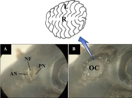
한국 고유종 돌마자의 후각기관(olfactory organ)은 주둥이 등 쪽에 좌우 한 쌍으로 위치해 있는데, 각 기관의 외부 형태 는 전비공(anterior nostril), 후비공(posterior nostril) 그리고 한 개의 비경계판(nasal flap)으로 구분된다(Fig. 1A). 두 비공 은 반원형을 나타내며 물은 입수공인 전비공(직경, 0.43~0.58 mm)으로 유입되어 출수공인 후비공(직경, 0.67~0.79mm)을 통해서 배출된다. 두 비공사이에 위치하는 비경계판은 아치형 의 형태로써 피부로부터 돌출되어있다. 후각기관의 내부는 1 개의 비강(olfactory chamber)이 존재하는데 비강은 여러 개 의 후판(lamella)과 가운데 후선(raphe)으로 구성된 로제트 구 조(rosette structure)를 보유한다(Fig. 1B). 각각의 후판(22~24
Fig. 1. Gross structure of the olfactory organ in Microphysogobio yaluensis. A, the external view showing the anterior and the posterior nostrils and a nasal flap; B, the internal view showing the rosette structure (the below inserted diagram) with in the olfactory chamber. AN, anterior nos
tril; L, lamella; NF, nasal flap; OC, olfactory chamber; PN, posterior nostril; R, raphe. Bars indicate 0.5 cm.
A B
개)은 중심부의 후선을 기준으로 가로와 방사형으로 배치되며 아래와 옆면의 가장자리는 비강의 내벽과 연결된다(Fig. 1B).
위와 같은 반원형의 비공과 비경계판으로 구성된 후각 기관 형태는 동굴 내에 서식하는 Sinocyclocheilus jii와 S.
furcodorsalis(Waryani et al., 2013), 하천의 늪지, 수로, 웅덩이 등에 서식하는 Cirrhinus reba(Ghosh and Chakrabarti, 2016), 기수 또는 담수의 강, 호수, 작은 습지를 선호하는 Catla catla (Kumari, 2008)와 같은 비교적 활동적이며 표층 또는 저서 표 층성의 생태적 습성을 갖는 다른 잉어목 어류들에서도 일부 보고된바 있다. 따라서 비공과 비경계판으로 구성되는 구조 적 형태는 잉어목 어류의 일반적인 특징으로 추측되나(Zeiske et al., 1992), 이에 따른 추가 조사가 더 필요할 것으로 보인 다. 또한, 돌마자의 후판은 가로와 방사형의 배열을 나타내는 데 이는 Yamamoto(1982)의 G type에 해당하며, 이러한 정렬 양상은 황어 Tribolodon hakonensis, 모래무지 Pseudogobio esocinus, 피라미 Zacco platypus와 잉어 Cyprinus carpio에 서도 확인된 바 있다(Yamamoto, 1982). 그러나 돌마자의 후
판 수는 22~24개로 Puntius sophore에서 16~18개(Ghosh and Chakrabarti, 2010), 잉어에서 25~26개(Chakrabarti and Hazra Choudhry, 2007), 모래무지에서 10개(Yamamoto, 1982), Labeo bata에서 24~26개(Ghosh and Chakrabarti, 2013)로 보 고된 다른 잉어목 어류들과 비교해 많은 수의 후판을 보유하 며 차이를 나타냈다. 일반적으로 많은 수의 후판은 후감각뉴 런(olfactory receptor neuron)이 발달하는 표면적의 증가와 연 관되기 때문에 어류가 생태적으로 후각에 높은 의존도를 보이 는 것으로 의미될 수 있다(Zeiske et al., 1976). 따라서 돌마자 의 많은 후판은 높은 후각 의존성을 나타내는 표현형적 증거 로 생각되며 이러한 특징은 분류학적 특성으로도 활용될 수 있을 것으로 판단된다.
조직학적 연구에서도 비강 내 로제트 구조는 후선과 여러 후판들로 구성되는 것을 재확인할 수 있다(Fig. 2A). 후선은 로제트 구조 중앙에 위치하며 각 후판들과 연결된다. 후판(두 께, 104.9~225.1μm)의 조직은 감각(sensory epithelium)과 비감각(nonsensory epithelium) 상피영역으로 구분된다(Fig.
Fig. 2. Histological characteristics of the rosette structure of Microphysogobio yaluensis, stained with hematoxylineosin(A, C, D) and alcian blue(pH 2.5)periodic acid Schiff(B). A, the rosette structure with several lamellae in cross section; B, the lamellae with the sensory and the nonsensory epitheliums; C, the outer margin of the lamella; D, the middle part of the lamella. Arrow, dendrite; arrowhead, mucous cell; broken arrow, blood capillary; BC, basal cell; CNC, ciliated nonsensory cell; CT, connective tissue; L, lamella; NSE, nonsensory epithelium; ORN, olfactory receptor neuron; R, raphe; SC, supporting cell; SE, sensory epithelium. The bars indicate 500μm in A, 200μm in B, 50μm in C, D, re
spectively.
A B
C D
2B). 감각상피는 위중층상피층으로써 후감각뉴런, 지지세포 (supporting cell), 기저세포(basal cell)들로 구성된다(Fig. 2D).
후감각뉴런은 기저막에서 상피층의 표면까지 축삭(axon)과 수상돌기(dendrite)들로 확장되는 양극성 신경세포이다. HE 염색에서 이들의 핵은 진한 보라색을 나타내며, 수상돌기는 세포 중심부와 가까운 쪽에서는 진한 보라색을 보이다가 상 피층 표면 쪽에 가까울수록 옅은 분홍색으로 염색되었다. 후 감각뉴런과는 다르게, 지지세포는 돌출된 세포내 기질을 갖지 않았으며 타원형의 핵과 원통형의 세포질을 나타낸다. 기저세 포는 상피층 맨 하부에 위치하며 2 type(편평형, 타원형)의 핵 모양을 보였는데 이들은 기저부 위 1개 층으로 정렬된다. 비 감각상피는 중층편평상피층으로 후판의 가장자리 부분을 따 라 발달하며 층상상피세포(stratified epithelial cell), 비감각섬 모세포(ciliated nonsensory cell), 점액세포들로 구성된다(Fig.
2B and C). 층상상피세포는 상피층 대부분을 구성하는 세포이 며 타원의 핵과 다소 부풀어 오른 세포질 특성을 나타낸다. 비 감각섬모세포는 상피층 최상부에 위치하는데 타원의 핵과 원 통형의 세포질을 나타내며 세포 표면에 많은 수의 섬모를 보 유하는 것이 특징이다. 이들 세포는 3~4개의 주변세포와 밀 접하게 위치하여 하나의 그룹화를 형성한다. 점액세포는 상피 층의 표면가까이에 위치하며 편평한 핵은 세포의 기저부에 발 달하고 세포질은 HE염색에 음성으로 반응한다.
감각상피의 후감각뉴런, 지지세포, 기저세포는 동물이 후 각의 기능을 수행하기 위한 필수적인 세포들이다. 따라서 대 부분의 경골어류들은 이러한 세포들을 감각상피에 보유하 게 된다(Hara, 1986). 돌마자의 비감각상피를 구성하는 층상 상피세포, 비감각섬모세포, 점액세포 또한 다른 잉어목 어류 의 후각기관에 관한 많은 연구에서 알려진 바 있다(Ghosh and Chakrabarti, 2016). 그러나 본 연구에서 경골어류의 일반 적인 특성과 비교되는 돌마자 후각기관의 특이적인 2가지 조 직학적 결과를 제시한다. 첫째, 돌마자의 비감각섬모세포는 후판가장자리의 비감각상피에서만 발달하며 2~3개의 세포 가 밀접하여 그룹화를 이룬다. 경골어류에서 비감각섬모세포 는 후각상피에서 비교적 흔하게 관찰되지만 감각상피와 비감 각상피의 발달과 정렬방식에 따라 다양한 위치와 세포조직화 를 나타낸다. 돌마자와는 다르게 Pantodon buchholtzi와 붕어 Carassius carassius에서 비감각섬모세포는 감각상피에서 공 통적으로 관찰된다. 하지만 Pantodon buchholtzi에서는 각각의 세포가 별개로 발생하는 반면에 Carassius carassius에서는 돌 마자와 같이 여러 개의 세포가 그룹화를 이룬다(Hansen et al., 1999; Hansen and Zielinski, 2005). 한편, 비감각섬모세포는 세 포 표면의 운동섬모 움직임에 의해 후점막에 있는 물과 점액 의 이동성에 도움을 주는 것으로 알려져 있다(Zeiske et al., 1992). 둘째, 돌마자의 감각상피 분포양상은 연속적 유형이다.
경골어류에서 후판위의 감각상피 분포유형은 1) 후판 외각의
비감각상피에 의해 둘러싸인 연속적 패턴, 2) 비감각상피에 의 해 분리되는 넓은 패턴, 3) 무작위 패턴, 4) 섬형 패턴 등 4가 지 유형으로 보고된 바 있다(Yamamoto, 1982). 최근까지의 연 구에서 돌마자의 연속적 유형은 다른 잉어목 어류에서 일반 적으로 확인되는 유형이며 말뚝망둥어속(Periophthalmus) 또 는 짱뚱어속(Boleophthalmus) 어류들의 섬형 또는 무작위 패 턴(Kuciel et al., 2013), 동갈치목(Beloniformes)의 송사리속 (Oryzias) 어류의 넓은 패턴(Kim et al., 2016)과는 차이를 보 인다.(Yamamoto, 1982). 그러나 이러한 분포양상은 같은 속 어류라 할지라도 종간 차이를 나타내기 때문에(Yamamoto, 1982) 한국산 모래주사속 5 종의 감각상피 분포양상에 관한 추가적인 비교연구가 필요할 것으로 생각된다.
조직화학적 연구에서, 점액세포는 한 가지 색(PAS, diastase/
PAS에서 적색; alcian blue pH 1.0 and 2.5, Methylation/sapon
ification/AB, Mild methylation/AB, Aldehyde fuchsin/AB에 서 파란색; toluidine blue에서 β-metachromasia, 연한 보라 색)과 두 가지 이상의 다양한 색(AB/PAS에서 파란색, 푸른 적색, 검정색, 빨간색)으로 각각 반응한다(Table 1). 이와 같 은 결과로부터 점액세포의 성분은 산성 및 중성의 황점액소 (sulfomucin)로 확인된다.
뮤신(mucin)은 당질과 단백질이 결합한 복합화합물로써 분 자의 이온 전하(net charge)에 따라 산성 및 중성으로 분류되 며(Forstner and Forstner, 1994), 그 중 산성의 뮤신은 다시 황 점액소와 시알로뮤신으로 구분될 수 있다(Filipe, 1979). 어류 에서 산성의 황점액소는 세포 또는 상피내 가스 교환과 분비, 산과 염기의 조절, 이온 조절, 삼투압 조절과 같은 기능을 수 행하며(Shephard, 1994), 특히 외부환경과 밀접하게 연관되어 지속적으로 물리 및 화학적 영향을 받게 되는 아가미, 소화관, 피부의 항균작용을 위해 생성되는 주된 점액성분으로 알려져 있다(Sharma and Schumacher, 1995; Jeong and Jo, 2007). 따라 Table 1. The nature of the olfactory mucous cells in Microphysogo- bio yaluensis, identified through each techniques
Employed techniques Mucous cell
Hematoxylin and eosin -
Weigerts iron -
Hematoxylin -
Masson trichrome -
PAS R
PAS after digestion in malt diastase/PAS R
Alcian blue (pH 1.0) B
Alcian blue (pH 2.5) B
Alcian blue/PAS B, BR, BN, R
Toluidine blue O β-meta
Methylation/saponification/Alcian blue B
Mild methylation/Alcian blue B
Aldehyde fuchsin/Alcian blue B
B, blue; BN, black; BR, blush red; R, red; β-meta, metachromasia; -, no rea
ction
서 돌마자에서 황점액소의 발생은 서식처 환경에서의 화학적 영향과 세균으로부터 후각기관내 신경세포를 포함한 다른 주 요 세포들을 보호하기 위해 생성되는 세포학적 적응으로 사료 된다. 최근까지 어류 내 황점액소에 관한 몇몇 보고가 이루어 져 있지만(Hirsch and Margolis, 1981; Kim et al., 2018), 여전 히 후각기관에서 황점액소에 관한 정확한 기능을 이해하기에 는 불충분하다. 그러므로 어류 후각기관내 뮤신의 역할에 관 한 조직 및 조직화학적 지속적인 후속 연구가 더 필요할 것으 로 생각된다.
요 약
한국 고유종 돌마자 Microphysogobio yaluensis의 후각기관 과 서식처 및 생태와의 연관성을 알아보기 위해 실체 현미경 과 광학현미경을 이용하여 구조 및 조직학적 특징을 조사하 였다. 후각기관은 외부적으로 전비공, 후비공 그리고 비경계 판으로 구성되어 있으며, 내부는 22~24개의 후판들과 비선으 로 구성되었다. 후판은 감각상피와 비감각상피로 이루어져 있 는데, 감각상피는 후감각신경, 지지세포, 기저세포들로 구성되 었으며, 비감각상피는 층상상피세포, 비감각섬모세포, 산성과 중성을 갖는 황점액소의 점액세포들로 구성되어 있다. 이러한 후각기관들의 특징들은 돌마자의 높은 후각능력과 의존성 그 리고 서식처 환경에 밀접한 관계가 있을 것으로 생각되며 분 류학적 연구에도 활용될 수 있을 것으로 생각된다.
사 사
본 논문은 2017년도 정부(교육부)의 재원으로 한국연 구재단의 일부 지원을 받아 수행된 기초연구사업임(No.
2017R1D1A1B03028268).
REFERENCES
Chakrabarti, P. and S.H. Choudhury. 2007. The fine structural or
ganization of the olfactory epithelium of Cyprinus carpio (Linnaeus): a scanning electron microscopic study. Folia Morphol., 66: 1014.
Filipe, M.I. 1979. Mucins in the human gastrointestinal epithelium:
a review. Invest Cell Pathol., 2: 195216.
Forstner, J. and G. Forstner. 1994. Gastrointestinal mucus. In: John
son L, editor. Physiology of the gastrointestinal tract, vol. 2, 3rd ed., pp. 12551283.(New York: Raven Press)
Ghosh, S.K. and P. Chakrabarti. 2009. Cellular architecture and functional aspects of the olfactory rosette of Wallago attu
(Bleeker). Turk. J. Fish. Aquat. Sci., 9: 187-190.
Ghosh, S.K. and P. Chakrabarti. 2010. Histological and scanning electron microscopic organization and functional aspects of the surface olfactory epithelium of the freshwater minor carp, Puntius sophore(Hamilton). Proceed. Zool. Soc., 63:
115119.(Springer, Verlag).
Ghosh, S.K. and P. Chakrabarti. 2011. Distribution and organization of different cells lining the olfactory epithelium of the In
dian minor carp, Labeo bata(Hamilton, 1822): a light and scanning electron microscopic analysis. Pak. J. Biol. Sci., 14: 736741.
Ghosh, S.K. and P. Chakrabarti. 2013. Studies on the morphology of the olfactory organ in the freshwater teleost, Labeo bata (Hamilton). Mesopot. J. Mar. Sci., 28: 163174.
Ghosh, S.K. and P. Chakrabarti. 2016. Histomorphological and microanatomical characteristics of the olfactory organ of freshwater carp, Cirrhinus reba(Hamilton). Arch. Polish Fish., 24: 201208.
Gurr, G.T. 1956. A practical manual of medical and biological stain
ing techniques. Interscience, New York, pp. 1-99.
Hansen, A. and Z. Eckart. 1998. The peripheral olfactory organ of the zebrafish, Danio rerio: an ultrastructural study. Chem.
senses, 23: 3948.
Hansen, A., H.P. Zippel, P.W. Sorensen and J. Caprio. 1999. Ultra
structure of the olfactory epithelium in intact, axotomized, and bulbectomized goldfish, Carassius auratus. Microsc.
Res. Tech., 45: 325338.
Hansen, A. and B.S. Zielinski. 2005. Diversity in the olfactory epi
thelium of bony fishes: development, lamellar arrangement, sensory neuron cell types and transduction components. J.
Neurocytol., 34: 183208.
Hara, T.J. 1986. Role of olfaction in fish behaviour. In: Pitcher, T.J.
(ed.), The behaviour of teleost fishes, Springer, US, pp.
152176.
Hirsch, J.D. and F.L Margolis. 1981. Isolation, separation, and anal
ysis of cells from olfactory epithelium. In Biochemistry of Taste and Olfaction, pp. 311332.(Academic Press, New York)
Huang, S.P., Y. Zhao, I.S. Chen and K.T. Shao. 2017. A New Spe
cies of Microphysogobio(Cypriniformes: Cyprinidae) from Guangxi Province, Southern China. Zoological Studies, 56:
Jeong, G.N. and U.B. Jo. 2007. Mucosubstance histochemistry of 8.
the epidermis in yellowtail, striped beakperch, brown spot
ted grouper, sea chub, and multicolorfin rainbowfish. Integr.
Biosci., 11: 183189.
Kim, H.T., Y.J. Lee and J.Y Park. 2016. An anatomical and histo
chemical study of the olfactory organ in rice-fish Oryzias sinensis(Pisces: Adrianichthyidae) in South Korea. Korean.
J. Ichthyol., 28: 223228.
Kim, H.T., Y.J. Lee and J.Y. Park. 2018. Functional anatomy and histology of the olfactory organ in Korean eel goby, Odon- tamblyopus lacepedii(Pisces: Gobiidae). Appl. Microsc.,
48: 1116.
Kim, I.S. and J.Y. Park. 2002. Freshwater fishes of Korea. Kyo-Hak Publishing Co. Ltd., Korea. pp. 388389.(in Korean).
Kuciel, M., K. Zuwala and U. Satapoomin. 2013 Comparative morphology(SEM) of the peripheral olfactory organ in the Oxudercinae subfamily(Gobiidae, Perciformes). Zool. Anz, 252: 424430.
Kumari, K. 2008. Morphology and morphometry of the olfactory rosette of a teleostean fish: Catla catla (Ham.). Our Nature, 6: 3037.
Masson, P. 1929. Some histological methods. Trichrome stainings and their preliminary technique. J. Tech. Methods, 12: 75- Motta, P.J. and K.M. Kotrschal. 1991. Correlative, experimental, 90.
and comparative evolutionary approaches in ecomorpholo
gy. Nether. J. Zool., 42: 400415.
National Institute of Biological Resources. 2013. Inventory and management of endemic species of Korea. National Insti
tute of Biological Resources. 147pp.(in Korean)
Park, J.Y., I.S. Kim and S.Y. Kim. 2003. Structure and histochemis
try of the skin of a torrent catfish, Liobagrus mediadiposa- lis. Environ. Biol. Fish., 66: 38.
Sharma, R. and U. Schumacher. 1995. Morphometric analysis of intestinal mucins under different dietary conditions and gut flora in rats. Digest. Dis. Sci., 40: 2532-2539.
Shephard, K.L. 1994. Functions for fish mucus. Rev. Fish. Biol.
Fisheries, 4: 401429.
Waryani, B., Y. Zhao, C. Zhang, R. Dai and A.R. Abbasi. 2013.
Anatomical studies of the olfactory epithelium of two cave fishes Sinocyclocheilus jii and S. furcodorsalis(Cyprin
iformes: Cyprinidae) from China. Pakistan J. Zool., 45:
10911101.
Yamamoto, M. 1982. Comparative morphology of the peripheral olfactory organs in teleosts. In: Hara, T.J.(ed.), Chemore
ception in fishes, pp. 39-59.(Elsevier, Amsterdam) Zeiske, E., J. Kux and R. Melinkat. 1976 Development of the ol
factory organ of oviparous and viviparous cyprinodonts (Teleostei). Zeitschrift für Zoologische Systematik und Evolutionsforschung, 14: 3440.
Zeiske E., B. Theisen and H. Breucher. 1992 Structure, develop
ment, and evolutionary aspects of the peripheral olfactory system. In: Hara TJ(ed.). Fish chemoreception, pp. 1339.
(Springer, Netherlands)