INTRODUCTION
The olfactory organ is a sensory system used for smell.
It enables fish to perform ecologically important behaviors such as prey searching, predator avoidance, reproduction, homing migration, habitat cognition, and fright reaction (Hara, 1986). Its morphology and histology varies in teleost fishes allowing them to adapt to diverse aquatic environ
ments. The olfactory organ displays diverse lamellar struc
tures from a lack of folded lamellae(e.g., whitespotted pygmy filefish Rudarius arcodes, yellow boxfish Ostracion tuberculatus, Japanese medaka Oryzias latipes, and Asian swamp eel Monopterus albus)(Kim, 2018) to many folded lamellae(120 in the whitespotted conger Conger myriaster
and 112 in Microdonophis erado)(Kasumyan, 2004).
Structural differences in the olfactory organ, such as the nostril’s position and shape, the number of nasal sacs, olfac
tory epithelium cell contents, and the olfactory receptor neuron,(ORN)’s dendrite shape, length and number are also found in taxonomically related and sympatric species (Kim, 2018).
Korean intertidal gobies show habitat preferences for bottom structure, water depth and volume, salinity, and spatial distribution(Kim et al., 2005). Their body special
ization and physiological habitat adaptation may also apply to olfactory organ morphological differences(Kim, 2018;
Kim et al., 2019). Three mudskippers, the shuffles mudskip
per Periophthalmus modestus(Kim et al., 2019), the blue spotted mudskipper Boleophthalmus pectinirostris(Kim, 2018), and the gigas goby Scartelaos gigas(Kim et al., 2014), all intertidal mudflat sympatric fishes, show olfac
—1 — http://www.fishkorea.or.kr
저자 직위: 김현태(박사 후 연구원), 박종영(교수)
* Corresponding author: Jong Young Park Tel: 82632703344, Fax: 82632703362, Email: [email protected]
Microscopic Characteristics of the Olfactory Organ in the Gluttonous Goby Chaenogobius gulosus (Pisces, Gobiidae), Compared to Sympatric Intertidal Gobies
By Hyun Tae Kim and Jong Young Park*
Department of Biological Science and Institute for Biodiversity Research, College of Natural Sciences, Jeonbuk National University, Jeonju 54896, Republic of Korea
ABSTRACT Using stereo, light, and scanning electron microscopes, we researched the anatomical and histological structure of Chaenogobius gulosus’s olfactory organ and compared it to those of sympatric gobies Luciogobius guttatus and Favonigobius gymnauchen. Results revealed the following common characteristics: i) tubular anterior nostril(AN) and flat posterior nostril(PN), ii) a single longitudinal lamella, iii) two accessory nasal sacs(ANS, ethmoidal and lacrimal), iv) abundant sensory epithelium lymphatic cells(LC), v) an eosinophil cell, and vi) a ciliary length a quarter of the knob diameter in the olfactory receptor neuron(ORN). Some characteristics are specific to C. gulosus and different from the other two gobies: i) 0.5~1.0mm AN and 0.2~0.5mm PN(vs. 0.2~0.3mm and 0.2~0.3mm in L. guttatus; 0.2~0.4mm and 0.1~0.3mm in F. gymnauchen), ii) two ANS(vs. absence in L. guttatus; two in F. gymnauchen), iii) abundant LC(vs. low in L. guttatus and F. gymnauchen), iv) low density non-sensory cilia on the lamellar surface(vs. high in L. guttatus; low in F. gymnauchen), and v) a quarter ciliary length to knob diameter ratio in the ORN(vs. mixture of a quarter to equal ratio in L. guttatus; two or three times in F. gymnauchen). From these results, we confirmed the C. gulosus olfactory organ has adapted anatomically and histologically to the sand-rock tidal zone.
Key words: Olfactory organ, nostril diameter, lymphatic cell, non-sensory cilia, a quarter ciliary length
and the sharpnosed sand goby Favonigobius gymnauchen (Kim et al., 2016). C. gulosus is an omnivorous species distributed along the western and southern coast of the Korean peninsula and northeastern Japan. It inhabits inter
tidal zones and estuaries with sandy, rocky, and gravel bot
toms(Kim et al., 2005). Other than some ecological studies (Kim et al., 2004; Baeck et al., 2010) and molecular bio
logical analyses(Hirase and Ikeda, 2014; Oh et al., 2016), there has not been a lot of morphological or histological C. gulosus research. This study investigates C. gulosus’s olfactory organ and analyzes structural differences between sympatric gobies.
MATERIALS AND METHODS
1. Specimen preparation
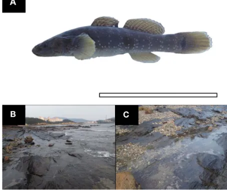
We collected 20 adult gobies, C. gulosus(Fig. 1A, 63 to 94mm standard length) using a scoop net(4×4mm mesh) from an intertidal pool at Gyeokpori, Byeonsanmyeon, Buangun, Chollabukdo, South Korea in April 2020(Fig.
1B). We took the fishes to the laboratory and anesthetized
Jeonbuk National University Institutional Animal Care and Use Committee.
2. Microscopic investigation
We anatomized and photographed the olfactory organs using a surgical blade, a stereo microscope(SM; Stemi DV4, Carl Zeiss, Germany), and a digital camera(TG3, Olympus, Japan).
For light microscopy, we washed the buffered formalin organs with tap water for 24 hours. Next, we passed the organs through an ascending alcohol series(70%, 80%, 90%, 95%, 100%), cleaned with xylene, and embedded them in paraffin wax. We cut the block into 5μm thick slices. We stained the tissue with hematoxylin and eosin (H&E) and Masson’s trichrome, and observed the samples using a light microscope(LM, Carl Zeiss, Germany). For the scanning electron microscopy, we washed the buffered GA organs with tap water for 20 minutes three separate times. Next, we postfixed the organs with 2% OsO4 in 0.1 M potassium phosphate buffer, dehydrated them in an as
cending alcohol series(50%, 60%, 70%, 80%, 90%, 95%, 100%), and immersed them in tertbutyl alcohol. We dried
Fig. 1. The photograph(A) of Chaenogobius gulosus and its habitat(B) and bottom view(C), 35°38′05″N, 126°27′59″E. The bar indicates 5cm.
A
B C
the organs using a freezedryer(VFD21S, Vacuum Device Co., Ltd., Ibaragi, Japan), coated them with OsO4 powder, and observed them under a scanning electron microscope (SEM; SUPRA40VP, Carl Zeiss, Germany).
RESULTS
1. Anatomy
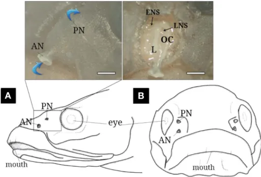
Externally, the C. gulosus olfactory organ is paired on both snout sides and composed of anterior(AN) and poste
rior(PN) nostrils(Fig. 2A and B). The AN(0.5 to 1.0mm diameter) is a tubular opening protruding outward. The PN (0.2 to 0.5mm diameter) lies flat against the skin surface.
Internally, the olfactory chamber(OC) is oval with only one longitudinal lamella and leads to the posterior ethmoidal and lacrimal accessory nasal sacs(ACS).
2. Histology
In a crosssectional view, the OC shows one longitudinal lamella covered by sensory epithelium(SE) on both sides (Fig. 3A and B). The ORN’s SE consists of basal cells(BC), supporting cells(SC), lymphatic cells(LC), and eosinophil cells(ELC)(Fig. 3C and D). The ORN is an elongated bi
polar neuron with a flattened nucleus located at the cell’s middle and colored dark red from the H&E stain and dark
violet from the Masson trichrome stain. Its cytoplasm ext
ends from the bottom to the superficial surface. The SC is an extending cell with cytoplasm that H&E stained weakly.
Its nucleus is larger than ORN. The round BC is cell situa
ted at the epithelia’s bottom. The LCs are small, dense, and abundant in the basal area. They are dark violet from the H&E stain and dark blue from Masson’s trichrome stain.
The ELCs are observable white blood cells in the SE’s middle layer and have two divided nuclei. They appear as a deep scarlet color in the cytoplasm.
Under the SEM, the longitudinal lamella is exposed inside the OC(Fig. 4A). Its surface shows the ORN’s den
drites, which consist of 3~5 short cilia on the knob(Fig.
4B).
DISCUSSION
The intertidal zone is a turbulent environment where the periodic tidal cycle causes constant physical and chemical changes in salinity, water volume, and degree of air expo
sure(Horn et al., 1999). Based on bottom sediment, these extreme areas can be subdivided into three types, rock, sand, and mud(Masselink and Gehrels, 2014). Our col
lection site was a sandrock tidal zone inhabited by three goby species: C. gulosus, L. guttatus, F. gymnauchen(Kim et al., 2005). To survive such extreme surroundings, gobies
Fig. 2. The schematic diagram of lateral(A) and front(B) views of Chaenogobius gulosus and its photographs(above) of the external(left) and inner(right) structures. The blue arrows indicate water flow. AN, anterior nostril; ENS, ethmoidal nasal sac; L, lamellae; LNS, lacrimal nasal sac;
OC, olfactory chamber; PN, posterior nostril. The bars indicate 1mm.
A B
possess the ability to tolerate rapid salinity changes and occasional body desiccation from air exposure. They can withstand ocean waves and often hide under substances (Horn et al., 1999). To better understand the relationship between C. gulosus and its environment, we studied how its olfactory organ adapted to the intertidal zone(viewed in Fig. 1B and C) and compared it with other gobiid fish
(Table 1).
The C. gulosus olfactory organ has the following charac
teristics: i) tubular AN and flat PN lying on the surface, ii) single longitudinal lamella, iii) two ANS(ethmoidal and lacrimal) cells, iv) SE with abundant LCs, v) ELC presence, and vi) ciliary length that is a quarter of the knob diameter.
Although all three gobies have tubeforming AN and flat
Fig. 3. Histological characteristics of the olfactory epithelium of Chaenogobius gulosus, stained with hematoxylin and eosin(A, C) and Masson’s trichrome(B, D). A and B, the single lamella covering the sensory epithelium; C and D, the sensory epithelium consisting of olfactory receptor neu
rons, basal cells, supporting cells, lymphatic cells and eosinophil and the connective tissue with blood capillaries and fibroblast cells. Broken arrow, red blood cell; BC, basal cell; CT, connective tissue; ELC, eosinophil; FBC, fibroblast cell; L, lamella; LC, lymphatic cell; OC, olfactory chamber;
ORN, olfactory receptor neuron; SE, sensory epithelium; SC, supporting cells. The bars indicate 200μm in A and B, 50μm in C and D, respectively.
C D
Fig. 4. The olfactory lamella and lamellar surface of Chaenogobius gulosus. A, the lamella inside the olfactory chamber; B, the olfactory receptor neuron with 3~5 short cilia. L, lamella; OC, olfactory chamber. The bars 200μm in A and 5μm in B.
A B
PN on their snouts, the AN and PN diameter sizes are differ ent. C. gulosus and F. gymnauchen’s AN are larger than their PN whereas L. guttatus’s AN and PN are similar.
Chernova(2008) divided the AN and PN diameter differ
ence into two categories: i) AN lager than PN and ii) AN similar to PN. These morphological variations occurred in fishes with similar ecological habits and can therefore be applied to systematic characterizations. Cox(2008) hypo
thesized that the sterlet’s wider PN compared to the pike is because the fish is more active and benefits from viscous environments. Therefore, C. gulosus and F. gymnauchen’s larger AN diameter may reflect more active swimming and smoother olfactory cavity ventilation than L. guttatus.
C. gulosus’s two ANS are the same as F. gymnauchen but higher than L. guttatus. Generally, ANS of teleost allows an entire olfactory organ volume change though its relaxa
tion and contraction by skeletal movements(Kasumyan, 2004). Kim(2019) states that P. modestus’s single ANS is olfactory evidence of terrestrial adaptation compared to the more aquatic mudskipper B. pectinirostris’s two. The two ANS of C. gulosus may be structural indicators of more act
ive olfactory ventilation than L. guttatus. This is supported by nonANS L. guttatus’s abundant nonsensory cilia, which help water flow over the lamella(Table 1; Doving et al., 1977).
C. gulosus contains abundant LCs at the OC’s basal area whereas L. guttatus and F. gymnauchen are lacking(Kim et al., 2016; Kim and Park, 2016). Most teleost fishes pro
duce lymphocytes or LCs to fight infective pathogens as part of a more advanced and acquired immune system (Press and Evensen, 1999). They have also been disco
vered in other immune cells, plasma cells and macro
phages, inner organs such as thymus, spleen, kidney, heart and skin(Press and Evensen, 1999). Kim(2018) stated that SE and NSE LCs in five estuary or coastal species are associated with brackish and seawater species survival. In our study, C. gulosus have many LCs. L. guttatus and F.
gymnauchen, however, did not. This may mean C. gulosus is more sensitive to the intertidal environment.
The ORN’s ciliary length varies by fish species: over 0.3~5μm in the goldfish Carassius carassius(Barbar and Boyde, 1968), 2~3μm in the Zebrafish Danio rerio(Han
sen and Eckart, 1998), 10μm in Acipenser baeri(Hansen and Zielinski, 2005), 5~6μm in the European river lam
prey L. fluviatilis(Thornhill, 1967), and 0.2~0.5μm in L.
reissneri(Kim and Park, 2020). Menco(1992) suggested the intertwined, longer cilia increase the interaction area and probability of external odor contact. Measuring exact ciliary length is difficult due to their curved and intertwined structure and position under the SEM. Instead, we mea
sured the ORN’s ciliary length by comparing its ratio to the knob. C. gulosus’s quarter ciliary length reflects a lower olfactory dependence than the other two species(Table 1).
With some counter staining techniques, a study on ana
tomy and histology of cell, epithelium, and components of fish olfactory organ well provide how fish has been adapt
ed to fullyaquatic habitat as well as semiaquatic life in aspect of its morphology and ecology(Kim et al., 2014, 2019; Kim and Park, 2020).
In conclusion, we confirm C. gulosus’s olfactory organ, compared to olfactory organs from two sympatric gobies, shows a morphological structure demonstrating 1) its sand and rock bottom, nonturbid water habitat 2) its active ven
tilation, and 3) its aggressive immunity.
Table 1. Anatomical and histological differences of the olfactory organ between three sympatric gobies living in the intertidal zone Chaenogobius gulosus
(this study) Luciogobius guttatus
(Kim and Park, 2016) Favonigobius gymnauchen (Kim et al., 2016) Anatomy
Diameter of AN(mm) Diameter of PN(mm) The number of ANS
0.5~1.0(n=20) 0.2~0.5(n=20) 2(ethmoidal and lacrimal)
0.2~0.3(n=13) 0.2~0.3(n=13)
0
0.2~0.4(n=9) 0.1~0.3(n=9) 2(ethmoidal and lacrimal) Histology
The density of LC *** * *
Ultrastructure of the OE surface The density of NSC
The ciliated ORN’s ciliary length of KD * A quarter
***
Mixture of a quarter to equal ratio * Two or three times minmax(n=number); AN, anterior nostril; ANS, accessory nasal sac; KD, knob diameter; LC, lymphatic cell; NSC, nonsensory cilia; ORN, olfactory receptor neuron; PN, posterior nostril; *, low or lacking; ***, high
Korea(NRF) funded by the Ministry of Education(2020R1 A6A3A0109608211).
REFERENCES
Baeck, G.W., C.I. Park, J.M. Jeong, M.C. Kim, S.H. Huh and J.M.
Park. 2010. Feeding habits of Chaenogobius gulosus in the coastal waters of Tongyeong, Korea. Korean J. Ichthyol., 22:
4148.
Barbar, V.C. and A. Boyde. 1968. Scanning electron microscopic studies of cilia. Z. Zellforsch, 84: 269284.
Chernova, N.V. 2008. Systematics and phylogeny of fish of the genus Liparis(Liparidae, Scorpaeniformes). J. Ichthyol., 48: 831
852.
Cox, J.P. 2008. Hydrodynamic aspects of fish olfaction. J. R. Soc.
Interface, 5: 575593. https://doi.org/10.1098/rsif.2007.1281.
Doving, K.B., M. DuboisDauphin, A. Holley and F. Jourdan. 1977.
Functional anatomy of the olfactory organ of fish and the cil
iary mechanism of water transport. Acta Zool., 58: 245255.
https://doi.org/10.1111/j.14636395.1977.tb00260.x.
Hansen, A. and B.S. Zielinski. 2005. Diversity in the olfactory epi
thelium of bony fishes: development, lamellar arrangement, sensory neuron cell types and transduction components. J.
Neurocytol., 34: 183208.
Hansen, A. and Z. Eckart. 1998. The peripheral olfactory organ of the zebrafish, Danio rerio: an ultrastructural study. Chem. Sens
es, 23: 3948.
Hara, T.J. 1986. Role of olfaction in fish behaviour. In: Pitcher, T.J.
(ed.), The behaviour of teleost fishes. Croom Helm, London, U.K., pp. 152176.
Hirase, S. and M. Ikeda. 2014. Divergence of mitochondrial DNA lineage of the rocky intertidal goby Chaenogobius gulosus around the Japanese Archipelago: reference to multiple Pleis
tocene isolation events in the Sea of Japan. Mar. Biol., 161:
565574. https://doi.org/10.1007/s0022701323595.
Horn, M.H., K.L. Martin and M.A. Chotkowski. 1999. Intertidal Fish
es: Life in Two Worlds. Academic Press, San Diego, U.S.A., 399pp.
Kasumyan, A.O. 2004. The olfactory system in fish: structure, func
tion, and role in behavior. J. Ichthyol., 44: 180223.
of the olfactory organ in the Korean sand goby Favonigobius gymnauchen(Pisces, Gobiidae). Korean J. Ichthyol., 28: 28
34.
Kim, H.T. and J.Y. Park. 2016. The anatomy and histoarchitecture of the olfactory organ in the Korean flat-headed goby Luciogo- bius guttatus(Pisces; Gobiidae). Appl. Microsc., 46: 5157.
https://doi.org/10.9729/AM.2016.46.1.51.
Kim, H.T. and J.Y. Park. 2020. Microscopic research on the olfactory organ of the Far Eastern brook lamprey Lethenteron reissneri (Pisces, Petromyzontidae). Appl. Microsc., 50: 17.
Kim, H.T., S.W. Yun and J.Y. Park. 2019. Anatomy, histology, and his
tochemistry of the olfactory organ of the Korean shuttles mud
skipper Periophthalmus modestus. J. Morphol., 280: 1485
1491. https://doi.org/10.1002/jmor.21044.
Kim, H.T., Y.J. Lee, J.S. Park and J.Y. Park. 2014. A study on the structure of peripheral olfactory organ in the Korean mud
skipper, Scartelaos gigas(Pisces, Gobiidae). Korean J. Ich
thyol., 26: 281287.
Kim, I.S., Y. Choi, C.L. Lee, Y.J. Lee, B.J. Kim and J.H. Kim. 2005.
Illustrated Book of Korean Fishes. Kyohak Publishing Co.
Ltd., Seoul, Korea, 615pp.
Kim, S.Y., C.B. Park, J.W. Kang, Y.C. Choi, S. Rho, H.J. Bawk, H.B. Kim and Y.D. Lee. 2004. Gonadal development and reproductive cycle of gluttonous goby Chasmichthys gulosus (Guichenot). Korean J. Ichthyol., 16: 261270.
Menco, B. 1992. Ultrastructural studies on membrane, cytoskele tal, mucous, and protective compartments in olfaction. Microsc.
Res. Tech., 22: 215224. https://doi.org/10.1002/jemt.1070 220303.
Masselink, G. and R. Gehrels. 2014. Coastal environments and global change. John Wiley and Sons, Chichester, U.K., 438pp.
Oh, J., T.W. Kim and S. Kim. 2016. The complete mitochondrial genome of Chaenogobius gulosus(Gobiidae, Perciformes) from the South Sea, Korea. Mitochondrial DNA A. DNA Mapp. Seq. Anal., 27: 42074208. https://doi.org/10.3109/19 401736.2015.1022742.
Press, C.M. and Ø. Evensen. 1999. The morphology of the immune system in teleost fishes. Fish Shellfish Immunol., 9: 309-318.
Thornhill, R.A. 1967. The ultrastructure of the olfactory epithelium of the lamprey Lampetra fluviatilis. J. Cell Sci., 2: 591602.
별망둑 the gluttonous goby Chaenogobius gulosus 후각기관의 해부 , 조직학적 특성 및 동소 망둑어과 출현종들과의 비교연구
김현태
·
박종영전북대학교 자연과학대학 생명과학과, 전북대학교 부설 생물다양성연구소
요 약 : 본 연구는 우리나라 서해의 모래와 암반 조간대에 서식하는 별망둑 Chaenogobius gulosus 후각기관 의 해부 및 조직학적 구조를 확인하고 그 특징을 동소종(미끈망둑 Luciogobius guttatus, 날개망둑 Favonigobius gymnauchen)들과 비교하였다. 별망둑은 튜브형 전비공, 표면과 평행하는 후비공, 세로배열의 한 개 후판, 두 개 비
낭, 후상피 하부에 풍부한 림프구, 호산구, 후감각뉴런에서 후돌기 직경의 1/4의 섬모 길이의 일반적 결과를 보여
주었다. 이러한 특징들 중 1) 0.5~1.0mm의 전비공 직경, 0.2~0.5mm의 후비공 직경(vs. 미끈망둑의 0.2~0.3mm, 0.2~0.3mm; 날개망둑의 0.2~0.4mm, 0.1~0.3mm의 전비공과 후비공 직경), 2) 감각상피에서의 상대적으로 풍부
한 림프구, 3) 비감각섬모의 부재(vs. 미끈망둑에서 높은 분포; 날개망둑에서 낮은 분포), 4) 후감각뉴런의 후돌기
대비 1/4의 섬모 길이(vs. 미끈망둑의 1/4에서 1 : 1 비율 혼재; 날개망둑의 2~3배)는 두 동소종들과 비교되는 특이
적인 결과였다. 결론적으로, 별망둑 후각기관의 해부 및 조직학적 특징들은 모래와 암반 조간대에서 다른 동소종들
보다 더 적극적인 면역반응과 활동적인 움직임에 적응된 결과로 간주된다.
찾아보기 낱말 : 후각기관, 비공 직경, 림프구, 비감각 섬모, 후감각뉴런 섬모 길이