IntroductIon
The olfaction of teleost evokes fundamental behaviors in ecological habits, and is conducted by the olfactory organ(Hara, 1986). The structure of the olfactory organ depending on the species varies in its position, number and detailed morphology(Yamamoto, 1982). Generally, this organ contains a rosette-like structure with one to numerous lamellae, developed on the floor of olfactory chamber or cavity(Hara, 1975). Instead of a rosette-like structure, the gobiid fishes are various in the number of the canals and sacs(Kuciel et al., 2011; Kim et al., 2014).
Such special structures of the intertidal fishes are not only related with each species’s adapted degree to the inherent microhabitat but also account explicitly for interspecific- systematic distance and relationship(Yamamoto, 1982;
Kuciel et al., 2013). Accordingly, the histology and anat- omy of the olfactory organ with distinct differences also
can be used as part of a taxonomic study(Kuciel et al., 2013).
The genus Favonigobius belonging to the family Gobi- idae in South Korea has known to be only one species, sand goby F. gymnauchen. It also live in brackish water and tidal zone, near to both Western and Southern coasts along South Korean peninsula, the Jeju Island, Japan, China and Philippines with a shallow sand bottom(Kim and Park, 2002; Kim and Nakaya, 2013). The characters of above the olfactory organ have accounted for an eco- logical relation between microhabitat and its structure (Hara, 1986). Nevertheless, the previous studies on F.
gymnauchen of Korea were reported in feeding habits (Huh and Kwak, 1998), survival, growth and oxygen con- sumption of rate(Kang et al., 2000), reproduction(Lee et al., 2000), spawning behavior and morphological de- velopment(Jin et al., 2003), but the olfactory research is still lacking. Thus, the aims of this study is to describe the anatomical and histological structures of the olfactory organ in the sand goby and then analyze relation between the structure and ecological habitat.
—28 — http://www.fishkorea.or.kr
* Corresponding author: Jong Young Park Tel: 82-63-270-3344, Fax: 82-63-270-3362, E-mail: [email protected]
The Anatomy and Histology of the Olfactory Organ in the Korean Sand Goby Favonigobius gymnauchen (Pisces, Gobiidae)
By Hyun Tae Kim, Hyeong Su Kim and Jong Young Park*
Department of Biological Science and Institute for Biodiversity Research, College of Natural Sciences, Chonbuk National University, Jeonju 54896, Korea
ABSTRACT the anatomy and histology of the olfactory organ in Favonigobius gymnauchen was investigated using a stereo microscopy, light microscopy and scanning electron microscopy. the paired olfactory organs in the dorsal snout are set in between the upper lip and the eyes. these organs are composed of two openings(anterior nostril with a tubular structure and posterior nostril), a single olfactory cavity, two nasal sac(ethmoidal and lacrimal sacs), olfactory nerve and olfactory bulb. the distributional pattern of the sensory epithelium is a only one type(continuous type). this epithelium is made up of the receptor cell, supporting cell and basal cell. the receptor cell has a only one type(ciliated receptor cell with 3~4 cilia). the non-sensory epithelium is built of the stratified epithelial cells and has mucous openings on the surface. Such an olfactory organ in F. gymnauchen may be considered to reflect its ecological habitat as a shallow water or tidal pool in the coastal zone.
Key words: Nostril, single olfactory cavity, two nasal sac, ciliated receptor cell, mucous opening
MaterIalS and MethodS
20 adult specimens of F. gymnauchen(32.8 to 55.8mm SL)(Fig. 1A) were caught in between July and August 2015 in the tide pools of Gyeokpo-ri, Byeonsan-myeon, Buan-gun, Jeollabuk-do, South Korea, 35°01N, 126°09E and Dongho-ri, Haeri-myeon, Jeollabuk-do, South Korea, 35°38N, 126°27E(Fig. 1B). A kick net was used to catch 20 fishes(10 specimens for each sex), of which 5 males and 5 females were fixed in neutral buffered formalin
solution(10%); remaining 10 were taken to the laboratory for a two fixation procedure(explained below). To check more clearly an internal structure, the olfactory organs of the fixed specimens were dyed using a stock solution of hematoxylin from Jakubowski(1967, 1975). And then they are anatomized under a stereoscopic microscopy (SM; Stemi DV4; Carl Zeiss, Germany), and filmed with a digital camera(TG-3, Olympus, Japan). A light micros- copy(LM; Axio imager A1; Carl Zeiss, Germany) and scanning electron microscopy(SEM; SUPRA40VP; Carl
Fig. 1. The photograph (A) and habitat (B) of Favonigobius gymnauchen, the tide pools of Gyeokpo-ri, Byeonsan-myeon, Buan-gun, Jeolla- buk-do, South Korea. The bar indicates 2cm.
A B
Fig. 2. Diagrams of the front(left) and side(right) view of the olfactory organ in the head of Favonigobius gymnauchen. (A), the external struc- ture with an anterior and posterior nostrils; (B), the internal structure with an olfactory cavity and two nasal sacs(ethmoidal and lacrimal sacs).
The solid red lines indicate the external outline of the olfactory organ. The arrows indicate the flowing of water. C, olfactory cavity; AN, anterior nostril; E, ethmoidal nasal sac; L, lacrimal nasal sac; PN, posterior nostril.
A B
eye
eye PN
AN AN
PN E
L
mouth
Zeiss, Germany) were used to identify the morphological features of the olfactory epithelium and sensory cells. For histological study under a LM, the dyed olfactory tissue (above identical method) was resected from the head of the specimen. It were dehydrated properly though an as- cending series of ethanol, were cleaned with xylene, and then were embedded in paraffin wax(Paraplast, Oxford) for 24 hours. Sections with the olfactory tissue were cut at 5μm, deparaffinized, stained with Harris hematoxylin- eosin(H-E)(Gurr, 1956) for a general histology. For a SEM observation, the living specimens were anaesthetiz- ed with MS-222, and fixed firstly in phosphate-buffered 2.5% glutaraldehyde(G.A.) solution with 0.1M phos- phate buffer(pH 7.4) for 24 hours. After that, the olfac- tory fragments were dissected out from the head, once again, fixed secondly in 2.5% G.A. solution for 24 hours, and are post-fixed in 1% osmium tetroxide(OsO4) with 0.1M phosphate buffer, and dehydrated gradually by eth- anol series, and dried by critical point drier with liquid CO2 and coated with osmium tetroxide by ion sputtering, and then were examined under a SEM. A cell type of de- termination was referred to Hara(1975) and Yamamoto (1982).
reSultS
1. Gross morphology
The paired olfactory organs of Favonigobius gymnau
chen, situated in each side of the snout, were comprised of two openings(anterior and posterior nostrils), an ol- factory cavity, two nasal sacs(ethmoidal and lacrimal sacs)(Fig. 2A, B), olfactory nerve and olfactory bulb(Fig.
3B). The anterior nostril as an incurrent opening is situat- ed at the initial part of the olfactory cavity. It also forms a short-tubular structure projecting from the epidermis (Fig. 2A). The posterior nostril as an excurrent opening
is set dorsally at the terminal part. It is an oval shape opened to the exterior(Fig. 2A). In major axis diameter, the anterior nostril is larger than that of the posterior nos- tril(Table 1). The olfactory cavity is not only connected with the exterior though the anterior nostril but also lead to the two nasal sacs. It also is a cylindrical shape in out- line of the lateral view(diagram in Fig. 2). The olfactory nerve is a nerve fiber that connects the olfactory cavity and the olfactory bulb(Fig. 3B). The olfactory bulbs are adjacent closely to the cerebrum of the brain(Fig. 3B). It is remarkably small in comparison with the contents(ce- rebrum, optic lobe and cerebellum) of the brain.
Major axis diameter(mm)
Anterior nostril Posterior nostril
N Mean±SD Range N Mean±SD Range
9 0.3±0.1 0.2~0.4 9 0.2±0.1 0.1~0.3
Thickness(μm) Sensory epithelium Non-sensory epithelium
N Mean±SD Range N Mean±SD Range
Initial part Central part Terminal part
66 6
77.2±6.9 61.7±2.2 61.9±5.2
67.4~86.1 57.8~63.8 56.7~71.0
6 6
36.2±8.7 14.0±4.1
22.8~45.0 9.6~20.4
Fig. 3. The gross morphological photograph (A) and diagram (B) of the olfactory organ in Favonigobius gymnauchen. The olfactory or- gan consists of anterior and posterior nostrils, an olfactory cavity, two nasal sacs(ethmoidal and lacrimal sacs), olfactory nerve and olfac- tory bulb. AN, anterior nostril; c, olfactory cavity; CBL, cerebellum;
CBR, cerebrum; E, ethmoidal nasal sac; L, lacrimal nasal sac; OB, olfactory bulb; OL, optic lobe; ON, olfactory nerve; PN, posterior nostril. The bar indicates 2mm.
A B
ANPN ON
OB OB
brain CBR
eye
OL CBL
2. histological structure
In a LM observation, histological characteristics of the olfactory epithelium are as follows: In the initial part with an anterior nostril, the sensory epithelium develops in both ventral and lateral floors, whereas the non-sensory epithelium constitutes the inner wall of the anterior nos- tril to the exterior(Fig. 4A). In the central part, the sen- sory epithelium develops in the entire floor of this part (Fig. 4B). In the terminal part with a posterior nostril, the sensory epithelium develops in both ventral and lateral floors. The non-sensory epithelium occurs in the inner wall of the posterior nostril(Fig. 4C). In thickness of the
olfactory epithelium, the sensory epithelium is about two times than that of the non-sensory epithelium(Table 1).
The sensory epithelium with a flat surface is a multi- cell layer. It consists of three types of cells: receptor cell, supporting cell and basal cell. They commonly contain nuclei stained prominently with hematoxylin as a dark- violet color. The receptor cells, which are bipolar neurons, have an elongated-oval nucleus. These nuclei are distrib- uted at the middle of an epithelial layer. The supporting cells, which are positioned at the upper of the layer, have an elliptical nucleus. The basal cells are located in the basal part of the layer and contain a flat oval nucleus(Fig.
4D).
Fig. 4. The anatomical diagram(above) in the side view and the photographs(below) of cross-serial sections stained with Harris hematoxylin-eo- sin in the olfactory cavity of Favonigobius gymnauchen. (A), the initial part with the anterior nostril and the sensory epithelium at both ventral and lateral floors; (B), the central part with the sensory epithelium in the entire floors; (C), the terminal part with the posterior nostril and the sensory epithelium at both ventral and lateral floors; (D), the sensory epithelium consisting of receptor cell, supporting cell and basal cell; (E), the non-sensory epithelium built of stratified epithelial cells. Asterisk, sensory epithelium; c, olfactory cavity; AN, anterior nostril; BC, basal cell;
broken blue line, outline of the non-sensory epithelium; LP, lamina propria; PN, posterior nostril; RC, receptor cell; red solid line, outline of the sensory epithelium; SC, supporting cell; SEC, stratified epithelial cell. The bars indicate 100μm in A, B, C and 20μm in D, E, respectively.
A B C
D E
AN eye
AN
A
B c
C PN
PN
SC SEC
BC
RC LP
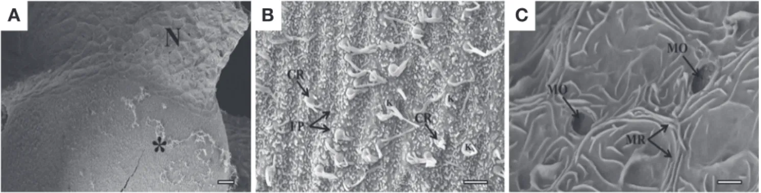
In a SEM observation, the olfactory epithelium is cate- gorized into two distinct types, the sensory and non-sen- sory epithelium, according to the existence of the recep- tor cells(Fig. 5A). The distributional pattern of the sen- sory epithelium is a continuous pattern. It also has a only one type of the receptor cell(ciliated cell) and numerous- fine protrusions. The ciliated receptor cell contains a knob on the apical surface of the cell body and with lying cilia (3~4 units in number, 0.5~2μm length)(Fig. 5B). In contrast, the non-sensory epithelium has mucous open- ings(1.5~2.0μm major axis diameter). The microridges are also arranged randomly on this epithelial surface(Fig.
5C).
dIScuSSIon
Even though the anterior nostril as a tube-like structure is common in many gobies fishes(genus Rhinogobius, Periophthalmus, Neogobius), there are considerable dif- ferences in its length, position and a shape of the entrance in the snout(Chen et al., 1999; Kuciel et al., 2013). In particular, the diverse position of this nostril is associated closely with each species’s microhabitat, as it is influenc- ed directly by environmental factors(Sarkar et al., 2014).
F. gymnauchen has a tubular anterior nostril set at the middle part of between the upper tip and the eye. This position is distinctive from some amphibious fishes(vs.
position at the tip of the upper lip in Boleophthalmus and Periophthalmus)(Kuciel et al., 2013). Above the amphi- bious fishes are active in an extremely little water at the low tide and suck in water into their olfactory organ (Murdy, 1989). Compared to such fishes, the middle po-
sition and tubular structure in F. gymnauchen may be affected by a water amount of the environment where it lives in.
As nasal sac has a function to help water ventilation via olfactory organ, therefore, it is called divisionally as accessory or ventilation sac(Zeiske et al., 1992). Such fish whose water ventilation occurs by nasal sacs is de- fined as cyclosmats(vs. isosmats by motile cilia)(Doving et al., 1977). So, F. gymnauchen with having two acces- sory nasal sacs(ethmoidal and lacrimal sacs) is pertinent to cyclosmats. In addition to that, this fish has almost no motile cilia on the olfactory floor. It may be a feature of reflection of which the ventilatory mechanism via the olfactory organ is more dependent on the nasal sacs than cilia.
The number of the nasal sac in fish’s snout varies in the gobiid fishes(Kuciel et al., 2013). In the subfamily Gobi- inae, Proterorhinus marmoratus and Neogobius fluviatilis have a only one nasal sac whereas N. melanostomus has two nasal sacs. Also, of the Oxudercinae, P. barbarus and P. argentilineatus have a only one nasal sac. Kuciel et al.
(2013) mentioned that the diversity in number of the na- sal sac is relevant to the adaptability on the land(only one nasal sac in P. barbarus vs. two nasal sacs in B. baddarti) and decrease of the olfactory dependence. Thus, as hav- ing two nasal sacs, F. gymnauchen seems that it depends on the olfaction, which such two nasal sacs regarded as the macrosmatic fish can also be used a taxonomic char- acter.
The olfactory epithelium of F. gymnauchen is largely divided into the sensory and non-sensory epithelium.
Among them, the sensory epithelium is a continuous type among the categories classified by Yamamoto and Ueda
Fig. 5. Scanning electron micrographs showing the surface structure of the olfactory epithelium of Favonigobius gymnauchen. (A), the olfactory epithelium showing two distinct types, the sensory and non-sensory cells; (B), the sensory epithelium with a ciliated receptor cells and fine pro- trusions; (C), the non-sensory epithelium with mucous openings and microridges. Asterisk, sensory epithelium; N, non-sensory epithelium; CR, ciliated receptor cell; FP, fine protrusion; MO, mucous opening; MR, microridge; K, knob. Bars indicate 20μm in (A) and 2μm(B), (C), respec- tively.
A B C
(1978). The distributional pattern of the sensory epithe- lium takes various types depending on the species(con- tinuous, large-zonal, irregular, small-zonal types) and therefore has been used as part of the taxonomic research (Yamamoto, 1982). These differences also show in the case of the gobiid fishes(Kuciel et al., 2013). P. barba
rus, variabilis, chrysospilos has an islet type whereas B.
boddarti, Scartelaos histophorus and Parapocryptes ric
tuosus has a continuous type. Such distinct types are re- garded to reflect differences of the ecological niches with varying levels(Kuciel et al., 2013).
Generally, the kind and composition of olfactory cells (receptor cell, supporting cell, basal cell) on the epithe- lium varies in between genus or species(Hara, 1994).
Among these cells, the number of the cilia in the receptor cell also has significant differences in teleosts(Yamamo- to, 1982). F. gymnauchen has a only one type, ciliated receptor cell with 3~4 cilia. Mudskipper P. barbarus has a ciliated receptor cell with 3~4 cilia whereas gold fish Carasius auratus with 5~7 cilia. Those variations may be in relation to feeding habits, niches, the selection of habi- tats and the time for foraging(Singh, 1994).
The covering of the mucus on the olfactory epithelium is well developed in a few of cases that fish is in adverse situations, exposed to acid rain, high level of UV light and silt-laden water(Muniz and Leivestad, 1980; Roberts and Bullock, 1980; Shephard, 1994). F. gymnauchen, which inhabit the intertidal zone with a sand bottom and periodic- tidal cycle, is possible to face above disturbances. In ac- cordance with above environment, this species has many mucous openings in the lateral floor of the olfactory cav- ity. Consequently, their occurrences may protect olfacto- ry receptor cell and epithelium against physical impact or chemical change in acting fields.
Based on this study, the olfactory organ of F. gymnau
chen is likely related with its ecological habitat as a shal- low water and tidal pool in the coastal or intertidal region.
referenceS
Chen, I.S., M. Kottelat and P.J. Miller. 1999. Freshwater gobies of the genus Rhinogobius from the Mekong basin in Thailand and Laos, with descriptions of three new species. Zool.
Stud., 38: 19-32.
Doving, K.B., M. Dubois-Dauphin, A. Holley and F. Jourdan. 1977.
Functional anatomy of the olfactory organ of fish and the ciliary mechanism of water transport. Acta Zool., 58: 245- 255.
Gurr, G.T. 1956. A practical manual of medical and biological stain-
ing techniques. Interscience, New York, pp. 1-99.
Hara, T.J. 1975. Olfaction in fish. Progr. Neurobiol., 5: 271-335.
Hara, T.J. 1986. Role of olfaction in fish behaviour. In: Pitcher, T.J.
(ed.), The behaviour of teleost fishes. Springer US, pp. 152- 176.
Hara, T.J. 1994. The diversity of chemical stimulation in fish olfac- tion and gustation. Rev. Fish. Biol. Fisheries, 4: 1-35.
Huh, S.H. and S.N. Kwak. 1998. Feeding habits of Favonigobius gymnauchen in the eelgrass(Zostera marina) bed in Kwang- yang Bay. Kor. J. Fish. Aquat. Sci., 31: 372-379.(in Kore- Jakubowski, M. 1967. A method for the manifestation of lateral-line an)
canals and their neuromasts in fishes. Copeia, 1: 234-235.
Jakubowski, M. 1975. Anatomical structure of olfactory organs pro- vided with internal nares in the Antarctic fish Gymnodraco acuticeps Boul.(Bathydraconidae). Bull. Acad. Pol. Sci.
Ser. Sci. Biol., 23: 115-120.
Jin, D.S., K.H. Han and J.W. Park. 2003. Spawning behavior and morphological development of larvae and juvenile of the nake-headed goby, Favonigobius gymnauchen(Bleeker).
Kor. J. Fish. Aquat. Sci., 36: 136-143.(in Korean)
Kang, J.C., P. Chin, J.S. Lee, Y.K. Shin and K.S. Cho. 2000. Effects of Salinity on Survival, Growth and oxygen consumption rates of the juvenile gobiid, Favonigobius gymnauchen.
Kor. J. Fish. Aquat. Sci., 33: 408-412.(in Korean)
Kim, B.J. and K. Nakaya. 2013. Fishes of Jeju Island, Korea. Na- tional Institute of Biological Resources, Incheon, 223pp.
Kim, H.T., Y.J. Lee, J.S. Park and J.Y. Park. 2014. A study on the structure of peripheral olfactory organ in the Korean mud- skipper, Scartelaos gigas(Pisces, Gobiidae). Kor. J. Ichthy- ol., 26: 281-287.
Kim, I.S. and J.Y. Park. 2002. Freshwater fishes of Korea. Kyo-Hak Publishing Co. Ltd., Korea. pp. 388-389.(in Korean).
Kuciel, M., K. Zuwala and M. Jakubowski. 2011. A new type of fish olfactory organ structure in Periophthalmus barbarous (Oxudercinae). Acta Zool., 92: 276-280.
Kuciel, M., K. Zuwala and U. Satapoomin. 2013. Comparative morphology(SEM) of the peripheral olfactory organ in the Oxudercinae subfamily(Gobiidae, Perciformes). Zool.
Anz., 252: 424-430.
Lee, J.S., J.W. Kim, J.C. Kang, Y.K. Shin and P. Chin. 2000. Repro- ductive cycle and gonadal development of the naked-head- ed Goby, Favonigobius gymnauchen(Teleostei: Gobiidae).
Kor. J. Fish. Aquat. Sci., 33: 219-224.(in Korean)
Muniz, I.P. and H. Leivestad. 1980. Toxic effects of aluminium on the brown trout, Salmo trutta L. In: Drablns, D., A. Tollan (eds.), Proc. Int. Conf. Ecol. Impact Acid Precip., Sande- fjord, Norway, pp. 11-14.
Murdy, E.O. 1989. A taxonomic revision and cladistic analysis of the Oxudercine Gobies(Gobiidae: Oxudercinae). Rec. Aust.
Mus., Suppl., 11: 1-93.
Roberts, R.J. and A.M. Bullock. 1980. The skin surface ecosystem of teleost fishes. Proc. R. Soc. Edinb., 79: 87-91.
ical variation of the olfactory apparatus in some Indian tele- osts with special reference to their ecological habitat. Folia Morphol. 73: 122-128.
Shephard, K.L. 1994. Functions for fish mucus. Rev. Fish. Biol.
Fisheries, 4: 401-429.
Singh, N. 1994. Scanning electron microscopic study of the olfac- tory epithelium of four coldwater hillstream teleosts from Garhwal hills(India). J. Biosci., 19: 91-102.
Yamamoto, M. 1982. Comparative morphology of the peripheral
ception in fishes. Elsevier, Amsterdam, pp. 39-59.
Yamamoto, M. and K. Ueda. 1978. Comparative morphology of fish olfactory epithelium-Ⅲ Cypriniformes. Bull. Jpn. Soc.
Sci. Fish. pp. 1201-1206.
Zeiske, E., B. Theisen and H. Breucker. 1992. Structure, develop- ment, and evolutionary aspects of the peripheral olfactory system. In: Hara, T.J.(ed.), Fish chemoreception. Springer Netherlands, pp. 13-39.
한국산 날개망둑 Favonigobius gymnauchen (Pisces, Gobiidae) 후각기관의 해부 및 조직학적 연구
김현태
·
김형수·
박종영전북대학교 자연과학대학 생물학과
요 약 : 한국산 날개망둑 Favonigobius gymnauchen 후각기관은 2015년 7월과 8월 사이 전라북도 부안군 변산
면 격포리의 조간대에서 채집된 개체들을 대상으로 실체현미경, 광학현미경 그리고 주사전자현미경을 이용하여 해
부학 및 조직학적 특징들을 조사하였다. 후각기관은 두부 주둥이 위 좌우로 한 쌍이 존재하며, 전비공과 후비공, 한
개의 비강, 두 개의 비낭, 후신경, 그리고 후구로 구성되었다. 비강 내 감각상피는 연결성 유형의 분포를 보였으며,
섬모성 감각세포만을 가지고 있었고, 감각세포, 지지세포, 기저세포들로 구성되었다. 비감각상피는 층상상피세포들
로 구성되었으며, 표면에 많은 점액공을 가지고 있었다. 따라서 이러한 후각기관의 특징들은 연안지역 조수 웅덩이
와 수심이 얕은 조간대의 생태적 서식처와 밀접한 관계가 있는 것으로 여겨진다.
찾아보기 낱말 : 비공, 비강, 비낭, 감각세포, 점액공