C
ANCERPREVENTION RESEARCH □ ORIGINAL ARTICLE □
81 책임저자:정주호, 130-701, 서울특별시 동대문구 회기동 1
경희대학교 고황의학연구소 Tel: 02-961-0298, Fax: 02-968-0560 E-mail: [email protected]
접수일:2008년 6월 11일, 게재승인일:2008년 6월 23일
Correspondence to:Joo-Ho Chung
Kohwang Medical Research Institute, School of Medicine, Kyung Hee University, 1, Hoegi-dong, Dongdaemun-gu, Seoul 130-701, Korea Tel: +82-2-961-0298, Fax: +82-2-968-0560
E-mail: [email protected]
Association between THSD4 Gene Polymorphisms and Hepatocellular Carcinoma in Korean Population
Su Kang Kim1, Sungwook Kang1, Bum Shik Kim2, Keon Sik Kim3 and Joo-Ho Chung1
1Kohwang Medical Research Institute, 2Department of Thoracic and Cardiovascular Surgery, 3Department of Anesthesiology and Pain Medicine, School of Medicine, Kyung Hee University, Seoul 130-701, Korea
Hepatocellular carcinoma (HCC) is a hypervascular tumor characterized by neovascularization, which plays an important role in the growth and progression of cancer. This study assessed the association of thrombospondin, type I, domain containing 4 (THSD4) polymorphisms with HCC in Korean population.
We selected three single nucleotide polymorphisms (SNPs) (rs961577, rs4777350, and rs2062651) in the THSD4 gene (heterozygosity>0.1). SNP genotyping was conducted using a customized Illumina BeadChip and direct sequencing in 189 HCC patients and 386 control subjects in Korean population. For the analysis of data, EM algorithm, SNPStats, SNPAnalyzer, and Helixtree programs were used. Multiple logistic regression analysis with the codominant, dominant, and recessive models was performed. Statistical analysis revealed that a significant association (rs2062651) was found between HCC and controls. Our genetic result suggests that THSD4 may be associated with the development of HCC. (Cancer Prev Res
13, 81-84, 2008)Key Words: Association, Hepatocellular carcinoma, Linkage disequilibrium, Polymorphism, Thrombos-
pondin, type I, domain containing 4 (THSD4)
INTRODUCTION
Thrombospondins (TSPs) are a family of multidomain ex- tracellular matrix glycoproteins, which consist of five members (TSP1, 2, 3, 4 and 5). TSP1 is synthesized and secreted by activated platelets, endothelial cells, macrophages, monotypes, fibroblasts, tumor cells, and vascular smooth muscular cells.
1∼3)TSP1 influences cell adhesion, motility, and growth.
4)Based on its effects on tumor, TSP family has attracted an interest as a potential regulator of tumor growth and metastasis. It has been reported that TSP1 expression suppresses growth or metastasis of some tumors, and inhibits angiogenesis.
5)However, another researcher reported that over expression of TSP1 in some tumors causes increased tumor progression.
6)Therefore, it is not clear at present to the relationship between
TSP1 expression and tumor progression. TSP1 is composed of three types of repeat sequences. These repeats mediate protein- protein interactions between TSP1 and transforming growth factor, matrix metalloproteinase 9, and the membrane protein CD36.
7)The activity of TSPs is contained in structural domain known as the TSP type repeat (TSR-1). A thrombospondin type 1 repeat may be involved in cell adhesion and angio- genesis.
8)Silverstein and Febbraio
9)reported that TSR domains may play an important role in regulating physiological and pathological angiogenesis. Very recently, thrombospondin, type I, domain containing 4 (THSD4) is known as a new gene in NCBI gene database. However, function of THSD4 has not defined.
Hepatocellular carcinoma (HCC) is characterized by a high
propensity for vascular invasion, and the angiogenic activity of
HCC correlates with the risk of vascular invasion.
10)Angio-
82 Cancer Prevention Research Vol. 13, No. 2, 2008
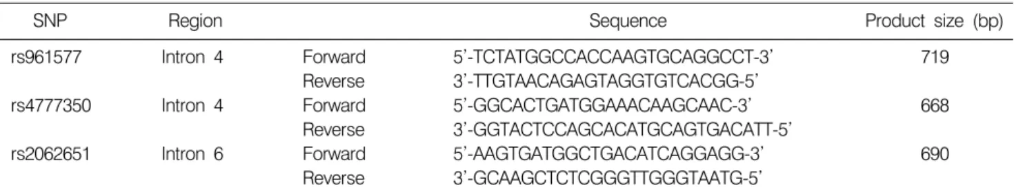
Table 1. Primer sequences for thrombospondin, type I, domain containing 4 (THSD4) gene polymorphisms
SNP Region Sequence Product size (bp)
rs961577 Intron 4 Forward 5’-TCTATGGCCACCAAGTGCAGGCCT-3’ 719
Reverse 3’-TTGTAACAGAGTAGGTGTCACGG-5’
rs4777350 Intron 4 Forward 5’-GGCACTGATGGAAACAAGCAAC-3’ 668
Reverse 3’-GGTACTCCAGCACATGCAGTGACATT-5’
rs2062651 Intron 6 Forward 5’-AAGTGATGGCTGACATCAGGAGG-3’ 690
Reverse 3’-GCAAGCTCTCGGGTTGGGTAATG-5’
SNP: Single nucleotide polymorphism, bp: base pair.
genesis in HCC has also been found to correlate with meta- stasis.
11)The process of angiogenesis is a complex multi step process initiated by the release of angiogenic factors from tumor cells.
Based on previous studies about thrombospondin, we presumed that polymorphisms of the THSD4 gene may be associated with HCC. Therefore, we carried out the association analysis of SNPs selected the THSD4 in Korean HCC patients.
MATERIALS AND METHODS
1. SubjectsAll case-control subjects used in the present study were recruited from the Kyung Hee University Medical Center and Keimyung University Dongsan Medical Center. In this study, 186 HCC (151 males and 38 females, average age at survey:
58.26 years) and 389 healthy controls (173 males and 213 females, average age at survey: 48.22 years) were included.
Written informed consent was obtained from each subject, and the study was approved by the Institutional Review Board of Kyung Hee University Medical Center. HCC was diagnosed either histological or by typical HCC imaging patterns, using angiography, computed tomography (CT), and/or magnetic resonance imaging (MRI), and sometimes serum AFP (above 400 ng/ml) analysis.
2. Genotyping
Genomic DNA was extracted from the whole blood of each subject using a commercially available Qiagen DNA Extraction kit (Qiagen, Tokyo, Japan). The SNPs of the THSD4 gene were selected on the basis of database (http://www.ebi.ac.uk/
ensemble/, http://ncbi.nlm.nih.gov/SNP). SNPs were excluded low heterozygosity (below 0.1) and minor allele frequency (below 0.05). The SNPs was also validated by HapMap
database. Finally, we selected three SNPs (rs961577, rs4777350, and rs2062651) in the THSD4 gene. SNP genotyping was conducted using the Golden-Gate SNP Genotyping Assay on an Illumina BeadStation 500 GX (Illumina Inc., San Diego, CA, USA) according to the manufacturer's protocol. In brief, DNA was first activated through a chemical reaction with activated biotin. Assay oligonucleotides (oligos) were added and hybridized to the DNA, and then PCR was performed. The fluorescent signals in the PCR products were imaged using the Illumina BeadArray Reader. The genotyping results were validated by direct sequencing. Genomic DNA was amplified using primer for each SNP (Table 1). The samples were sequenced using an ABI Prism 377 automatic sequencer (PE Applied Biosystems, Foster City, CA, USA). Sequence data were analyzed using the SeqManII software (DNASTAR Inc., Madison, WI, USA).
3. Statistical Analysis
For the analysis of Hardy-Weinberg equilibrium, chi-square test was used. Statistical analysis was performed using SNPStats, Helixtree (Golden Helix Inc., MT, USA), and SNPAnalyzer (ISTECH Inc., Goyang, Korea). The linkage disequilibrium (LD) block was tested using Haploview version 3.32.
12)Significance level was set at 0.05. Power analysis was performed using G*Power computer software.
13)To avoid static error, Bonferroni correction was also used, giving a significance level of 0.05/κ (κ=3, number of SNPs).
RESULTS
No deviation of Hardy-Weinberg equilibrium was found in
genotype distributions of three polymorphisms between HCC
and control (data not shown). Table 2 shows a single
association result between HCC and controls using SNPstats.
Su Kang Kim, et al:THSD Polymorphism and Hepatocellular Carcinoma 83
Fig. 1. Linkage disequilibrium (LD) block among thrombo- spondin, type I, domain containing 4 (THSD4) gene poly- morphisms. LD was visualized by Haploview.
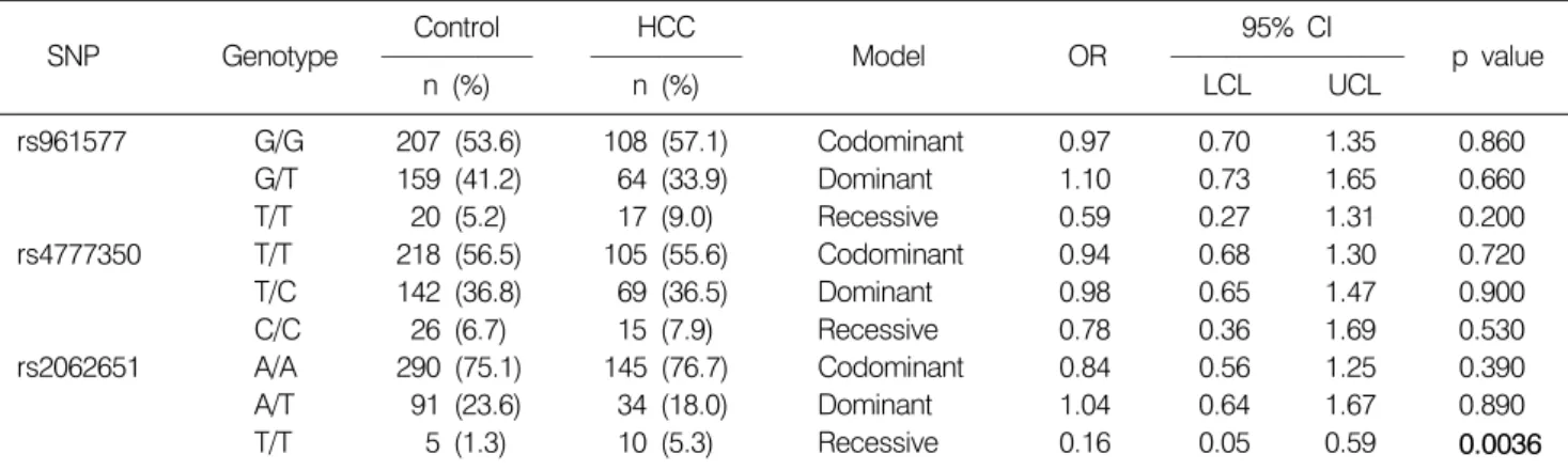
Table 2. Association between THSD4 gene polymorphisms and HCC in Korean population
Control HCC 95% CI
SNP Genotype Model OR p value
n (%) n (%) LCL UCL
rs961577 G/G 207 (53.6) 108 (57.1) Codominant 0.97 0.70 1.35 0.860
G/T 159 (41.2) 64 (33.9) Dominant 1.10 0.73 1.65 0.660
T/T 20 (5.2) 17 (9.0) Recessive 0.59 0.27 1.31 0.200
rs4777350 T/T 218 (56.5) 105 (55.6) Codominant 0.94 0.68 1.30 0.720
T/C 142 (36.8) 69 (36.5) Dominant 0.98 0.65 1.47 0.900
C/C 26 (6.7) 15 (7.9) Recessive 0.78 0.36 1.69 0.530
rs2062651 A/A 290 (75.1) 145 (76.7) Codominant 0.84 0.56 1.25 0.390
A/T 91 (23.6) 34 (18.0) Dominant 1.04 0.64 1.67 0.890
T/T 5 (1.3) 10 (5.3) Recessive 0.16 0.05 0.59 0.0036
Genotype distributions are shown as number. Odds ratio (OR), 95% confidence interval (CI), and p values were from logistic regression analyses with the codominant, dominant, and recessive models controlling age and gender as covariates. THSD4:
thrombospondin, type I, domain containing 4, n: number, SNP: single nucleotide polymorphism, HCC: hapatocellular carcinoma, LCL: lower confidence limit, UCL: upper confidence limit.
There was significant difference in the SNP (rs2062651) of THSD4 gene between HCC and control [recessive model, p=
0.0036, odds ratio (OR)=0.16, 95% confidence interval (CI)=
0.05−0.59] (Table 2). This association of the SNP was still significant after Bonferroni correction (p<0.05). LD and common haplotype block were analyzed by Haploview software version 3.2. When LD data were calculated for all SNP pairs between the control and case groups, however, LD block was not constructed between three SNPs in THSD4 genes (Fig. 1).
In sample number analysis, we calculated sample powers for the significant SNP to confirm the effects between HCC and
control. In our case-control study, we had 0.801 power for detecting significance, assuming an α-level of 0.05 (rs2062651, effect size=0.129). Thus, this case-control study was suffi- ciently powerful for determining a positive association.
DISCUSSION
Present study was aimed to determine whether SNPs within THSD4 gene correlate with susceptibility to HCC in Korean population. We first found that one (rs2062651) SNP was significantly associated with HCC. The rs961577 is located on intron 4. GG, GT, and TT genotype frequencies are reported to be 0.217, 0.517, and 0.267 in European, 0.400, 0.556, and 0.044 in Chinese, 0.644, 0.311, and 0.044 in Japanese, and 0.237, 0.373, and 0.390 in Sub-Saharan African, respectively (http://www.ncbi.nlm.nih.gov/SNP). GG, GT, and TT geno- type frequencies in Korean population were 0.536, 0.412, and 0.052. The rs4777350 is also located on intron 4. TT, TC, and CC genotype frequencies are reported to be 0.345, 0.483, and 0.172 in European, 0.400, 0.489, and 0.111 in Chinese, 0.636, 0.341, and 0.023 in Japanese, and 0.712, 0.254, and 0.034 in Sub-Saharan African, respectively (http://www.ncbi.
nlm.nih.gov/SNP). TT, TC, and CC genotype frequencies in
Korean population were 0.565, 0.368, and 0.067. The
rs2062651 is located on intron 6. AA, AT, and TT genotype
frequencies are reported to be 0.133, 0.433, and 0.433 in
European, 0.556, 0.422, and 0.022 in Chinese, 0.800, 0.156,
and 0.044 in Japanese, and 0.200, 0.483, and 0.317 in
84 Cancer Prevention Research Vol. 13, No. 2, 2008
Sub-Saharan African, respectively (http://www.ncbi.nlm.nih.
gov/SNP). AA, AT, and TT genotype frequencies in Korean population were 0.751, 0.236, and 0.013. The frequencies of three SNPs is similar to those in the Japanese.
Function of THSD4 has not been known until now.
However, it has been reported that TSP family play a role in cardiovascular pathology,
1)and TSP1 is a potent endogenous inhibitor of both physiologic and pathologic angiogenesis.
5,14)TSP1 is a large trimeric glycoprotein that interacts with extracellular matrix components and with surface receptors.
15)Normally found at low concentrations in the circulation and soft tissues, TSP1 expression is altered in a number of pathologic diseases.
16)In cancer, TSP1 expression generally decreases with malignant progression, resulting from regulation of its expression by a number of oncogene and tumor suppressor gene products.
5)In contrast, TSP1 overexpressing tumors typically grow slower, exhibit less angiogenesis, and have fewer metastasis.
17)Angiogenesis in HCC has also been found to correlate with metastasis.
18)To our knowledge, no previous study has examined an association between THSD4 and HCC. Our study first showed a significant association between SNP of the THSD4 gene and HCC.
In conclusion, we found a significant association between THSD4 polymorphism and HCC. The result suggests that THSD4 may be contributed to the etiology of HCC in Korean population. Further works on different THSD4 SNPs and/or larger sample size will be required to elucidate an exact role for THSD4 in HCC.
ACKNOWLEDGMENTS
This study was supported by the Governance Program of Kyung Hee University.
REFERENCES
1) Stenina OI, Topol EJ, Plow EF. Thrombospondins, their polymorphisms, and cardiovascular disease. Arterioscler Thromb Vasc Biol 27, 1886-1894, 2007.
2) Iruela-Arispe ML, Liska DJ, Sage EH, Bornstein P. Differen- tial expression of thrombospondin 1, 2, and 3 during murine development. Dev Dyn 197, 40-56, 1993.
3) O'Shea KS, Dixit VM. Unique distribution of the extra- cellular matrix component thrombospondin in the developing
mouse embryo. J Cell Biol 107, 2737-2748, 1988.
4) Lawler J, Sunday M, Thibert V, Duquette M, George EL, Rayburn H, Hynes RO. Thrombospondin-1 is required for normal murine pulmonary homeostasis and its absence causes pneumonia. J Clin Invest 101, 982-992, 1998.
5) Roberts DD. Regulation of tumor growth and metastasis by thrombospondin-1. Faseb J 10, 1183-1191, 1996.
6) Tuszynski GP, Nicosia RF. The role of thrombospondin-1 in tumor progression and angiogenesis. Bioessays 18, 71-76, 1996.
7) Lawler J, Detmar M. Tumor progression: the effects of thrombospondin-1 and -2. Int J Biochem Cell Biol 36, 1038- 1045, 2004.
8) Vogel T, Guo NH, Krutzsch HC, Blake DA, Hartman J, Mendelovitz S, Panet A, Roberts DD. Modulation of endo- thelial cell proliferation, adhesion, and motility by recom- binant heparin-binding domain and synthetic peptides from the type I repeats of thrombospondin. J Cell Biochem 53, 74- 84, 1993.
9) Silverstein RL, Febbraio M. CD36-TSP-HRGP interactions in the regulation of angiogenesis. Curr Pharm Des 13, 3559- 3567, 2007.
10) Poon RT, Ng IO, Lau C, Zhu LX, Yu WC, Lo CM, Fan ST, Wong J. Serum vascular endothelial growth factor predicts venous invasion in hepatocellular carcinoma: a prospective study. Ann Surg 233, 227-235, 2001.
11) Yao DF, Wu XH, Zhu Y, Shi GS, Dong ZZ, Yao DB, Wu W, Qiu LW, Meng XY. Quantitative analysis of vascular endothelial growth factor, microvascular density and their clinicopathologic features in human hepatocellular carcinoma.
Hepatobiliary Pancreat Dis Int 4, 220-226, 2005.
12) Barrett JC, Fry B, Maller J, Daly MJ. Haploview: analysis and visualization of LD and haplotype maps. Bioinformatics 21, 263-265, 2005.
13) Faul F, Erdfelder E, Lang AG, Buchner A. G*Power 3: a flexible statistical power analysis program for the social, behavioral, and biomedical sciences. Behav Res Methods 39, 175-191, 2007.
14) Lawler J. Thrombospondin-1 as an endogenous inhibitor of angiogenesis and tumor growth. J Cell Mol Med 6, 1-12, 2002.
15) Chen H, Herndon ME, Lawler J. The cell biology of throm- bospondin-1. Matrix Biol 19, 597-614, 2000.
16) Esemuede N, Lee T, Pierre-Paul D, Sumpio BE, Gahtan V.
The role of thrombospondin-1 in human disease. J Surg Res 122, 135-142, 2004.
17) Isenberg JS, Ridnour LA, Perruccio EM, Espey MG, Wink DA, Roberts DD. Thrombospondin-1 inhibits endothelial cell responses to nitric oxide in a cGMP-dependent manner. Proc Natl Acad Sci U S A 102, 13141-13146, 2005.
18) Sahani DV, Holalkere NS, Mueller PR, Zhu AX. Advanced hepatocellular carcinoma: CT perfusion of liver and tumor tissue--initial experience. Radiology 243, 736-743, 2007.