Accepted: 2017.12.05 Published: 2018.05.26
1844 5 — 31
Association Between Polymorphisms of
Interleukin 1 Family Genes and Hepatocellular Carcinoma
ABCD 1
Ki Hong Tak
ABCD 2
Gyeong Im Yu
BCD 2
Mi Young Lee*
ABCDEF 2
Dong Hoon Shin*
* These 2 authors (Mi Young Lee and Dong Hoon Shin) contributed equally
Corresponding Authors: Mi Young Lee, e-mail: [email protected] (research promoting), Dong Hoon Shin, e-mail: [email protected]
Source of support: This work was supported by a Research-Promoting grant from Keimyung University Dongsan Medical Center in 2008 and 2012
Background:
Hepatocellular carcinoma (HCC) is one of the most common malignancies occurring worldwide and is most fre- quent type of liver cancer. The risk for developing HCC increases with the severity of inflammation and fibro- sis. The members of the interleukin-1 (IL-1) family are primarily proinflammatory cytokines due to their ability to stimulate the expression of genes associated with inflammation and autoimmune diseases. Several studies have suggested that some proinflammatory cytokines, such as the IL-1 family (IL-1a, IL-1b, and IL-1 receptor antagonist) are involved in the pathogenesis of HCC.
Material/Methods: This study aimed to determine whether polymorphisms in the IL-1 family of genes are associated with HCC. We analyzed 178 HCC patients and 397 controls to investigate the association between polymorphisms in IL-1a, IL-1b, and IL-1 receptor antagonist (IL-1RA) genes and HCC in the Korean population. All subjects were geno- typed for the selected SNPs in IL-1a, IL-1b, and IL-1RA genes by Golden-Gate SNP Genotyping Assay.
Results:
Statistical analysis revealed a significant association at IL-1b between HCC and controls. Three individual poly- morphisms (rs1143633, rs3917356, and rs1143627) were found to be associated with HCC. The SNPs of IL-1b gene (rs1143633A>G and rs1143627T>C) protected against HCC in the dominant model (p=0.027, OR=0.59, 95% CI=0.37–0.94; p=0.019, OR=0.56, 95% CI=0.34–0.91). The SNP of IL-1b gene (rs3917356G>A) increased the risk of HCC in the recessive model (p<0.001, OR=2.58, 95% CI=1.53–4.33), whereas other SNPs in IL-1a and IL-1RA showed no significant association between HCC patients and controls.
Conclusions:
These results suggest that IL-1b in the IL-1 family contributes to HCC susceptibility.
MeSH Keywords: Carcinoma, Hepatocellular • Interleukin-1 • Polymorphism, Single Nucleotide
Full-text PDF: https://www.medscimonit.com/abstract/index/idArt/907524
Authors’ Contribution:
Study Design A Data Collection B Statistical Analysis C Data Interpretation D Manuscript Preparation E Literature Search F Funds Collection G
1 Department of Occupational and Environmental Medicine, Sungseo Hospital, Daegu, South Korea
2 Department of Preventive Medicine, School of Medicine and Institute for Cancer Research, Keimyung University, Daegu, South Korea
Background
Hepatocellular carcinoma (HCC) is one of the most common malignancies occurring worldwide. Almost 90% of primary ma- lignant hepatic tumors lead to liver cancer among adults [1].
The distribution of HCC varies across the world; the highest HCC incidence is in South-East Asia and Sub-Saharan Africa, where prevalence of chronic hepatitis B (HBV) and hepatitis C (HCV) virus infection are high. These viruses are the most frequent etiological factors of HCC [2–4]. Also, increased body mass index (BMI) and diabetes with subsequent development of non-alcoholic steatohepatitis are significant risk factors for HCC. Other non-viral causes of HCC include iron overload syndromes, alcohol drinking, smoking, oral contraceptive use, and aflatoxin exposure, which are common in the developing world [5]. Male sex is another risk factor for HCC, since men are more susceptible to diseases than women, and the imbal- ance in the ratio of males to females in a population varies from 2: 1 up to 4: 1 [6,7].
The risk for developing HCC increases with the severity of in- flammation and fibrosis [8,9]. HCC develops slowly in a back- ground of chronic inflammation triggered by exposure to in- fectious agents (e.g., HCV and HBV), toxic compounds (e.g., alcohol), or metabolic impairment. Although the molecular mechanism links that connect inflammation and cancer are not thoroughly understood, a key role for cytokines such as interleukin-6 (IL-6) and interleukin-1 (IL-1) in HCC has been established in experimental animal models [10].
IL-1a and IL-1b are members of the IL-1 family. These cyto- kines are pleiotropic cytokines and are synthesized by a variety of cell types, including activated macrophages, keratinocytes, stimulated B lymphocytes, and fibroblasts. These cytokines are involved in various immune responses, inflammatory process- es, and hematopoiesis, and they increase the expression of adhesion factors on endothelial cells and enable transmigra- tion of leukocytes. IL-1 increases angiogenesis and can pro- mote tumor invasiveness and metastasis [11,12]. Also, IL-1a stimulates cytotoxic cells, and proteolytic activation of prote- ase by neutrophils induced by the IL-1 family enhances angio- genesis and tumorigenesis [13].
The IL-1 family of genes encoding the 3 proteins, IL-1a, IL-1b, and IL-1 receptor antagonist (IL-1 RA) were mapped to chromo- some 2q13-21 [14]. Association polymorphisms of IL-1 family genes and development of gastric cancer [15,16], breast can- cer [17,18], and gallbladder cancer [19] were reported.
Specific polymorphisms associated with susceptibility to var- ious diseases have been studied with great interest. Among these polymorphisms, single-nucleotide polymorphisms (SNPs)
are very common throughout the genome. Therefore, they are considered appropriate tools for identifying individual suscep- tibility to common disease.
Recently, associations of numerous functional gene polymor- phisms of proinflammatory and profibrogenetic factors with HCC have been found. The objective of the present study was to determine whether polymorphisms in the IL-1 family genes are associated with HCC in the Korean population.
Material and Methods
Subjects and clinical data
We recruited 178 patients (150 males and 28 females) di- agnosed with HCC at Keimyung University Dongsan Medical Center. Each patient was diagnosed by aspiration cytology or biopsy. The tumor-node-metastasis system was used to stage the tumors. Tumor metastasis and portal vein were determined by computed tomography and angiography, respectively. As a control group, we recruited 397 patients (166 males and 231 females) who had visited our Health Promotion Center for routine check-ups from September to December 2004. All control group subjects were free of hypertension, dyslipid- emia, diabetes, and liver diseases. The average age of controls was 47.84±10.56 years old (Table 1). All study subjects were Korean and provided written informed consent. The study was approved by our Institutional Review Board (IRB No. 03-09).
SNPs selection and genotyping
Twenty-one SNPs in the IL-1 family (IL-1a, IL-1b, and IL-1RA) genes of proinflammatory cytokines were selected based on database searches (http://ncbi.nlm.nih.gov/SNP) for this study.
The IL-1 family of genes are located on chromosome 2q14. The 21 SNPs were selected in the IL-1 family genes based on het- erozygosity (above 0.1). The selected SNPs of the IL-1 family genes were located on chromosome 2q14, which included IL- 1a and IL-1b (2 in the exon region, 1 in the 5’near gene, and 6 in the intron region).
Peripheral blood samples were collected from each subject and then stored at –20°C. The genomic DNAs of subjects were ex- tracted from the peripheral blood samples with a commercial DNA extraction kit (Macherey Nagel, Germany). Genotyping of all SNPs was carried out using Golden-Gate SNP Genotyping Assay.
Statistical analysis
First, we assessed whether genotype frequencies were in Hardy-
Weinberg equilibrium. The results were considered statistically
significant at p values less than 0.05. The allele frequencies and genotype distribution were compared between HCC pa- tients and controls using the chi-square test. Odds ratios (OR) and 95% confidence intervals (CI) were calculated for effect of genotypes and allele frequencies at the 5% level of significance.
For the logistic regression analysis of genetic data, SNPStats and SPSS 17.0 were used [20]. Logistic regression analysis was performed to investigate the association between SNPs of the IL-1 family of genes and HCC adjusted by age and sex. A link- age disequilibrium (LD) block of polymorphisms was analyzed using Haploview 3.32 [20,21]. The haplotypes and their fre- quencies were calculated using the EM algorithm.
Results
Demographic and clinical characteristics of study subjects Table 1 shows demographic and clinical characteristics of study subjects. Male subjects showed higher prevalence of HCC than females (150 vs. 28, age range 31–80 years). Type of tumor was diffuse in 13 patients (7.3%), massive in 42 patients (23.6%), massive and diffuse in 1 patient (0.6%), massive and nodu- lar in 2 patients (1.1%), nodular in 101 patients (56.7%), and simple nodular in 19 patients (10.7%). Metastasis was found in 25 patients (14.0%), and portal vein involvement was found in 62 patients (34.8%). Stage of HCC was stage I in 18 patients
Variable Total (n=178)
Age 59.5 (31–80)
Gender Female 28 (15.7)
Male 150 (84.3)
Cause
Hepatitis B virus 133 (74.7)
Hepatitis C virus 15 (8.4)
Alcoholic liver disease 21 (11.8)
Autoimmune liver disease 2 (1.1)
Type
Diffuse 13 (7.3)
Massive 42 (23.6)
Massive + diffuse 1 (0.6)
Massive + nodular 2 (1.1)
Nodular 101 (56.7)
Simple nodular 19 (10.7)
Metastasis Yes 25 (14.0)
No 152 (85.4)
Stage
I 18 (10.1)
II 50 (28.1)
III 50 (28.1)
IV-A 34 (19.1)
IV-B 25 (14.0)
Size (cm) <5 101 (56.7)
³5 75 (42.1)
PVT Negative 115 (64.6)
Positive 62 (34.8)
Albumin (g/dL) 3.5 (1.7–5.1)
AST (U/L) 65.5 (21–923)
ALT (U/L) 40 (5–654)
Table 1. Demographics and clinical variables of hepatocellular carcinoma patients.
Age, albumin, AST and ALT are shown as median (range); Gender, type, metastasis, stage, size, PVT (portal vein thrombosis) are shown
as frequency (percentage).
(10.6%), stage II in 50 patients (28.1%), stage III in 50 patients (28.1%), stage IV-A in 34 patients (19.1%), and stage IV-B in 25 patients (14.0%). Tumor size was over 5 cm in 75 patients (42.1%). Albumin, AST, and ALT ranges were 1.7–5.1, 21–923, and 5–654, respectively. The allele frequency in subjects fol- lowed Hardy-Weinberg equilibrium (p>0.05).
Logistic regression analysis of IL-1 family genes SNPs between HCC and controls
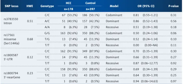
Table 2 shows the genotype distributions in IL-1a gene in HCC patients and controls. All genotype distributions were consis- tent with the Hardy-Weinberg principle (p>0.05). There were no significant differences between HCC patients and controls in rs3783550, rs17561, rs1800587, or rs1800794 (Table 2).
Table 3 shows the genotype distributions in IL-1b in HCC pa- tients and controls. Rs1143633A>G and rs1143627T>C showed protective effects in the dominant model (A/A vs. A/G + G/G;
T/T vs. T/C + C/C) (p=0.027, OR=0.59, 95% CI=0.37–0.94;
p=0.019, OR=0.56, 95% CI=0.34–0.91), and rs3917356G>A in- creased the risk of HCC in the recessive model (G/G + G/A vs.
AA) (p<0.001, OR=2.58, 95% CI=1.53–4.33), whereas all oth- er loci (rs1071676, rs1143637, rs1143634, rs3136558, and rs143630) showed no significant associations.
There were no significant differences among HCC patients and controls in IL-1RA gene polymorphism (rs2234679, rs928940, rs439154, rs419598, rs315952, and rs315951) (Table 4).
Logistic regression analysis of IL-1 family genes SNPs with clinical characteristics in HCC patients
Table 5 shows the relationship of SNP of IL-1b gene with clin- ical characteristics in males with HCC. Among tested SNPs of IL-1 family genes, rs3917356 showed a significant association with portal vein thrombosis (PVT) (OR=2.17, 95% CI=1.08–1.35, p=0.029) and metastasis (OR=2.70, 95% CI=1.09–6.67, p=0.032).
However, all other loci did not show any significant associa- tions with clinical characteristics in males with HCC.
Discussion
It is believed that immune system-mediated chronic inflam- mation of the liver can lead to HCC development because the former induces continuous cell death, resulting in cell prolif- eration and increased frequency of genetic alterations [22,23].
The proinflammatory cytokines of the IL-1 family, most no- tably IL-1b and IL-18, also have very important roles in anti- microbial host defense. IL-1a and IL-1b, which bind and acti- vate the same receptor, 8630372, activate the release of other
SNP locus HWE Genotype HCC Control
Model OR (95% CI) P-value
n=178 n=397
rs3783550
Intron 0.51
C/C 67 (53.2%) 186 (50.1%) Codominant 0.81 (0.55–1.21) 0.31 A/C 51 (40.5%) 157 (42.3%) Dominant 0.86 (0.52–1.43) 0.56 A/A 8 (6.3%) 28 (7.5%) Recessive 0.51 (0.19–1.38) 0.18 rs17561
missense (Ser114Ala)
0.68
G/G 163 (92.6%) 350 (88.2%) Codominant 0.50 (0.24–1.06) 0.06 T/G 13 (7.4%) 45 (11.3%) Dominant 0.52 (0.24–1.14) 0.10 T/T 0 (0.0%) 2 (0.5%) Recessive 0.00 (0.00–NA) 0.11
rs1800587
5’-UTR 0.12
C/C 162 (91.5%) 349 (87.9%) Codominant 0.70 (0.35–1.39) 0.30 T/C 14 (7.9%) 45 (11.3%) Dominant 0.66 (0.31–1.39) 0.27 T/T 1 (0.6%) 3 (0.8%) Recessive 0.87 (0.06–12.77) 0.92
rs1800794 5’-nearGene
0.23
C/C 158 (91.9%) 350 (88.6%) Codominant 0.69 (0.34–1.39) 0.29 T/C 13 (7.6%) 43 (10.9%) Dominant 0.64 (0.30–1.39) 0.25 T/T 1 (0.6%) 2 (0.5%) Recessive 0.94 (0.06–14.63) 0.97
Table 2. Logistic regression analysis and genotype frequency of polymorphisms in IL-1a gene between HCC and control.HWE is Hardy-Weinberg equilibrium. Genotype distributions are shown as number (%). Odds ratio (OR), 95% confidence interval (CI),
and p-values were from logistic regression analysis with codominant, dominant, and recessive models controlling age and gender
as covariates. SNP is single nucleotide polymorphism. Total number of each SNP is different, because genotypes of some SNPs are
unreadable.
proinflammatory cytokines such as TNF and IL-6, and induce a Th (helper T cell) 17 bias in the cellular adaptive responses [24].
In vivo, IL-1 is largely responsible for the acute-phase response, which includes fever, acute protein synthesis, anorexia, and somnolence [25]. IL-18 is essential for the induction of inter- feron gamma (IFN-g) and Th1 responses. Through these mech- anisms, cytokines of the IL-1 family are a crucial component of host defense against infections.
Many proinflammatory cytokines, such as IL-1b and IFN-g poly- morphisms have been associated with an increased risk of HCC [26–28]. Many studies have been conducted to analyze
SNPs as genetic markets as a result of their high density and even distribution in the human genome.
Here, we investigated whether the IL-1 family (IL-1a, IL-1b, and IL-1RA) of genes polymorphisms are related to risk of HCC by genotyping 21 selected SNPs in the Korean population. In this study, we found that 3 SNPs of IL-1b gene were association with HCC. In our investigation of the association between IL-1 family genes and HCC progression, only rs3917356 showed a significant association with portal vein thrombosis (OR=2.17, 95% CI=1.08–1.35, p=0.029) and metastasis (OR=2.70, 95%
CI=1.09–6.67, p=0.032). However, all other loci did not show any significant associations with clinical characteristics in male HCC.
SNP locus HWE Genotype HCC Control
Model OR (95% CI) P-value
n=178 n=397
rs1071676
3’-UTR 0.25
G/G 174 (97.8%) 376 (94.7%) Codominant 0.32 (0.09–1.14) 0.06 C/G 4 (2.2%) 20 (5.0%) Dominant 0.32 (0.09–1.14) 0.06
C/C 0 (0.0%) 1 (0.2%) Recessive 0.00 (0.00–NA) 0.57
rs1143637
intron 0.25
G/G 174 (97.8%) 376 (94.7%) Codominant 0.32 (0.09–1.14) 0.06 A/G 4 (2.2%) 20 (5.0%) Dominant 0.32 (0.09–1.14) 0.06
A/A 0 (0.0%) 1 (0.2%) Recessive 0.00 (0.00–NA) 0.57
rs1143634 synonymous (Phe105Phe)
0.23
C/C 174 (97.8%) 377 (95%) Codominant 0.32 (0.09–1.14) 0.06
T/C4 (2.2%) 19 (4.8%) Dominant 0.32 (0.09–1.14) 0.06
T/T
0 (0.0%) 1 (0.2%) Recessive 0.00 (0.00–NA) 0.57
rs1143633
intron 0.72
A/A
71 (43.3%) 141 (35.9%) Codominant 0.75 (0.53–1.06) 0.10
A/G73 (44.5%) 194 (49.4%) Dominant 0.59 (0.37–0.94)
0.027 G/G20 (12.2%) 58 (14.8%) Recessive 0.97 (0.50–1.91) 0.94
rs3136558
intron 0.73
T/T
48 (28.1%) 118 (30.0%) Codominant 1.18 (0.86–1.62) 0.31
T/C86 (50.3%) 190 (48.4%) Dominant 1.23 (0.75–2.01) 0.41
C/C37 (21.6%) 85 (21.6%) Recessive 1.26 (0.73–2.19) 0.41
rs1143630
intron 0.26
C/C
117 (70.1%) 261 (66.2%) Codominant 1.00 (0.66–1.51) 1.00
A/C42 (25.1%) 118 (29.9%) Dominant 0.92 (0.57–1.50) 0.75
A/A8 (4.8%) 15 (3.8%) Recessive 1.58 (0.50–4.95) 0.44
rs3917356
intron 0.22
G/G
52 (32.1%) 121 (30.8%) Codominant 1.30 (0.96–1.76) 0.09
A/G56 (34.6%) 183 (46.6%) Dominant 0.83 (0.51–1.36) 0.47
A/A54 (33.3%) 89 (22.6%) Recessive 2.58 (1.53–4.33)
<0.001rs1143627
5’-UTR 0.48
T/T
56 (33.5%) 105 (26.6%) Codominant 0.88 (0.65–1.20) 0.42
T/C61 (36.5%) 190 (48.1%) Dominant 0.56 (0.34–0.91)
0.019 C/C50 (29.9%) 100 (25.3%) Recessive 1.33 (0.80–2.23) 0.27
Table 3. Logistic regression analysis and genotype frequency of polymorphisms in IL-1b gene between HCC and control.HWE is Hardy-Weinberg equilibrium. Genotype distributions are shown as number (%). Odds ratio (OR), 95% confidence interval (CI),
and p-values were from logistic regression analysis with codominant, dominant, and recessive models controlling age and gender
as covariates. SNP is single nucleotide polymorphism. Total number of each SNP is different, because genotypes of some SNPs are
unreadable.
SNP locus HWE Genotype HCC Control
Model OR (95% CI) P-value
n=178 n=397
rs2234679
5’-UTR 0.79
G/G 148 (84.6%) 331 (83.6%) Codominant 0.73 (0.40–1.34) 0.31 C/G 27 (15.4%) 62 (15.7%) Dominant 0.74 (0.40–1.35) 0.32
C/C 0 (0.0%) 3 (0.8%) Recessive 0.00 (0.00–NA) 0.56
rs928940
intron 0.25
G/G 42 (34.1%) 114 (31.1%) Codominant 0.95 (0.67–1.34) 0.76 T/G 53 (43.1%) 170 (46.5%) Dominant 0.84 (0.49–1.45) 0.54 T/T 28 (22.8%) 82 (22.4%) Recessive 1.05 (0.57–1.93) 0.89 rs439154
5’nearGene 0.38
A/A 77 (47.8%) 166 (42.5%) Codominant 0.81 (0.58–1.14) 0.22 A/G 60 (37.3%) 171 (43.7%) Dominant 0.66 (0.41–1.05) 0.07 G/G 24 (14.9%) 54 (13.8%) Recessive 1.04 (0.53–2.03) 0.90 rs419598
synonymous (Ala39Ala)
0.72
T/T 155 (88.6%) 345 (87.1%) Codominant 0.69 (0.35–1.37) 0.28 T/C 20 (11.4%) 50 (12.6%) Dominant 0.69 (0.35–1.37) 0.28
C/C 0 (0.0%) 1 (0.2%) Recessive 0.00 (0.00–NA) 0.76
rs315952 synonymous (Ser112Ser)
0.67
C/C 48 (38.1%) 122 (33.5%) Codominant 0.88 (0.62–1.25) 0.49 T/C 55 (43.6%) 174 (47.8%) Dominant 0.83 (0.49–1.41) 0.50 T/T 23 (18.2%) 68 (18.7%) Recessive 0.86 (0.45–1.65) 0.65 rs315951
3’-UTR 0.17
C/C 74 (46.0%) 141 (36.4%) Codominant 0.74 (0.54–1.01) 0.06 G/C 61 (37.9%) 174 (45.0%) Dominant 0.65 (0.41–1.03) 0.07 G/G 26 (16.1%) 72 (18.6%) Recessive 0.81 (0.51–1.28) 0.37
Table 4. Logistic regression analysis and genotype frequency of polymorphisms in IL-1RA gene between HCC and control.HWE is Hardy-Weinberg equilibrium. Genotype distributions are shown as number (%). Odds ratio (OR), 95% confidence interval (CI), and p-values were from logistic regression analysis with codominant, dominant, and recessive models controlling age and gender as covariates. SNP is single nucleotide polymorphism. Total number of each SNP is different, because genotypes of some SNPs are unreadable.
rs3917356
OR (95% CI) p
G/G+G/A A/A
Albumin (g/dL)
<3.5 43 (64.2) 24 (35.8) 1
³3.5 53 (63.9) 30 (36.1) 1.01 (0.52–1.98) 0.97
AST (U/L) 65.5 44 (63.8) 25 (36.2) 1
³65.5 52 (66.7) 26 (33.3) 0.88 (0.45–1.74) 0.71
ALT (U/L) <40 44 (62.0) 27 (38.0) 1
³40 52 (65.8) 27 (34.2) 0.85 (0.43–1.65) 0.62
PVT Negative 68 (70.8) 28 (29.2) 1
0.029
Positive 28 (52.8) 25 (47.2) 2.17 (1.08–1.35)
Stage I + II 31 (62.0) 19 (38.0) 1
III + IV 64 (64.6) 35 (35.4) 0.89 (0.44–1.80) 0.75
Metastasis Yes 85 (67.5) 41 (32.5) 1
0.032
No 10 (43.5) 13 (56.5) 2.70 (1.09–6.67)
Cause Hepatitis 78 (62.4) 47 (37.6) 1
Alcohol 14 (70.0) 6 (30.0) 0.71 (0.26–1.87) 0.51
Size (cm) <5 50 (61.7) 31 (38.3) 1
³5 45 (67.2) 22 (32.8) 0.79 (0.40–1.56) 0.49
Table 5. Relationship of SNP of IL-1b gene with clinical characteristics in male with HCC.
Variables are shown as frequency (percentage). Albumin, AST, and ALT are divided by median value. Odds ratio (OR), 95% confidence
interval (CI), and p-values were from logistic regression analysis controlling age as covariates.
Rs1143633A>G and rs1143627T>C revealed protective effects in the dominant model (p=0.027, OR=0.59, 95% CI=0.37–0.94;
p=0.019, OR=0.56, 95% CI=0.34–0.91), and rs3917356G>A was found to be a risk factor of HCC in the recessive model (p<0.001, OR=2.58, 95% CI=1.53–4.33). Rs1143633 and rs3917356 are located in an intron of the IL-1b gene and do not lead to an amino acid change in the IL-1b protein. However, rs1143627 is associated with the transcription of IL-1b [29].
It is an attractive hypothesis that the IL-1b rs1143627 T al- lele enhances IL-1 production in the liver and induces injury to hepatocytes, which may result in the development of HCC.
In this study, rs1143627 T allele and C allele were found to be risk and protective factor, respectively, and this result corre- sponds with the previous hypothesis. In another study, IL-1b gene was reported to be a possible candidate gene for in- creased risk of persistent HCV and HBV infection and fibrosis.
IL-1b rs16944 C allele is a genetic marker for HCC development in chronic hepatitis B patients in the Thai population, with the possibility of an association with high IL-1b production in the liver [30], and rs1143627 T allele or rs16944/rs1143627 hap- lotype C-T is associated with the presence of HCC with chronic
HCV infection in the Japanese population. The IL-1b rs1143627 T allele and MMP-3 5A allele are cooperative risk factors for poor prognosis in HCC patients, suggesting that these gene polymorphisms are potential markers for predicting the prog- nosis of HCC patients [31].
Conclusions
Our genetics results suggest an association between SNPs (rs1143627, rs1143633, rs3917356) of IL-1b gene in the IL-1 family and the risk of HCC in the Korean population. These SNPs of IL-1b gene might be used as a marker to identify a subgroup at higher risk of HCC in Korean, although further re- search is necessary to explain the relationship between the expression levels of IL-1a, IL-1b, and IL-1RA and SNPs of IL- 1b gene in HCC patients.
Conflict of interest None.
References:
1. Brunot A, Le Sourd S, Pracht M et al: Hepatocellular carcinoma in elderly patients: Challenges and solutions. J Hepatocell Carcinoma, 2016; 3: 9–18 2. Sherman M: Hepatocellular carcinoma: Epidemiology, risk factors, and
screening. Semina Liver Dis, 2005; 25: 143–54
3. Ghouri YA, Mian I, Rowe JH: Review of hepatocellular carcinoma: Epidemiology, etiology, and carcinogenesis. J Carcinog, 2017; 16: 1
4. Marrero CR, Marrero JA: Viral hepatitis and hepatocellular carcinoma. Arch Med Res, 2007; 38: 612–20
5. Blonski W, Kotlyar DS, Forde KA: Non-viral causes of hepatocellular carci- noma. World J Gastroenterol, 2010; 16: 3603–15
6. Montella M, D’Arena G, Crispo A et al: Role of sex hormones in the development and progression of hepatitis B virus-associated hepatocellu- lar carcinoma. Int J Endocrinol, 2015; 2015: 854530
7. El-Omar EM, Carrington M, Chow WH et al: Interleukin-1 polymorphisms as- sociated with increased risk of gastric cancer. Nature, 2000; 404: 398–402 8. Parikh P, Ryan JD, Tsochatzis EA: Fibrosis assessment in patients with chron-
ic hepatitis B virus (HBV) infection. Ann Transl Med, 2017; 5: 40 9. Carmona I, Cordero P, Ampuero J et al: Role of assessing liver fibrosis in
management of chronic hepatitis C virus infection. Clin Microbiol Infect, 2016; 22: 839–45
10. Bandiera S, Billie Bian C, Hoshida Y et al: Chronic hepatitis C virus infec- tion and pathogenesis of hepatocellular carcinoma. Curr Opin Virol, 2016;
20: 99–105
11. Vornov E, Shouval DS, Krelin Y et al: IL-1 required for tumor invasiveness and angiogenesis. Proc Natl Acad Sci USA, 2003; 100(5): 2645–50 12. Apte RN, Dotan S, Elkabets et al: The involvement of Il-1 in tumorigene-
sis, tumor invasiveness, metastasis and tumor-host interactions. Cancer Metastasis Rev, 2006; 25: 387–408
13. Afonina IS, Muller C, Martin SJ et al: Proteolytic processing of interlukin- 1family cytokines: Variations on a common theme. Immunity, 2015; 42:
991–1004
14. Nicklin MJ, Weith A, Duff GW: A physical map of the region encompassing the human interleukin-1 alpha, interleukin-1 beta, and interleukin-1 recep- tor antagonist genes. Genomics, 1994; 19: 382–84
15. Chen X, Xu Y, Cao X et al: Association of Il-1 family-related polymorphisms with gastric cancer risk and the role of mir-197 in Il-1f5 expression. Medicine, 2015; 94(47): 1–8
16. Panic N, Mastrostefano E, Leoncini E, et al: Susceptibility to Helicobacter pylori infection: Results of an epidemiological investigation among gastric cancer patients. Molecular Biology Reports, 2014; 41: 3637–50 17. Lee KM, Park SK, Hamajima N et al: Genetic polymorphisms of in-
terleukin-1 beta (Il-1B) and Il-1 receptor antagonist (IL-1Rn) and breast can- cer risk in Korean women. Breast Cancer Res Treat, 2006; 96: 197–202 18. Jin T, Cao W, Zuo X et al: IL-1RN gene polymorphisms are associat-
ed with breast cancer risk in a Chinese Han population. J Gene Med, 2017 [Epub ahead of print]
19. Vishnoi M, Pandey SN, Choudhuri G et al: IL-1 gene polymorphisms and genetic susceptibility of gallbladder cancer in a north Indian population.
Cancer Genet Cytogenet, 2008; 186(2): 63–68
20. Kim SK, Chung JH, Kwon OY: Promoter polymorphism (-174, G/C) of inter- leukin-6 and arterial thromboembolic events: A meta-analysis. Med Sci Monit, 2016; 22: 4345–53
21. Kim SK, Seok H, Park HJ et al: Association between secretoglobin Family 3A member 2 (SCGB3A2) gene polymorphisms and asthma in a Korean pop- ulation. Med Sci Monit, 2018; 24: 1880–85
22. Sprinzl MF, Galle PR: Immune control in hepatocellular carcinoma devel- opment and progression: role of stromal cells. Semin Liver Dis, 2014; 34:
376–88
23. Visvanathan K, Lewin SR: Immunopathogenesis: Role of innate and adap- tive immune responses. Semin Liver Dis, 2006; 26: 104–15
24. Dixon AL, Liang L, Moffatt MF et al: A genome-wide association study of global gene expression. Nat Genet, 2007; 39: 1202–7
25. Dinarello CA: IL-18: A TH1-inducing, proinflammatory cytokine and new member of the IL-1 family. J Allergy Clin Immunol, 1999; 103: 11–24 26. Bei CH, Bai H, Yu HP et al: Combined effects of six cytokine gene poly-
morphisms and SNP-SNP interactions on hepatocellular carcinoma risk in Southern Guangxi, China. Asian Pac J Cancer Prev, 2014; 15: 6961–67 27. Bortolami M, Kotsafti A, Cardin R et al: Fas/FasL system, IL-1beta expres-
sion and apoptosis in chronic HBV and HCV liver disease. J Viral Hepat, 2008; 15: 515–22
28. Moriyama Y, Nishiguchi S, Tamori A et al: Tumor-suppressor effect of inter- feron regulatory factor-1 in human hepatocellular carcinoma. Clin Cancer Res, 2001; 7: 1293–98
29. Melzer D, Perry JR, Hernandez D et al: A genome-wide association study iden- tifies protein quantitative trait loci (pQTLs). PLoS Genet, 2008; 4: e1000072
30. Hirankarn N, Kimkong I, Kummee P et al: Interleukin-1beta gene polymorphism associated with hepatocellular carcinoma in hepatitis B vi- rus infection. World J Gastroenterol, 2006; 12: 776–79
31. Wang Y, Kato N, Hoshida Y et al: Interleukin-1beta gene polymorphisms associated with hepatocellular carcinoma in hepatitis C virus infection.
Hepatology, 2003; 37: 65–71