Brief Report
Vol. 31, No. 4, 2019 489
Received May 9, 2018, Revised March 16, 2019, Accepted for publication March 23, 2019
Corresponding author: Bark-Lynn Lew, Department of Dermatology, Kyung Hee University Hospital at Gangdong, 892 Dongnam-ro, Gangdong-gu, Seoul 05278, Korea. Tel: 82-2-440-7329, Fax: 82-2-440-7336, E-mail:
ORCID: https://orcid.org/0000-0003-4443-4161
This is an Open Access article distributed under the terms of the Creative Commons Attribution Non-Commercial License (http://creativecommons.
org/licenses/by-nc/4.0) which permits unrestricted non-commercial use, distribution, and reproduction in any medium, provided the original work is properly cited.
Copyright © The Korean Dermatological Association and The Korean Society for Investigative Dermatology
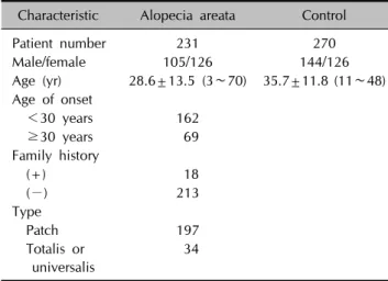
Table 1. Clinical characteristics of study groups Characteristic Alopecia areata Control
Patient number 231 270
Male/female 105/126 144/126
Age (yr) 28.6±13.5 (3∼70) 35.7±11.8 (11∼48) Age of onset
<30 years 162
≥30 years 69
Family history
(+) 18
(−) 213
Type
Patch 197
Totalis or universalis
34
Values are presented as number only or mean±standard deviation (range).
https://doi.org/10.5021/ad.2019.31.4.489
Association between EGF and EGFR Gene Polymorphisms and Susceptibility to Alopecia Areata in the Korean
Population
Yong-Yon Won, Sik Haw1, Joo-Ho Chung2, Bark-Lynn Lew, Woo-Young Sim
Department of Dermatology, College of Medicine, Kyung Hee University, Seoul, 1Department of Dermatology, Inje University College of Medicine, Busan, 2Department of Pharmacology and Kohwang Medical Research Institute, College of Medicine, Kyung Hee University, Seoul, Korea
Dear Editor:
The roles of epidermal growth factor (EGF) and epidermal growth factor receptor (EGFR) in the pathogenesis of alo- pecia areata (AA) are unknown. However, several reports have suggested an association between EGF signaling and AA. In mice, EGF blocked hair follicle induction by down- regulation of signaling pathways such as Wnt, Sonic hedge- hog, and bone morphogenetic protein pathways1. In hu- man hair follicle culture, EGF and EGFR showed a ca- pacity for inhibiting hair shaft elongation and changing the morphology to catagen growth pattern by suppressing mi- totic regulators including RCC2 and Stathmin12,3. A pre- vious study reported that the use of EGFR inhibitors can cause skin inflammation and exacerbation of autoimmune diseases, and that these immune-related effects of EGFR inhibitors are due to their direct effects on the expression of the major histocompatibility complex class I and/or class II molecules4.
The role of single nucleotide polymorphisms (SNPs) of EGF and EGFR on the pathogenesis of AA has not yet been studied; however, our study suggested that EGF and EGFR could be associated with the pathogenesis of AA. The
study aimed to determine whether variations in EGF and EGFR contribute to risk of AA in Korean population. There- fore, we investigated the significance of EGF and EGFR gene polymorphisms in the susceptibility to AA and to un- derstand the pathogenesis of AA.
The study included patients who had AA and healthy con- trol subjects and visited Kyung Hee University Hospital at Gangdong. The controls were recruited after they had been determined to be mentally and physically healthy in a general health check-up program. In this study, 231 pa- tients with AA (105 males and 126 females, average age:
28.6±13.5 years) (Table 1) and 270 healthy controls (144 males and 126 females, average age: 35.7±11.8 years) were included. Informed consent was obtained from each subject, and the study was approved by the Institutional Review Board of Kyung Hee University Hospital at Gang- dong (KHNMC IRB 2008-022). Genomic DNA was iso-
Brief Report
490 Ann Dermatol
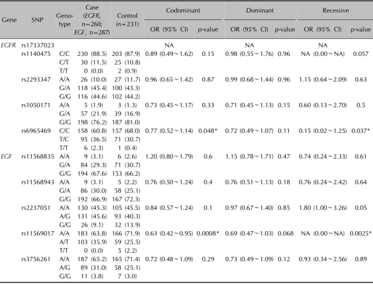
Table 2. Logistic analysis of EGF, EGFR polymorphisms in patients with alopecia areata, and in normal control subjects
Gene SNP Geno-
type
Case (EGFR, n=260;
EGF, n=287)
Control (n=231)
Codominant Dominant Recessive
OR (95% CI) p-value OR (95% CI) p-value OR (95% CI) p-value
EGFR rs17337023 NA NA NA
rs1140475 C/C 230 (88.5) 203 (87.9) 0.89 (0.49∼1.62) 0.15 0.98 (0.55∼1.76) 0.96 NA (0.00∼NA) 0.057 C/T 30 (11.5) 25 (10.8)
T/T 0 (0.0) 2 (0.9)
rs2293347 A/A 26 (10.0) 27 (11.7) 0.96 (0.65∼1.42) 0.87 0.99 (0.68∼1.44) 0.96 1.15 (0.64∼2.09) 0.63 G/A 118 (45.4) 100 (43.3)
G/G 116 (44.6) 102 (44.2)
rs1050171 A/A 5 (1.9) 3 (1.3) 0.73 (0.45∼1.17) 0.33 0.71 (0.45∼1.13) 0.15 0.60 (0.13∼2.70) 0.5 G/A 57 (21.9) 39 (16.9)
G/G 198 (76.2) 187 (81.0)
rs6965469 C/C 158 (60.8) 157 (68.0) 0.77 (0.52∼1.14) 0.048* 0.72 (0.49∼1.07) 0.11 0.15 (0.02∼1.25) 0.037*
T/C 95 (36.5) 71 (30.7) T/T 6 (2.3) 1 (0.4)
EGF rs11568835 A/A 9 (3.1) 6 (2.6) 1.20 (0.80∼1.79) 0.6 1.15 (0.78∼1.71) 0.47 0.74 (0.24∼2.33) 0.61 G/A 84 (29.3) 71 (30.7)
G/G 194 (67.6) 153 (66.2)
rs11568943 A/A 9 (3.1) 5 (2.2) 0.76 (0.50∼1.24) 0.4 0.76 (0.51∼1.13) 0.18 0.76 (0.24∼2.42) 0.64 G/A 86 (30.0) 58 (25.1)
G/G 192 (66.9) 167 (72.3)
rs2237051 A/A 130 (45.3) 105 (45.5) 0.84 (0.57∼1.24) 0.1 0.97 (0.67∼1.40) 0.85 1.80 (1.00∼3.26) 0.05 A/G 131 (45.6) 93 (40.3)
G/G 26 (9.1) 32 (13.9)
rs11569017 A/A 183 (63.8) 166 (71.9) 0.63 (0.42∼0.95) 0.0008* 0.69 (0.47∼1.03) 0.068 NA (0.00∼NA) 0.0025*
A/T 103 (35.9) 59 (25.5) T/T 0 (0.0) 5 (2.2)
rs3756261 A/A 187 (65.2) 165 (71.4) 0.72 (0.48∼1.09) 0.29 0.73 (0.49∼1.09) 0.12 0.93 (0.34∼2.56) 0.89 A/G 89 (31.0) 58 (25.1)
G/G 11 (3.8) 7 (3.0)
Values are presented as number (%) or OR (95% CI). SNP: single nucleotide polymorphism, OR: odds ratio, CI: confidence interval, NA: not applicable. *p<0.05.
lated from peripheral blood using a genomic DNA iso- lation reagent kit (Core BioSystem, Seoul, Korea).
Five SNPs (rs11568835 [promoter], rs11568943 [exon], rs2237051 [exon], rs11569017 [exon], and rs3756261 [promoter]) for EGF and another five SNPs (rs17337023 [exon], rs1140475 [exon], rs2293347 [exon], rs1050171 [exon], and rs6965469 [promoter]) for EGFR with a hetero- zygosity greater than 0.3 among SNPs located in the pro- moter or exon (http://www.ncbi.nlm.nih.gov/SNP) were selected. All 10 selected SNPs were included using Hardy –Weinberg Equilibrium test (HWE, p>0.05). The geno- types were determined by direct sequencing. The samples were sequenced using an ABI Prism 3730XL Analyzer (PE Applied Biosystems, Foster City, CA, USA). Sequence data were analyzed using the SeqManII software (DNASTAR Inc., Madison, WI, USA).
The HWE for the two SNPs was assessed using SNPStats
(http://bioinfo.iconcologia.net/index.php)5. Multiple logis- tic regression models with three alternative models (co- dominant, dominant, and recessive)6 were calculated for the odds ratio, 95% confidence interval, and correspond- ing p-values, with control for gender as a covariable.
SNPStats, HelixTree software (Golden Helix Inc., Bozeman, MT, USA), and SNPAnalyzer (ISTECH Inc., Goyang, Korea) were used.
Baseline characteristics of patients and controls are sum- marized in Table 1. Among five SNPs, one SNP (rs11569017) of EGF showed significant difference between the AA group and control group (Table 2). One SNP (rs6965469) of EGFR showed a significant difference between the AA group and control group (Table 2). There were no sig- nificant differences in expression of any of the SNPs of EGF and EGFR between early-onset AA and late-onset AA (data not shown). Moreover, none of the SNPs of EGF and
Brief Report
Vol. 31, No. 4, 2019 491 EGFR showed significant differences between patients
with and without familial history (data not shown). There was no significant difference associated with any of the SNPs of EGF. On the contrary, one SNP (rs17337023) of EGFR showed significant differences between patchy-type AA and alopecia totalis (AT) (data not shown).
The present study is, to the best of our knowledge, the first to investigate a potential influence of the EGF and EGFR polymorphisms in patients with AA. There are amount of studies to association between EGF and EGFR gene poly- morphism and internal malignancies. However there are only a few studies about the relationship between EGF and EGFR polymorphisms and autoimmune disease. One study from Taiwan reported the association between EGFR and rheumatoid arthritis (RA)7. In this study, 188 patients with RA and 128 controls were enrolled. The study reveal- ed new information on EGFR polymorphisms (rs17337023) with regard to the association between susceptibility to development of RA and polymorphisms. The rs17337023 SNP was also found to be associated with systemic lupus erythematosus (SLE)8, endometriosis, leiomyomas9, and malignant oral keratinocytes10, in previous studies. In our study, the rs17337023 SNP also showed significant differ- ences between patchy-type AA and AT, suggesting that rs17337023 is an important SNP that is extensively in- volved in the phenotype of AA as well as in other auto- immune diseases.
Polymorphism of EGF may cause compensatory expres- sion of EGF. The elevation of EGF inhibits the induction of anagen phase and decreases hair shaft elongation. The polymorphism of EGFR may cause functional deceleration of protein-like EGFR inhibitor and thus, modulate the ex- pression of immune molecules. In this manner, the SNPs of EGF and EGFR polymorphisms may be correlated with the pathogenesis of AA.
To our knowledge, this is the first study to demonstrate that EGF and EGFR polymorphisms are involved in the pathophysiology of AA or AA phenotypes. The genotype frequency of rs11569017 in EGF and rs6965469 in EGFR was significantly increased in patients with AA compared with the corresponding frequencies in healthy controls. In addition, rs17337023 polymorphism of EGFR may con- tribute to the clinical type of AA (patchy type or AT).
Previous studies on RA and SLE have reported no sig- nificant clinical features according to different genotypes7,8. In conclusion, EGF and EGFR polymorphisms may con- tribute to the increased susceptibility to AA, and may be associated with the phenotype of AA in the Korean popu- lation. In particular, the rs17337023 SNP of EGFR is a no- table SNP that is involved in the phenotype of AA as well as in other autoimmune diseases.
ACKNOWLEDGMENT
This work was supported by the National Research Foun- dation of Korea(NRF) grant funded by the Korea govern- ment(MSIT)(No. 2017R1C1B5017740).
CONFLICTS OF INTEREST
The authors have nothing to disclose.
ORCID
Yong-Yon Won, https://orcid.org/0000-0002-5358-4359 Sik Haw, https://orcid.org/0000-0001-6908-5864 Joo-Ho Chung, https://orcid.org/0000-0003-3172-2471 Bark-Lynn Lew, https://orcid.org/0000-0003-4443-4161 Woo-Young Sim, https://orcid.org/0000-0001-5300-6869
REFERENCES
1. Hoffmann R1, Eicheler W, Huth A, Wenzel E, Happle R.
Cytokines and growth factors influence hair growth in vitro.
Possible implications for the pathogenesis and treatment of alopecia areata. Arch Dermatol Res 1996;288:153-156.
2. Richardson GD, Bazzi H, Fantauzzo KA, Waters JM, Crawford H, Hynd P, et al. KGF and EGF signalling block hair follicle induction and promote interfollicular epidermal fate in developing mouse skin. Development 2009;136:2153-2164.
3. Bichsel KJ, Hammiller B, Trempus CS, Li Y, Hansen LA. The epidermal growth factor receptor decreases Stathmin 1 and triggers catagen entry in the mouse. Exp Dermatol 2016;25:
275-281.
4. Pollack BP, Sapkota B, Cartee TV. Epidermal growth factor receptor inhibition augments the expression of MHC class I and II genes. Clin Cancer Res 2011;17:4400-4413.
5. Solé X, Guinó E, Valls J, Iniesta R, Moreno V. SNPStats: a web tool for the analysis of association studies. Bioinformatics 2006;22:1928-1929.
6. Lewis CM. Genetic association studies: design, analysis and interpretation. Brief Bioinform 2002;3:146-153.
7. Lo SF, Wan L, Lin HC, Huang CM, Chen SY, Liu SC, et al.
Association of rheumatoid arthritis risk with EGFR genetic polymorphisms in Taiwan's Han Chinese population. Rheu- matol Int 2012;32:2301-2306.
8. Huang CM, Tsai CH, Chen CL, Chang CP, Lai CC, Tsai FJ.
Epidermal growth factor receptor (EGFR) gene Bsr I poly- morphism is associated with systemic lupus erythematosus.
Lupus 2004;13:773-776.
9. Hsieh YY, Chang CC, Tsai FJ, Lin CC, Tsai CH. T homo- zygote and allele of epidermal growth factor receptor 2073 gene polymorphism are associated with higher susceptibi-
Brief Report
492 Ann Dermatol
Received April 6, 2018, Revised August 4, 2018, Accepted for publication August 29, 2018
Corresponding author: Atousa Hakamifard, Department of Infectious Diseases, School of Medicine, Isfahan University of Medical Sciences, Isfahan 8174675731, Iran. Tel: 98-91-32291573, Fax: 98-31-36604918, E-mail: [email protected]
ORCID: https://orcid.org/0000-0001-9456-2239
This is an Open Access article distributed under the terms of the Creative Commons Attribution Non-Commercial License (http://creativecommons.org/
licenses/by-nc/4.0) which permits unrestricted non-commercial use, distribution, and reproduction in any medium, provided the original work is properly cited.
Copyright © The Korean Dermatological Association and The Korean Society for Investigative Dermatology lity to endometriosis and leiomyomas. Fertil Steril 2005;83:
796-799.
10. Shintani S, Matsuo K, Crohin CC, McBride J, Tsuji T, Donoff
RB, et al. Intragenic mutation analysis of the human epi- dermal growth factor receptor (EGFR) gene in malignant human oral keratinocytes. Cancer Res 1999;59:4142-4147.
https://doi.org/10.5021/ad.2019.31.4.492
Comments on “Cutaneous Abscess as a Complication of Bisphosphonate-Related Osteonecrosis of the Jaw” by Yang et al.
Atousa Hakamifard
Department of Infectious Diseases, School of Medicine, Isfahan University of Medical Sciences, Isfahan, Iran
Dear Editor:
Yang et al.1 recently reported an interesting case entitled,
“Cutaneous Abscess as a Complication of Bisphosphonate- Related Osteonecrosis of the Jaw”.
Bisphosphonate-related osteonecrosis of the jaw (BRONJ) represents a rare complication of bisphosphonate treat- ment. This condition is characterized by necrosis of the maxilla and mandible2. Actinomyces species (spp.) are gram-positive, non-acid fast, filamentous bacteria and most- ly are facultative anaerobe. These species are commensals of the mucosa of mouth, colon and vagina. The key step in pathogenesis of actinomycosis is mucosal disruption by trauma, surgical procedures, or foreign bodies3,4. Actinomyces spp. are considered to be important agents involved in the pathogenesis of BRONJ. One of the clinical manifestations of actinomycosis is oral cervicofacial disease which can present as large abscess with or without mandibular osteo- myelitis, ulcer or mass lesion. The diagnosing of cervicofa- cial actinomycosis is by histological examination and cul- ture of abscess pus or suspected bone specimen, if osteo- myelitis is considered. The microscopic study for visual-
ization of gram positive, non-acid-fast, thin, branching fila- ments are helpful. Culture for isolation may take up to 2 to 4 weeks and this note should be considered. The demon- stration of sulfur granules in pus or pathologic section of surgical specimens is also helpful and diagnostic. The treatment of choice is penicillin with or without surgical therapy especially for abscess drainage and resection of necrotic bone in cases of osteomyelitis and osteonecrosis.
The agents that should be avoided for treatment are metro- nidazole, aminoglycosides, anti-staphylococcal penicillin such as cloxacillin and first generation of cephalosporin5. Hence; the diagnosis of cervicofacial actinomycosis should always be considered in any painless mass at jaw and also in the differential diagnosis of any lesion in neck and head for appropriate management.
Yang et al.1 described bacterial culture of skin tissue re- vealed gram positive cocci (Streptococcus constellatus).
Also they reported that fungal and mycobacterial cultures were both negative, but authors have not mentioned that study of the specimen in this case for detecting actino- myces had performed or not. The diagnosis of actino-