- 29 -
Benign Diseases Widening the Unilateral Ethmoidal Infundibulum: : :Clinical and Radiological Characteristics :
Kyung Chul Lee, M.D., Jeong Woo Lee, M.D., Sang Ho Kim, M.D., Jae Ho Ban, M.D., Kee Hwan Kwon, M.D. and Sung Min Jin, M.D.
ABSTRACT
Background and Objectives:The ethmoidal infundibulum is a cleft-like space situated like a funnel before the maxillary ostium. In this cleft, unilateral inflammations and benign diseases occur sometimes. In this study, we are trying to evaluate benign diseases widening the unilateral ethmoidal infundibulum and their chief complaints, and to identify the radiologic patterns of them in contrast-enhanced computerized tomography (CT) and magnetic resonance imaging (MRI).
Subjects and Methods:Sixty eight cases with unilateral ethmoidal infundibular widening on contrast enhanced CT were evaluated with MRI between Jan, 1995 and Dec, 1999. Fifty- six cases of pathologically proved inflammatory or benign diseases were analyzed prospectively by medical records, CT and MRI. Results:Benign diseases widening the unilateral ethmoidal infundibulum included antrochoanal polyp, maxillary sinus mucocele, prolapsed antral mucosa (a variant of maxillary sinus mucocele), noninvasive fungal sinusitis, nasal polyp with sinusitis and inverted papilloma. They showed soft tissue density in the unilateral maxillary sinus and infundibulum with uncinate process erosion on CT. They were differentiated by the signal intensity on T2 weighted MRI and enhancement patterns on T1 weighted MRI. Conclusion:The preoperative diagnosis of benign diseases widening the unilateral ethmoidal infundibulum depends on endoscopic biopsy generally, but MR imaging may be helpful for differential diagnosis of them.
KEY WORDS:Ethmoidal infundibulum·MRI.
INTRODUCTION
The ethmoidal infundibulum is a 3-dimensional, cleft-like space bounded medially by the uncinate pr- ocess, laterally by the lamina papyracea and poste- riorly by the ethmoidal bulla. If inflammation occurs, it easily extends to the paranasal sinuses through the hiatus semilunaris, the frontal recess, and the maxillary sinus ostium.1) Benign diseases widening the bilateral ethmoidal infundibulum are common in nasal polyp with sinusitis,2) but the study of unilateral benign di- seases is still rare.
In this study, we are trying to evaluate benign dis- eases widening the unilateral ethmoidal infundibulum and their chief complaints, and to identify the radi- ologic patterns of them in contrast-enhanced CT and MRI.
SUBJECTS AND METHODS Subjects
Among patients from January 1995 to December 1999 who visited our department with unilateral nasal symptoms, there were 68 cases where unilateral ethmoi- dal infundibulum widening was detected by contrast- enhanced CT. With the consent of our radiology depar- tment, MRI was carried out to these patients for free.
Excluding 12 cases where no surgery took place, this study was based on 56 cases of pathologically proven inflammatory or benign diseases. Noninvasive fungal sinusitis with calcification that can be identified by CT exactly and malignant diseases already well researched Department of Otolaryngology, Kangbuk Samsung Hospital,
School of Medicine, Sungkyunkwan University, Seoul, Korea Address correspondences and reprint requests to Kyung Chul Lee M.D., Department of Otolaryngology, School of Medicine, Sungkyunkwan University, Kangbuk Samsung Hospital, 108 Pyungdong, Jongrogu, Seoul 110-746, Korea
Tel: 82-2-2001-2268, Fax: 82-2-2001-2273, E-mail: [email protected]
Accepted for publication on July 24, 2001
through MRI have been excluded from this study. The sex distribution was 38 male and 18 female patients with a variable age distribution from 8 to 69 years old (average 44 years old).
Methods
Unilateral ethmoidal infundibular widening that is detected on CT is where the lesion causes erosion of the unilateral uncinate process and the ethmoidal in- fundibular widening is more obvious compared to the contralateral side.
A prospective analysis was carried out on benign diseases widening the unilateral ethmoidal infundi- bulum by medical records, contrast-enhanced CT, T1 weighted MRI, the signal intensity of T2 weighted MRI and the enhancement pattern of Gadolinium (Gd)-enhanced T1 weighted MRI.
RESULTS
Benign diseases widening unilateral ethmoidal infundibulum
Benign diseases widening the unilateral ethmoidal infundibulum include antrochoanal polyp, maxillary sinus mucocele, prolapsed antral mucosa (a variant of maxillary sinus mucocele), noninvasive fungal sinu- sitis, nasal polyp with sinusitis and inverted papilloma (Table 1).
Chief complaints and nasal endoscopic findings
Chief complaints are nasal obstruction followed by rhinorrhea, posterior nasal drip, headache, buccal pain, periorbital pain and hyposmia in order of frequency (Fig. 1).
Nasal endoscopic findings of antrochoanal polyp, prolapsed antral mucosa (variant of maxillary sinus mucocele), noninvasive fungal sinusitis, nasal polyp with sinusitis, and inverted papilloma, were single or multiple polypoid masses protruding into the nasal cavity. However, only in maxillary sinus mucocele, were variable bulgings noticed in the lateral nasal wall of the middle meatus.
Contrast-enhanced CT findings
Benign diseases widening the unilateral ethmoidal infundibulum showed soft tissue density in the unila- teral maxillary sinus and infundibulum with uncinate process erosion on CT. Especially, in maxillary sinus mucocele, maxillary sinus expansion and bony wall destruction was identified.
However, differential diagnosis was difficult only with contrast-enhanced CT.
Table 1. Benign diseases widening the unilateral ethmoidal infundibulum (N=56)
No. of patients (%)
Antrochoanal polyp 10 (18)
Maxillary sinus mucocele 10 (18) Noninvasive fungal sinusitis 10 (18) Nasal polyp with sinusitis 9 (16)
Inverted papilloma 9 (16)
Prolapsed antral mucosa 8 (14)
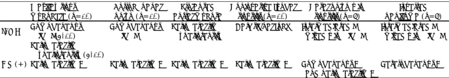
Table 2. Summary of MR findings in benign diseases widening the unilateral ethmoidal infundibulum Maxillay sinus
mucocele (n=10)
Antrochoanal polyp (n=10)
Prolapsed antral mucosa
Noninvasive fungal sinusitis (n=10)
Nasal polyp with sinusitis (n=9)
Inverted papilloma (n=9) T2WI Homogeneous
high S(5/10)
Homogeneous high S
Peripheral rim hyperintensity
Dark signal foci Intermediated S mixed with high S
Intermediated S mixed with high S Peripheral rim
hyperintensity (5/10)
Gd (+) Peripheral rim E Peripheral rim E Peripheral rim E Peripheral rim E Homogeneous E
and peripheral rim E Heterogeneous E T2WI:T2 weighted imaging, Gd (+):Gadolinium enhanced T1WI, S:signal intensity, E:enhancement
Fig. 1. Chief complaints of benign diseases widening the unil- ateral ethmoidal infundibulum.
Nasal obstruction Rhinorrhea Posterior nasal drip Headache Buccal pain Periorbital Pain Hyposmia
MR findings (Table 2)
All benign diseases widening the unilateral ethmoi- dal infundibulum showed low signal intensity and intermediate signal intensity in T1 weighted MRI.
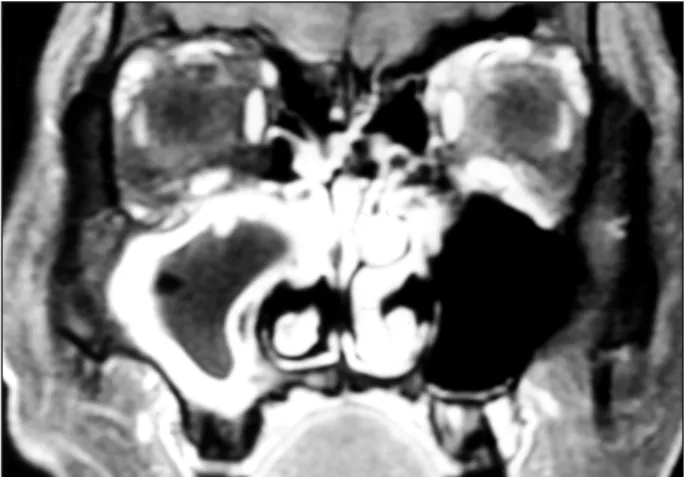
Peripheral rim enhancement was identified in Gd- enhanced T1 weighted MRI in all maxillary sinus mucocele (Fig. 2).
In all cases of antrochoanal polyp, homogeneous high signal intensity was identified on T2 weighted MRI (Fig. 3). Whereas in the case of prolapsed antral mucosa (a variant of maxillary sinus mucocele), peri- pheral rim hyperintensity was accompanied in most cases with central intermediated or high heterogeneous signal intensity (Fig. 4).
Noninvasive fungal sinusitis showed calcified les- ions of dark low signal intensity on T2 weighted MRI
in all cases (Fig. 5).
Nasal polyp with sinusitis and inverted papilloma both showed a mixed pattern of high and interm- ediate signal intensity on T2 weighted MRI in all cases. Also, Gd-enhanced T1 weighted MRI showed homogeneous enhancement with peripheral rim en- hancement of the maxillary sinus in nasal polyps (Fig. 6), whereas inverted papilloma showed hetero- geneous enhancement (Fig. 7).
DISCUSSION
Benign diseases widening the unilateral ethmoidal infundibulum can take place in two cases which are when lesions of the maxillary sinus cause erosion of the uncinate process and expand to the nasal cavity or when the lesion develops close to the ehtmoidal infun- dibulum and causes erosion of the uncinate process
Fig. 2. Maxillary sinus mucocele. Coronal T1 Gd-enhanced ma- gnetic resonance imaging (MRI) shows peripheral rim enha- ncement in the left nasal cavity and maxillary sinus.
Fig. 3. Antrochoanal polyp. Coronal T2 weighted MRI shows homogeneous high signal intensity in the left nasal cavity and maxillary sinus.
Fig. 4. Prolapsed antral mucosa:a variant of maxillary sinus mucocele. Coronal T2 weighted MRI shows peripheral rim hyperintensity in the right maxillary sinus.
Fig. 5. Noninvasive fungal sinusitis. Coronal T2 weighted MRI shows dark signal foci in the right nasal cavity.
and expands to the maxillary sinus. Preoperative dia- gnosis of these diseases with endoscopic biopsy is possible, however, we wish to discover whether or not MRI can be of any help in this matter.
On MRI, maxillary sinus mucocele can reveal varia- ble findings based on the characteristics of the cystic component. Especially, in Gd-enhanced T1 weighted MRI, maxillary sinus mucocele reveals peripheral rim enhancement whereas, malignant diseases reveal ho- mogeneous enhancement. Therefore, MRI can be very beneficial in differentiating the two when they either exist separately or together.3) In this study, peripheral enhancement was reveled on Gd-enhanced T1 weig- hted MRI in all cases.
Erosion of the uncinate process and infundibular wid- ening can be more clearly identified in antrochoanal polyp when protrusion has occurred through the na-
tural ostium rather than the maxillary accessory ostium.
Also, on T2-weighted MRI, cystic components in the maxillary sinus reveal homogeneous high signal in- tensity and the solid components in the nasal cavity reveal a high signal intensity on T2 weighted MRI and peripheral rim enhancement on Gd-enhanced T1 weig- hted MRI.4) In our study, homogeneous high signal intensity was also identified on T2-weighted MRI in all cases as well.
Prolapsed antral mucosa was first introduced in 1985 by Murcia et al., as a disorder revealing similar findings on CT as antrochoanal polyp where the antral mucosa protrudes into the middle meatus.5) This dis- order is a variant of maxillary sinus mucocele that prolapses through the maxillary natural ostium without maxillary sinus expansion or bony erosion generally seen in maxillary sinus mucocele. It reveals peripheral hyperintensity with heterogeneous signal intensity of central intermediated or high signal intensity on T2 weighted MRI.6) Similar findings were mostly identi- fied in our study as well.
75% of all noninvasive fungal sinusitis are diagno- sed with CT, however, the false positive and false negative diagnostic rate are each 12%. Bacterial sinu- sitis with thick bacterial pus or hemorrhage can be misdiagnosed as noninvasive fungal sinusitis on CT.
Therefore, pathologic and bacteriologic diagnosis is necessary. Also, the specificity of T2 weighted MRI is higher than CT due to the low signal intensity revealed by the calcium or ferromagnetic elements in the fungus ball.7) In our study, all lesions revealed a dark and low signal intensity on T2 weighted MRI, making differ- entiation with other diseases a lot more convenient.
While nasal polyp with sinusitis reveal peripheral rim enhancement on Gd-enhanced T1 weighted MRI, inverted papilloma reveals solid heterogenerous enh- ancement on Gd-enhanced T1 weighted MRI.8) In our study, while all nasal polyps revealed homogeneous enhancement with peripheral rim enhancement of the maxillary sinus mucosa on Gd-enhanced T1 weighted MRI, inverted papilloma revealed heterogenerous en- hancement, which made differentiation among the two possible. Also, inflammatory sinus mucosa revealed a high signal intensity on T2 weighted MRI and 95% of all neoplasms, excluding a few minor salivary neopl- asm and neurogenic tumors that revealed high signal intensity, all revealed an intermediate signal intensity.
Fig. 6. Nasal polyp with sinusitis. Coronal T1 Gd-enhanced MRI shows peripheral rim enhancement of the right maxillary sinus.
Nasal polyp shows homogeneous enhancement.
Fig. 7. Inverted papilloma. Coronal T1 Gd-enhanced MRI shows heterogeneous enhancement in the right nasal cavity and maxillary sinus.
Therefore, it can be concluded that T2 weighted MRI is more useful than CT for the differential diagnosis of these disorders.9)
CONCLUSION
Benign diseases widening the unilateral ethmoidal infundibulum include antrochoanal polyp, maxillary sinus mucocele, prolapsed antral mucosa (a variant of maxillary sinus mucocele), noninvasive fungal sinusitis, nasal polyp with sinusitis and inverted papilloma.
The preoperative diagnosis of benign diseases wid- ening the unilateral ethmoidal infundibulum depends on endoscopic biopsy generally, but MR imaging may be helpful for the differential diagnosis of such benign diseases.
REFERENCES
1) Messerklinger W. The ethmoidal infundibulum and its inflammatory illness. Arch Otorhinolaryngol 1979;222(1):11-22.
2) Liang EY, Lam WW, Woo JK, Van Hasselt CA, Metreweli C. Ano- ther CT sign of sinonasal polyposis: truncation of the bony middle turbinate. Eur Radiol 1996;6(4):553-6.
3) Lanzieri CF, Shah M, Krauss D, Lavertu P. Use of gadolinium-en- hanced MR imaging for differentiating mucoceles from neoplasms in the paranasal sinuses. Radiology 1991;178(2):425-8.
4) Youn EK, Chung EC, Lee YU. CT and MR evaluation of choanal polyps. Korean Radiol Soc 1998;39:283-8.
5) Nino-Murcia M, Rao VM, Mikaelian DO, Som P. Acute sinusitis mimicking antrochoanal polyp. Am J Neuroradiol 1985;7:513-6.
6) Lee KC, Lee SC. Prolapsed antral mucosa: variant of Maxillary sinus mucocele. Korean J Otolaryngol 2000;43:620-5.
7) Zinreich SJ, Kennedy DW, Malat J, Curtin HD, Epstein JI, Huff LC, et al. Fungal sinusitis: diagnosis with CT and MR imaging. Radi- ology 1988;169(2):439-44.
8) Yousem DM, Felllows DW, Kennedy DW, Bolger WE, Kashima H, Zinreich SJ. Inverted papilloma: evaluation with MR imaging. Ra- diology 1992;185(2):501-5.
9) Som PM, Shapiro MD, Biller HF, Sasaki C, Lawson W. Sinonasal Tumors and inflammatory tissues: Differentiation with MR imaging.
Radiology 1988;167(3):803-8.