J Rhinol 20(1), 2013
- 54 -
상악동에 발생한 기질화 혈종 3례
울산대학교 의과대학 울산대학교병원 이비인후과학교실
이호민·이정민·김재기·이태훈
Three Cases of Organizing Hematoma in the Maxillary Sinus
Ho Min Lee, MD, Jung Min Lee, MD, Jae Ki Kim, MD and Tae-Hoon Lee, MD
Department of Otolaryngology-Head and Neck Surgery, Ulsan University College of Medicine, Ulsan University Hospital, Ulsan, Korea
ABSTRACT
Organizing hematomas are rare benign tumors and appear as masses that are composed of neovascularization with organizing fibrotic tissue in a hematoma. Although histologically benign, this condition may be clinically progressive. Radiological findings can range from a benign appearance to a more aggressive process, including bony erosion. Organizing hematoma of the maxillary sinus is a rare clinical disease. To our knowledge, only a few cases have previously been published, and most were without bleeding history and disorders. Until now, all of the reports about this lesion have discussed the clinical course, and no previous report has closely considered patho- logic findings and pathogenesis. We report three cases of organizing hematoma of the maxillary sinus presenting with an enlarging maxillary sinus mass.
KEY WORDS : Oraganizing Hematoma · Maxillary Sinus · Pathology.
서 론
기질화 혈종은 여러 가지 원인으로 인해 혈종이 형성된 후 그 안에 혈관이 신생되고 섬유화가 진행되는 기질화 과 정을 거치면서 종괴의 형태로 나타나는 드문 양성 질환이
다.
1) 2)어느 부위에나 생길 수 있지만, 주로 연부조직, 두개
내, 척수내, 근골격계, 부신, 폐, 인두주위공간 및 비부비동 내에서의 발생이 보고되고 있다.
상악동에서 발생한 기질화 혈종은 1996년 처음 보고된 이래 현재까지 보고된 증례가 많지 않다. 그 중 대부분은 임상적인 특징과 경과에 대해서 설명한 것들이 대부분이고 기질화 혈종의 병리학적인 소견과 이를 통한 질병의 발생 과정에 대한 연구보고는 드물다.
기질화 혈종은 악성질환과 감별해야 할 질환으로 환자의 병력이나 증상과 같은 임상적인 단서를 가지고 의심할 수 있지만 조직검사를 통해 확진이 가능하다. 저자들은 상악 동 내에 국한되어있는 기질화 혈종 환자 3례를 경험하였고 각 증례의 임상양상, 내시경 및 영상학적 검사, 그리고 병 리조직학적 소견에 대해 보고하고자 한다.
증 례 1
31세 여자 환자로 2주 전부터 시작된 반복적인 우측 비 출혈, 양측 비폐색감을 주소로 개인 이비인후과 의원을 방문 후 만성 부비동염과 비용종이 의심되어 본원으로 전 원되었다. 외상이나 부비동 수술, 혈관질환 등의 병력은 없었고, 내원시 시행한 비내시경 소견상 우측 중비도를 채우고 있는 적색의 종물이 쉽게 출혈하는 경향을 보였 다. 굴곡형 내시경상 좌측 비강, 비인두, 구인두, 후두의 이상 소견은 보이지 않았다. 부비동 전산화 단층촬영상
논문접수일 : 2012년 6월 13일 / 심사완료일 : 2013년 2월 5일교신저자 : 이태훈, 울산시 동구 전하동 290-3 울산대학교병원 이비인후과
전 화 : (052) 250-7180, · 전 송 : (052) 234-7182 E-mail : [email protected]
www.ksrhino.or.kr
이호민 등 : 상악동 기질화 혈종
/ 55
상악동을 채우고 있는 불균일한 연조직 음영이 비중격까 지 위치하고 있으나 다른 부비동이나 안구 등 주변 조직 으로의 침범은 보이지 않았다(Fig 1). 출혈성 용종 의심 하에 외래에서 조직검사를 시행하였고, 출혈양은 많지 않 아서 쉽게 지혈이 되었다.
조직 검사 결과 기질화된 혈전 및 육아종성 조직이 관 찰되어 기질화 혈종으로 진단하였고 부비동 내시경 수술 을 시행하였다. 출혈은 많지 않았고 술 후 합병증은 없었 고 재발은 없었다. 조직의 육안소견은 표면에 결절이 보이 고 암갈색, 노란색, 붉은색 부위가 혼재된 모양이었다(Fig.
2). 병리조직학적으로 혈종 주위 섬유막이 잘 형성되어있
고, 종괴 내부에는 혈관에서 누출된 많은 적혈구와 섬유세 포들 및 섬유화 과정이 관찰되었다. 팽창된 혈관은 관찰되 지 않았다(Fig. 3).
증 례 2
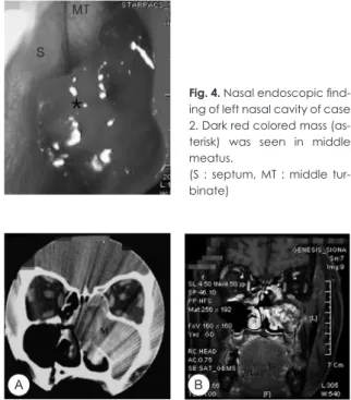
62세 여자 환자로 1년 전부터 시작된 혈성 객담을 주소 로 개인 이비인후과 의원을 방문하여 시행한 부비동 전산 화 단층촬영상 좌측 상악동 악성 종양으로 의심되어 본원 으로 전원되었다. 환자는 좌측 비폐색감과 간헐적 좌측 비 출혈, 후각 저하도 호소하고 있었다. 과거력상 당뇨로 경구 혈당 강하제를 8년 전부터 복용하고 있는 것 외에 혈액응 고장애 등 다른 질병은 없었다. 내원시 시행한 비내시경 소 견에서 좌측 중비도를 채우고 있는 암적색의 쉽게 출혈하 는 종물이 보였다(Fig. 4). 부비동 전산화 단층촬영 및 자기 공명영상촬영에서 상악동을 채우고 있는 불균일한 연조직 음영이 비중격을 우측으로 편위시키고 있었으나 그외 다른 주변 조직으로의 침범은 보이지 않았다(Fig. 5).
외래에서 시행한 조직 검사에서 기질화 혈종으로 진단되 었고 부비동 내시경 수술과 Caldwell-Luc 접근술을 함께
Fig. 1. Preoperative coronal CT of case 1 scan shows a large soft tissue mass filled the right maxillary sinus with associated expansion of the sinus. Note extension of the mass into na- sal cavity, widening of OMU, loss of bony contour of medial wall of the sinus.(M: mass)
Fig. 4. Nasal endoscopic find- ing of left nasal cavity of case 2. Dark red colored mass (as- terisk) was seen in middle meatus.
(S : septum, MT : middle tur- binate)
Fig. 5. A. Preoperative coronal CT of case 2 scan shows a large soft tissue mass filled the left maxillary sinus with associated ex- pansion of the sinus. Note extension of the mass into nasal cav- ity, loss of bony contour of medial wall of the sinus, and the nasal septal deviation to the right (M: mass). B. Preoperative enhanced coronal MRI T1WI of case 2 also shows a well en- hanced soft tissue mass filled the left maxillary sinus with associ- ated expansion of the sinus.
Fig. 2. Gross specimen of organizing hematoma removed from the maxillary sinus of case 1 showing heterogenous color of yel- low, brown and red.
A B
Fig. 3. Microscopic view of organizing hematoma of case 1
showing old hematoma, organizing thrombi including prolifera-
tion of fibroblast and fibrin(F), extravasated red blood cells (ar-
row). The dilated vessel was not found in the hematoma.(He-
matoxyline and Eosin staining, X 100)
56 / J Rhinol 20(1), 2013
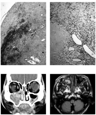
시행하였다. 역시 출혈은 많지 않았고 술 후 일시적인 협부 종창 이외의 합병증 및 재발은 없었다. 조직은 육안적으로 표면에 많은 결절과 함께 암갈색의 잘 부스러지는 종물로 보였고, 병리조직학적으로 적혈구와 섬유소가 혼재된 혈 종, 섬유아세포(fibroblast)와 신생혈관의 증식 등 기질화 가 활발히 진행중인 혈종의 형태를 보였고 팽창된 혈관들 이 관찰되었다(Fig. 6).
증 례 3
72세 남자 환자로 4개월 전부터 시작된 반복적인 우측 비 출혈을 주소로 본원 외래를 방문하였다. 특별한 외상의 병 력이나 부비동 수술, 혈관질환 등의 병력은 없었고, 내원시 시행한 비내시경 소견상 우측 중비도를 채우고 있는 적색 의 쉽게 출혈하는 종물이 보였다.
부비동전산화 단층촬영 및 자기공명 영상촬영에서 상악 동을 채우고 있는 불균일한 연조직 음영이 상악동을 팽창 시키고 비강내로 돌출되어 있었으며, 우측 안와하벽의 부 분적 결손도 관찰되었다(Fig. 7).
부비동 내시경 수술 중 출혈은 많지 않았고 술 후 합병증 및 재발은 없었다. 조직의 육안소견은 표면에 결절이 보였
고 암갈색, 붉은색 부위가 혼재된 불규칙적인 모양이었다.
혈종 내부에 섬유화가 진행되어 유리질화와 다양한 크기의 팽창된 혈관들이 관찰되었다. 그리고 일부에서는 팽창된 혈관의 괴사와 비교적 신선한 형태의 적혈구가 관찰되어 재출혈을 의심케 하였다(Fig. 8).
고 찰
기질화 혈종은 종양이 성장하면서 인접 구조물이나 골조 직 등을 파괴하여 다양한 임상증상을 유발할 수 있다. 주 로 본 증례들처럼 비폐색이나 반복적인 비출혈이 생기지만 상악동 내에 국한되거나 점막하 출혈인 경우에는 비출혈이 없을 수도 있다. 그 외에 후각저하, 두통, 협부 종창 등이 있 을 수 있고 크기가 점점 커지게 되면 안구돌출, 얼굴 감각 저하, 치통, 두통 등의 증상이 동반되기도 한다.
3)진단을 위해 시행하는 비내시경 검사에서는 출혈성향 이 강한 종물로 보여 육안적으로는 화농성 육아종, 혈관 종, 악성 흑색종 및 악성 종양과 감별하기 어렵다. 부비동 전산화 단층촬영상 균일한 양상의 종물이 상악동 벽을 파 괴하거나 안와하벽을 침범할 수 있고, 드물지만 협부조 직을 침범할 수도 있다. 자기공명 영상촬영에서는 종괴
Fig. 6. Microscopic view of organizing hematoma of case 2 showing old hematoma with proliferation of neovascularization. There were red blood cells with fibrin(F) and fibroblast. The dilated vessels were found in the hematoma(arrow).(Hematoxyline and Eosin staining, A : X 40, B : X 100)
Fig. 7. A. Preoperative coronal CT of case 3 scan shows a large soft tissue mass filled the right maxillary sinus with associated ex- pansion of the sinus. Note extension of the mass into right orbital floor(M: mass). B. Preoperative axial T1-weighted brain MRI of case 3 shows a large soft tissue mass filled the right maxillary sinus with associated expansion of the sinus(M: mass).
Fig. 8. Microscopic view of organizing hematoma of case 3
showing old hematoma with hylalinization. The dilated vessels
were found in the hematoma with necrosis(arrow). There were
dilated vessels which were rebleeding with fresh red blood
cells(arrowhead).(Hematoxyline and Eosin staining, A : X 40, B
: X 100)
이호민 등 : 상악동 기질화 혈종
/ 57
가 T1, T2 강조 영상에서 낮은 신호 강도에서부터 높은 신호 강도까지 다양하게 보이고 조영 증강되는 소견을 보 인다. 이처럼 방사선학적 검사는 술 전 검사에서 최종진 단을 내리는 데는 큰 도움이 되지 못하고, 결국 확진은 조 직검사로 가능하게 된다.
4)기질화 혈종은 조직학적으로 섬유막(fibrous capsule)으 로 잘 둘러싸인 혈종 내에 섬유화에 의한 무정형성 섬유질 (amorphous fibrin mass)과 적혈구, 신생혈관 등이 혈종 과 혼재되어 나타나는 소견을 보인다. 부비동 기질화 혈종 은 출혈이 부비동의 환기 및 배액 장애로 인해 혈종으로 된 후, 혈관이 생기고 섬유화가 진행되는 기질화(organiza- tion) 과정을 거쳐서 종물의 형태로 나타나게 된다.
3)어떠 한 기전으로 혈종에 신생혈관이 생기고 자라는 가에 대해 서는 아직 확실하지 않다.
Ozaki 등은 기질화 혈종의 발생 과정을 Negative spiral theory를 통해 주장하였는데 폐쇄된 공간에서 혈관종이나 안면 손상, 염증 등 다양한 출혈 원인에 의해서 생긴 혈괴가 차례로 괴사, 섬유화, 유리질화의 과정을 거치게 되고 이의 치유 과정에서 신생 혈관이 생성된다. 하지만, 신생혈관의 말단에서는 혈액흐름이 느리고 그 결과 혈관이 팽창되고 재출혈이 발생한다는 것이다.
5) 6)본 증례들에서 증례 1의 경우에는 섬유화가 잘 관찰되었으나 팽창된 혈관은 관찰되 지 않았다. 증례 2의 경우에는 팽창된 혈관이 잘 관찰되었 고, 증례 3의 경우에는 팽창된 혈관과 함께 일부 혈관의 괴 사 및 재출혈의 소견을 보였다. 즉, 증례 1의 경우는 재출혈 이 일어난 후 섬유화의 과정을 거치는 단계로, 증례 2의 경 우는 유리질화를 거쳐 신생혈관 생성과 혈관 팽창이 진행 되는 단계로, 그리고 증례 3의 경우는 팽창된 혈관에서 다 시 재출혈이 발생하고 괴사되는 단계로 볼 수 있다.
기질화 혈종은 양성종양이지만 제거하지 않으면 국소팽 창하고 주변 골조직을 파괴할 수 있으므로 수술적 제거가 반드시 필요하다. 수술은 비강 내에 국한된 경우 혈종의 크
기가 작아 대부분 부비동 내시경 수술로 충분히 완전 절제 가 가능하지만, 경우에 따라 정상 점막과 기질화 혈종의 구 분이 불명확할 수 있고, 상악동 내 점막과 박리가 쉽게 되 지 않는 위치에 존재할 수도 있다. 이러한 경우에는 증례 2 에서처럼 부비동 내시경 수술과 함께 Caldwell-Luc 접근 을 함께 시행하는 것이 바람직할 것이다.
7)중심 단어:기질화 혈종·상악동·병리조직학.
저자역할(Author Contributions)
이호민, 이정민, 김재기, 이태훈은 본 연구에서 모든 자료에 접근 할 수 있으며, 자료의 완전성과 자료 분석의 정확성에 책임을 지고 있습니다. 연구 기획 : 이호민, 이정민, 김재기, 이태훈. 자료 해석 및 분석 : 이호민, 이정민, 김재기, 이태훈. 논문 초안 : 이호민. 연 구 총괄 : 이태훈
REFERENCES
1) Ito M, Tajima A, Sato K, Ishii S. Calcified cerebellopotine angle hematoma mimicking recurrent acoustic neuroma. ClinNeurol- Neurosurg 1988;90:65-70.
2) Lee YY, Moser R, Bruner JM, Tassel P. Organized intracerebral hematoma with acute hemorrhage: CT patterns and pathologic cor- relations. AJR Am J Roentagenol 1986;147:111-8.
3) Tabaee A, Kacker A. Hematoma of the maxillary sinus presenting as a mass –a case report and review of literature. Int J PediatrOto- rhinolaryngol 2002;65:153-7.
4) Nishiguchi T, Nakamura A, Mochizuki K, Tokuhara Y, Yamane H, et al. Expansile organized maxillary sinus hematoma: MR and CT findings and review of literature. AJNR Am J Neuroradiol 2007;28:1375-7.
5) Ozaki M, Sakai M, Ikeda H. Hemagioma of the nasal cavity and sinuses-a report of twenty cases. Otolaryngol Head Neck Surg 1977;49:53-8.
6) Omura G, Watanabe K, Fujishiro Y, Ebihara Y, Nakao K, et al.
Organized hematoma in the paranasal sinus and nasal cavity- Imaging diagnosis and pathologic findings. AurisNasus Larynx 2010;37:173-7.
7) Ha MS, Song YJ, Han KY, Yeo NK. A case of organizing hema- toma of the nasal septum. Korean J Otolaryngol 2010;53:324-6.