191 Left atrial (LA) thrombus is known as a common complica-
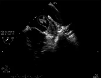
tion of rheumatic mitral stenosis. It is more common in the presence of atrial fibrillation (AF). Incidence of LA clot forma- tion in sinus rhythm is 2.4–13.5%1)2) and the incidence is as high as 33% in patients with mitral stenosis in AF. Because transthoracic echocardiography may not detect the left atrial appendage (LAA) thrombus, transesophageal echocardiogra- phy (TEE) remains the gold standard for identifying the LAA thrombus. We report a case of LAA thrombus mimicking like a hydatid cyst. A 38 years old male with severe mitral stenosis due to rheumatic heart disease presented with progressive wors- ening of dyspnea (New York Heart Association class II–III) over a period of two years. The electrocardiogram revealed sinus rhythm with evidence of LA enlargement. There was no past history of AF and he was not on any anticoagulants. The transthoracic echocardiogram revealed thickened mitral leaf- lets with mitral orifice area of 1.0 cm2 and mean mitral valve gradient of 18 mm Hg. The LA was enlarged and there was grade IV spontaneous echo contrast in LA. On TEE, there was large cystic mobile formation noted in the LA, originating from LAA and protruding into LA body. Nodular structures were visible within the cystic formation (Fig. 1, Supplementa- ry movie 1). In four-chamber view at 0°, it was appearing like a blooming flower (Fig. 2, Supplementary movie 2). Throm- bus is identified by the homogenous echo texture and visual- ised in more than one plane. It can appear in various forms and shapes depending on the organisation of clot.
TEE is a useful tool to detect LA thrombus in patients with mitral stenosis. The clot in LA can occur in various forms from cystic lesions to mass like appearences. One should look for LAA clot in cases of rheumatic mitral stenosis before subject- ing for balloon mitral valvotomy whether patients are in AF or sinus rhythm. In the presence of high grade of spontanous echo contrast, LA clot should be always suspected.3)
pISSN 1975-4612/ eISSN 2005-9655 Copyright © 2015 Korean Society of Echocardiography www.kse-jcu.org http://dx.doi.org/10.4250/jcu.2015.23.3.191
IMAGES IN CARDIOVASCULAR ULTRASOUND J Cardiovasc Ultrasound 2015;23(3):191-192
• Received: March 1, 2015 • Revised: April 1, 2015 • Accepted: July 22, 2015
• Address for Correspondence: Shivashankara Tarikere Hemaraju, Department of Cardiology, Sri Jayadeva Institute of Cardiovascular Research and Sciences, Jaya Nagar 9th Block, BG Road, Bangalore 560069, Karnataka, India Tel: +91-9241675034, Fax: +91-8026534477, E-mail: [email protected]
• This is an Open Access article distributed under the terms of the Creative Commons Attribution Non-Commercial License (http://creativecommons.org/licenses/by-nc/3.0) which permits unrestricted non-commercial use, distribution, and reproduction in any medium, provided the original work is properly cited.
Blooming Flower in Misty Left Atrium
Shivashankara Tarikere Hemaraju, MD, Amjad Ali, MD, and Manjunath Cholenahally Nanjappa, MD
Department of Cardiology, Sri Jayadeva Institute of Cardiovascular Research and Sciences, Bangalore, India
KEY WORDS: Left atrial thrombus · Mitral stenosis · Transesophageal echocardiography.
Fig. 1. Cystic mass arising from left atrial appendage with nodular structures inside it.
Fig. 2. Arrow showing nodular structures within cystic formation mimicking hydatid cyst in left atrium.
Journal of Cardiovascular Ultrasound 23 | September 2015
192
Supplementary movie legends
Movie 1. Cystic mass arising from left atrial appendage with nodular structures inside it.
Movie 2. Nodular structures within cystic formation mim- icking hydatid cyst in left atrium.
References
1. Conradie C, Schall R, Marx JD. Echocardiographic study of left atrial thrombi in mitral stenosis. Clin Cardiol 1993;16:729-31.
2. Manjunath CN, Srinivasa KH, Panneerselvam A, Prabhavathi B, Ravindranath KS, Rangan K, Dhanalakshmi C. Incidence and predictors of left atrial thrombus in patients with rheumatic mitral stenosis and sinus rhythm: a transesophageal echocardiographic study. Echocardiography 2011;28:457-60.
3. Daniel WG, Nellessen U, Schröder E, Nonnast-Daniel B, Bednar- ski P, Nikutta P, Lichtlen PR. Left atrial spontaneous echo contrast in mitral valve disease: an indicator for an increased thromboembolic risk. J Am Coll Cardiol 1988;11:1204-11.