- 85 -
Mast Cells and Allergic Rhinitis
Joong Saeng Cho, M.D.
ABSTRACT
That mast cells play a role in acute allergic inflammation by releasing various inflammatory mediators, including histamine, leukotrienes (LT), such as LTC4 and LTD4, and prostaglandins (PG), such as PGD2, is well known. Additionally, mast cells contribute to the development of allergic inflammation also through the release of multifunctional cytokines. The incidence of intraepithelial mast cells (IEMC) is found to be greater in nasal mucosa exposed to an allergen, and the cells are thought to play an important role in producing the immediate allergic reaction. Lamina propira mast cells (LPMC) are known to be the dominant source of TH2 cytokine and are responsible for development of the late phases of an allergic reaction They may upregulate the expression of adhesion molecules on the endothelial cells and induce basophil and eosinophil recruitment. Based on these con- sideration it can be proposed that mast cell is a initiating cell of allergic reaction in target organ and IEMC and LPMC have capacity to make major contribution to both immediate or late phase reaction of allergic rhinitis.
KEY WORDS:Allergic rhinitis·Mast cells·Chemical mediators·Cytokines·Adhesion molecules.
INTRODUCTION
In an atopic individual, specific IgE is bound as Fc fragm- ents to Fc receptors on the surface of mast cells. The binding of antigen to the cell surface-bound IgE initiates the process of mast cell activation and the secretion of chemical mediators.
These mediators are responsible for most of the early events that characterize allergic reactions in the various organs. It has recently been established that mast cells contribute to the chronic inflammatory events of allergic disease by secreting cytokines.
The nose is a potential site for allergic inflammation. Alle- rgic rhinitis (AR) is an IgE-mediated atopic disease charact- erized by symptoms of sneezing, rhinorrhea and nasal obstruction, an increased level of serum specific IgE and nasal eosinophilia.
Previous studies have shown that there is an increase in the number of mast cells in the epithelial compartment of the nasal mucosa of patients with allergic rhinitis and that the increase correlates with the severity of the disease. However, the resp- onsibility for the initiation and perpetuation of allergic infla- mmation does not lie with any single cellular element and occurs as result of a cascade of events involving a variety of cells.
This article discusses the characteristics of nasal mast cells and their role in allergic rhinitis. The recent literature on mast cells is reviewed as well.
Biology of mast cells and related cell types
Mast cells and basophils are effector cells in allergic diseases but are also involved in a variety of IgE-independent biologic responses. It is now thought that basophils are terminally di- fferentiated leukocytes that have matured in the bone marrow from precursors closely related to those of eosinophils, and that they are released into the blood stream as fully matured cells. In contrast, mast cells leave the bone marrow and circ- ulate in the blood only as progenitors, and it is not until they enter the tissue that they undergo their terminal differentiation into mature mast cells. Basophils do not normally reside in the tissue but are usually found in the circulation. Even in a nor- mal physiologic state, mast cells can be detected in various ti- ssues, can increase or decrease with various biologic responses, and may vary in their phenotype characteristics according to the anatomic site.1)
A recent advance in the understanding of mast cell biology is the finding that, in rodents, mature mast cells assume one of two phenotypes. Mast cells found in the mucosa of the ga- strointestinal tract contain chondroitin sulfate as their major granule proteoglycan. Such“mucosal”mast cells contain li- ttle histamine. The second phenotype has been found in the lung and in the serosa of body cavities. These“connective ti- ssue”mast cells contain heparin as their major granule prot- eoglycan and produce large quantities of histamine. Mast cells Department of Otolaryngology, College of Medicine, Kyung
Hee University, Seoul, Korea
Address correspondence and reprint requests to Joong Saeng Cho, M.D., Department of Otorhinolaryngology, College of Me- dicine, Kyung Hee University, Hoeki-dong #1, Dongdaemoon- ku, Seoul 130-702, Korea
Tel:82-8-958-8482, Fax:82-8-958-8470 Accepted for publication on October 15, 1998
may also be cultured from rodent bone marrow in the presence of IL-3 and IL-4. Such cultured mast cells resemble mucosal mast cells based on granule content of chondroitin sulfate and low levels of histamine. Moreover, the presence of mucosal mast cells in vivo appears to depend on T cells, the presumed source of IL-3 and IL-4 since they are absent in athymic mi- ce.2) Bone marrow-derived mucosal mast cells can be changed to a connective tissue mast cell phenotype by co-culture with fibroblasts.2) Recent repopulation experiments2)3) in mast cell- deficient mice further suggest that the mucosal and connective tissue phenotypes are not fixed and that bi-directional changes may be possible in suitable microenvironments. However, it is likely that in normal development there is a maturational sequence from bone marrow precursor to mucosal type mast cell to connective tissue mast cell. The key point is that the pr- ecise nature of the mast cell and the mediators it can produce vary with its anatomic location.
In humans, the factors that regulate mast cell growth and de- velopment are less well defined. Human T cell-independent connective tissue mast cells and T cell-dependent mucosal mast cells appear to share similar patterns.3) In addition, mast cells phenotypes are not as clearly differentiated in humans as they are in mice. The major differences between the types of human mast cells are in the composition of the serine protea- ses found in the granules (trypsin-like or chymotrypsin-like in substrate specificity) and in the ultrastructural morphology of the granules. Nevertheless, it does appear that in humans as well as in mice the pattern of mediators produced by mast cells varies with anatomic location.
The event that initiates immediate hypersensitivity is the binding of antigen to IgE on the mast cell or basophil surface.
Mast cells are activated by the cross-linking of FcεRI mole- cules, which is thought to occur by the binding of multivalent antigens to the attached IgE molecules. Experimen-tally, ant- igen binding can be mimicked by polyvalent anti-IgE or by anti-FcεRI antibodies. The activation of mast cells results in three distinct biologic responses:4) 1) the mast cells undergo regulated secretion in which the preformed contents of their granules are released by exocytosis;2) the mast cells enzy- matically synthesize lipid mediators derived from precursors stored in the cell membranes;and 3) the mast cells initiate transcription, translation, and secretion of cytokines. Basophils also undergo degranulation and synthesize lipid mediators;it is not yet known whether basophils synthesize cytokines.
The mechanisms of granule exocytosis are partly understood, largely from studies3)5) of rat mast cell and basophil leukemia cell lines. The cross-linking of FcεRI results in the activation of a G protein that, in turn, activates membrane-bound phos- pholipase C to catalyze the breakdown of phosphatidyl inositol bisphosphate into inositol triphosphate (IP3) and diacylglyc- erol (DAG). IP3 causes elevation of cytoplasmic calcium, and
DAG activates protein kinase C. The effect of the elevated calcium is less clear, but the binding of calcium to calmodulin leads to the activation of myosin light chain kinase, which ph- osphorylates myosin light chain at distinct amino acid residues from protein kinase C.
Mast cells may be activated by mechanisms other than the cross-linking of FcεRI.6) For example, mast cells or baso- phils respond to interleukin-8 (IL-8) and other mononuclear phagocyte-derived cytokines produced as part of natural im- munity, to as yet undefined T cell-derived cytokines produced as part of cell-mediated immunity, and to complement-derived anaphylatoxins, such as C5a, produced during humoral immune responses. Mast cells may also be recruited into inflammatory reactions and activated by neutrophil granule contents or by neurotransmitters such as norepinephrine and substance P.
These latter agents are potentially important as links between the nervous system and the immune system.
Preformed mediators from mast cells
The mediator most readily associated with the mast cell is the simple dopamine, histamine. It is synthesized in the mast cell granule by the decarboxylation of histidine, mainly by histidine decarboxylase, dopa decarboxylase. Histamine is st- ored in the acid proteases. Once in the extracellular environ- ment, histamine exerts potent effects, which include contraction of the bronchial smooth muscle, increased mucus production, vasodilatation and contraction of post-capillary venular end- othelial cells, which increases vasopermeability.
The backbone of the crystalline mast cell granule is prote- oglycan which, in human mast cells is mainly heparin, which constitutes some 75% of the granule proteoglycan, with a mi- xture of chondroitin sulphates. The mast cell protease, present in all mast cells regardless of sub-type (Table 1), is tryptase.
Tryptase is a tetrameric serine protease of around 130 kDa which is stored in a fully active form in the granule.7) It is a mitogen for fibroblasts8) and in a human epithelial cell line may induce its proliferation, stimulate it to release the granu- locyte chemoattractant IL-8 and up-regulate its expression of ICAM-1.
Chymase, which is present only in the MCTC subset, is a monomeric protease of 30 kDa which is stored in the same secretory granules as tryptase.9) Like tryptase, chymase is st- ored within the granule in its fully active form, so that it needs
Table 1. Distribution of neutral proteases in mast cells sub-types
MCT MCTC
Tryptase Tryptase
Chymase
Carboxypeptidase
Gathepsin G
no further processing before release. The enzymatic activities of chymase (Table 2) include the degradation of neurotensin,10) substance P or VIP11) and the cleavage of angiotensin I to an- giotensin II. Chymase may also contribute to the purported role of mast cells in tissue remodelling by cleaving type IV coll- agen. Actions pertinent to mucosal inflammation include the activation of interleukin-1β (IL-1β) to IL-1, the degradation of IL-4 and the stimulation of secretion from cultured subm- ucosal gland cells.
Newly generated mediators from mast cells
The immunological activation of mast cells induces the li- beration of arachidonic acid within the membrane. This pho- spholipid is then rapidly oxidized down one of two pathways:
the cyclo-oxygenase pathway, to form PGD2 or the lipoxygenase pathway, to form LTC4. These are the only two eicosanoids made by the human mast cell. PGD2 is a potent bronchocons- trictor agent which is rapidly degraded to another bronchoco- nstrictor agent, 9α, 11β-PGF2. In addition to its bronchoc- onstrictor effect, PGD2 is chemokinetic for human neutrophils, augments LTB4-induced neutrophilia in the epithelium and is a powerful inhibitor of platelet aggregation.
Leukotriene C4 is produced by a variety of inflammatory cells including mast cells and eosinophils. In the extracellular environment, glutamine is removed from the glutathione res- idue of LTC4 by γ-glutamine transpeptidase to yield LTD4, from which glycine is then removed by LTD dipeptidase to yield LTE4. The physiological effects of the leukotrienes inc- lude potent contraction of smooth muscles, contraction of ar- terial smooth muscles, enhanced permeability of post-capillary venules and enhanced mucus secretion, most of which, in the airways, are mediated by LTD4.12)
Biological properties of human mast cell cytokines
It is now established that mast cells are a source of several multifunctional cytokines. Each of the cytokines so far ident- ified as human mast cell products are likely to be involved in
the pathogenesis of allergic mucosal inflammation. IL-4 act- ivates B cells for Ig secretion through up-regulation of cell- surface MHC class II antigen,13) CD23 and CD4014) and plays a pivotal role in the isotype switching of B cells to IgE syn- thesis. IL-4 specifically increases expression of vascular cell adhesion molecule-1 (VCAM-1) involved in the very late antigen (VLA)-4-dependent recruitment of T cells15) and eo- sinophils,16) and induces the expression of low-affinity IgE receptors (FCRII, CD23) on monocytes. In addition, IL-4 induces fibroblast chemotaxis and collagen secretion.17) Pos- sibly the most important effect of IL-4 comes from its ability to induce the development of the TH2 phenotype of T cells, an action that produces IL-4, IL-5 and IL-6, as reviewed by Romagnani.18) The presence of IL-4 at the onset of an immu- nological response may therefore dictate whether a cell-med- iated or humoral response develops.
The effects of IL-5 in humans are almost exclusively limited to eosinophils. IL-5 is a growth and differentiation factor19) and activator for eosinophils,20) and in addition prevents their programmed cell death to prolong survival.21) IL-5 promotes eosinophil adhesion to vascular endothelium through a CD11/
CD18-dependent mechanism,22) and primes eosinophils for chemotaxis in response to other mediators as well as being directly chemotactic itself.23) As a consequence, IL-5 is cons- idered to be a pivotal cytokine in allergen- and parasite-med- iated eosinophilic responses.
IL-6 activates a wide range of cellular processes, including those of T-cells, and the stimulation of Ig production in B cells, and thus it enhances IL-4-dependent IgE synthesis. IL-6 is also the most important cytokine responsible for the prod- uction of acute-phase proteins by hepatocytes during inflam- matory responses. IL-8 belongs to the intercrine family of cytokines and is secreted by a wide variety of cells, including T cells, macrophage/monocytes, endothelium and epithelial cells.24) It is a potent chemoattractant for neutrophils and is also chemotactic for eosinophils after priming with IL-3, IL-5 or GM-CSF.25)
TNF-α (cachectin) is another cytokine implicated in the pathogenesis of asthma. When administered by inhalation or intravenously in animals, TNF-α increases airway responsi- veness.26) It is a chemoattractant for neutrophils and monocytes, increases microvascular permeability, and enhances both the release of mast cell mediators27) and the cytotoxicity eosinop- hils.28) In addition, it has the capacity to up-regulate the leuk- ocyte endothelial cell adhesion molecules (CAM) E-selectin, VCAM-1 and ICAM-1 involved in the recruitment of neutr- ophils, eosinophils, monocytes and T cells into inflammatory zones. TNF-α also stimulates fibroblast proliferation and the secretion of matrix proteins, collagenase and cytokines, incl- uding IL-6.29)
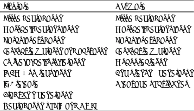
Table 2. Biological properties of mast cell protease
Tryptase Chymase Tissue degradation Tissue degradation
Mast cell degranulation Mast cell degranulation Vascular exudation Vascular exudation Leukocyte migration and activation Leukocyte migration Epithelial cell proliferation Mucus secretion ICAM-1 up-regulation Angiotensin ii generation IL-8 release Cleavage of cytokines Bradykinin generation
Degradation of VIP and CGRP
Heterogeneity of mast cells in allergic rhinitis
Since Enerback’s reports30) on the heterogeneity of rat in- testinal and peritoneal mast cells, which classify them into mucosal mast cells and connective tissue mast cells, further studies have showed the heterogeneity of the cells. Human mast cells are classified into at least two phenotypically dist- inct subpopulations based on the basics of the type of neutral protease they contain:MCT (those mast cells that contain only tryptase) and MCTC (those mast cells that contain both tryp- tase and chymase). MCT are found predominantly located at mucosal surfaces, increase in number in allergic disease and are reduced in number in acquired and chronic immunodefi- ciency syndromes. In contrast, MCTC are found predominantly in submucosal and connective tissues, and are not increased in number in areas of heavy lymphatic infiltration. Furthermore, the tissue-specific heterogeneity of mast cells, their heteroge- neity in response to various antagonists and their heterogeneity in cytokine expression are well documented.31)
Although Kitamura32) has demonstrated tissue-specific he- terogeneity in mice and switching from one subtype to another, there is no such clear evidence in human beings. Recently Ba- rdding et al.,33) reported heterogeneity in mast cells based on cytokine expression. Pawankar34) compared the expression of cytokines in nasal epithelial (IEMC) and lamina propria mast cells (LPMC), and also found heterogeneity in the extent of cytokine expression between IEMC and LPMC. She also ob- served that the expression of FCRI was differentiated in that the nasal mast cells of AR patients expressed increased levels of FCRI as compared to the nasal mast cells of chronic rhi- nitis (CR) patients and that the expression of FCRI in mast cells was upregulated by IL-4. These findings suggest that, even within the same tissue, local microenvironmental factors play a crucial role in modulating the immunophenotypic characte- ristics of mast cells.
Chemical mediators in allergic rhinitis
The reagent on an anaphylatic mechanism refers to the events that follow the combination of antigen with IgE mole- cules specific for it on the surface of mast cells. This involves the release of various mediators, like histamine, leukotriene, chemotactic factors and platelet activating factors. In atopic patients, nasal insuffulation with an allergen to which they are sensitive induces immediate itching, sneezing, and rhinorrhea, with nasal blockage developing over 30 to 40 minutes after the challenge and persisting for up to two hours depending on the strength of the immunological challenge. During this immediate nasal response, the lavage of the nasal cavity demonstrated increased local concentration of histamine, kinins, prostagla- ndins D2, LTs (C4, D4 and E4) and tryptase.35)36) The findings
of increased levels of both PGD2 and tryptase in the lavage suggests a mast cell origin. And Gomez et al.,37) has observed these results also by nasal biopsy, after inducing mast cell gr- anulation in the nose by antigen challenge and forcing the el- evation of the mediator after the challenge in the nasal tissue.
Allergen nasal challenge in patients, especially pediatric pa- tients, is also reported to be associated with late nasal reaction.
There is a less constant finding in the nose than in the lower airways after an allergen challenge. The level of histamine, Ki- nins, and LTC4 has been found to recover and elevate during this late response. But there is no evidence of elevation of PGD2.38) Leukotrienes are derived from the metabolic oxida- tion of arachidonic acid by the lipogenase pathway in which the unstable 5-hydroperoxyeicosatetraenoic acid (HPETE) is converted initially to LTA4, which depending on the cell con- cerned, either metabolites to LTC4, LTD4 or LTE4. The latter process occurs in various granulocytes and it is possible that at least two cell types are required for the full expression of leukotriene synthesis. Histamine, and these peptide leukotrienes, which enhance vascular permeability, lead to fluid extravasa- tion and tissue edema.
Cytokines from mast cells in the nasal mucosa of allergic rhinitis
In both AR and CR patients, cells expressing IL-1, IL-2, IL- 3, IL-4, IL-5, IL-6, IL-7, IL-8, IL-10, IL-13, GM-CSF, IFN-γ and TNF-α can be detected in the nasal mucosa. When the expressions of the cytokines are compared in the nasal mucosa between AR and CR, up-regulation of the expression of IL-4 gene clusters is found in AR patient.34) These include IL-4, IL-5, IL-6, IL-13 and GM-CSF. By contrast, an increased nu- mber of cells expressing IL-8, IFN-γ and TNF-α are noted in the nasal mucosa of CR patients.
A variety of inflammatory cells such as mast cells, eosino- phils, T lymphocytes and macrophages as well as resident ep- ithelial cells contribute as sources of these cytokines (Table 3). However, mast cells and T lymphocytes constitute the pr- edominant source of TH2-type cytokines, excluding IL-5, which is expressed by lymphocytes, mast cells and eosinophils. Brad- ding et al.,33) report that in their study mast cells accounted for
>90% of IL-4- and IL-6-expressing cells and 50% of IL-5- expressing cells in In contrast, Ying et al.,39) report that CD4+
T lymphocytes were the predominant source of IL-4 in AR, accounting for >70% of IL-4-expressing cells. Altho-ugh it is well agreed that mast cells are an important source of IL-4, IL- 5 and IL-6, this discrepancy in the relative contribution of mast cells as cytokine-expressing cells may be the consequence of the different experimental techniques used. Whatever the case, the numerical predominance of cytokine-expressing cells can- not be equated to the amount of cytokine produced, and further
studies are necessary to define the precise amounts of cytokines secreted by each cell type.
Expression of very late antigens in nasal mast cells
Several types of cell surface molecules are involved in the recruitment of inflammatory cells into the specific sites of inflammation;40) lymphocytes and mast cells in the tissues are surrounded by other cells like fibroblasts and mucosal cells as well as by extracellular matrix protein (e.g. collagen, fib- ronectin and laminin). Therefore, it may be thought that the interaction of these inflammatory cells with fibroblasts and extracellular matrix is important in the perpetuation of chronic inflammation. It has been demonstrated that β1 integrins, such as very late antigen-4 (VLA-4) and very late antigen-5 (VLA-5), and the vitronectin receptor integrins of cytokine
expression in mast cells play a role in cell survival. Pawankar34) has detected the expression of VLA-4 and VLA-5 in the nasal mast cells of AR patients. Furthermore, they also found up- regulation of the expression of VLA-4 and VLA-5 in nasal mast cells of these patients.
VCAM-1 and intercellular adhesion molecule-1 are count- erreceptors of ligands for β1 and β2 integrins, respectively.
Studies have shown increased expressions of ICAM-1 and VCAM-1 in the nasal mucosa of AR. A striking feature of the inflammation is the selective accumulation of activated eosi- nophils and basophils, without increased numbers of neutro- phils. Lee et al.,41) have demonstrated that ICAM-1 expresses in the nasal mucosa of AR and that the expression was not up- regulated after stimulation with antigen;in contrast VCAM-1 in the nasal mucosa may result in the accumulation of activated eosinophils and basophils without increasing the number of neutrophils. It is known that IL-4 and IL-13 can up-regulate VCAM-1 expression in endothelial cells;thus mast cells and lymphocytes may indirectly contribute to the recruitment of eosinophils and basophils through the IL-4- and IL-13-induced up-regulation of VCAM-1 expression in the nasal mucosa.
FcεRI-expressing cells in the nasal mucosa of al- lergic rhinitis
The cross-linking of allergen-specific Ig E bound to the high -affinity Ig E receptors (FcεRI) on the surface of mast cells with multivalent allergens results in the release of inflamma- tory mediators, which produces clinical symptoms. The expr- ession of FcεRI in mast cells is therefore important for the development of allergic diseases. However, the precise relation
Table 3. Cytokine profle in the nasal mucosa
Type of cell Allergic rhinitis Chronic rhinitis Epitherial cells IL-, IL-1, IL-6, IL-8,
GM-CSF, TNF-α
IL-α, IL-1, IL-8 TNF-α Lymphocytes IL-, IL-1, IL-3, IL-4, IL-5
IL-6, IL-7, IL-8, IL-10 IL-13, GM-CSF, TNF-α
IL-α, IL-β, IL-2, IL-3, IL-5, IL-7, IL-8, IL-10, GM-CSF, IFN-γ, TNF-α
Mast cells IL-3, IL-4, IL-5, IL-6, IL-7, IL-8, IL-10, IL-13, TNF-α
IL-8, TNF-α
Eosinophills IL-5, IL-6, GM-CSF
Macrophages IL-α, IL-1β IL-α, IL-1β
Fig. 1. Possible roles of nasal mast cells in early-phase and late-ph- ase allergic reactions.
between the FcεRI and atopy is unclear. It has been reported that eosinophils and monocytes also express FcεRI.42)43) Mo- rphologically, FcεRI+ cells in the nasal mucosa of AR pa- tients are either mast cells or basophils. However, basophils co- nstitute <6% of the total number of FcεRI+ cells. Occasi- onally a few FcεRI+ cells with monocyte-like morphologic features are detected. Neither eosinophils nor macrophages de- monstrate in the nasal mucosa of AR patients. In a comparison of the number of FcεRI+ cells in the nasal mucosa of AR and CR patients, an increased number of FcεRI+ cells are demonstrated in the nasal mucosa of AR patients.34)
Role of nasal mast cells in allergic rhinitis
After the cross-linking of allergen-specific IgE on the sur- face of mast cells with multivalent antigen, a series of the re- actions occurs (Fig. 1). First, several morphologic changes, including swelling of the cytoplasmic granules, widening of the fenestra on the cell surface, and subsequent solublization of its granule contents, occur in the mast cells. Histamine, tr- yptase, PGD2, and LTC4 are among the mast-cell products that can be elaborated from the cells immediately after exposure to allergens.44) Histamine induces vasodilatation, increased va- scular permeability, and increased glandular secretion in the ipsilateral and contralateral sides through neural reflexes. Sn- eezing results from the action of histamine on H1 receptors present on the sensory nerve endings of the trigeminal nerve.
Of particular importance is the action of histamine on the su- bepithelial blood vessels, which causes vasodilatation, hype- remia, and mucosal edema. Prostaglandins such as PGD2 also cause edema by vasodilatation and increased vascular perme- ability. The precise role of tryptase is not yet known. The ph- ysiologic effects of newly synthesized leukotrienes including LTC4, LTD4, and LTE4 are mediated by increased vascular pe- rmeability, vasodilatation, and induction of mucous secretion.
It is well known that the number of IEMC (mucosal mast cells) increases in the nasal mucosa on exposure to allergens and that these cells play a crucial role in allergic inflammation through the allergen- and IgE-dependent release of these in- flammatory mediators. And LPMC are known to be the pred- ominant source of TH2-type cytokines in AR. It is therefore proposed that both IEMC and LPMC play important but div- erse roles in perpetuating allergic inflammation.
In short, it can be thought that IEMC contribute to the development of the immediate or acute phases of allergic re- action through the release of inflammatory mediators inclu- ding tryptase, histamine, PGD2, and LTs, whereas LPMC are rsponsible for the development of the late phase of allergic reaction by up-regulating the expression of adhesion molecules including VCAM-1 and ICAM-1 (IL-4, IL-13, and TNF-α) on endothelial cells and by contributing to basophil and eosinophil
recruitment (IL-4, IL-13, and TNF-α). LPMC may also be important in promoting chronic allergic inflammation by reg- ulating the growth, differentiation, and IgE isotype switching of B cells (IL-4, IL-6, and IL-13);by maintaining the levels of VCAM-1 and ICAM-1 expressions on endothelial cells (IL- 4, IL-13, and TNF-α);and by promoting eosinophil devel- opment and survival (IL-3 and IL-5). In view of the substan- tial number of B cells present in the nasal mucosa and based on the evidence that mast cells in AR patients induce IgE synthesis in B cells, it is speculated that mast cells promote chronic all- ergic inflammation by amplifying and maintaining IgE synt- hesis locally in the target organ.
REFERENCES
1) Galli SJ. New insights into ‘the riddle of mast cells’ : Microenvir- onmental regulation of mast cell development and phenotypic he- terogeneity. Lab Invest 1990;62:5-33.
2) Ishizaka KT, Ishizaka MM. Physicochemical properties of human reaginic antibody. IV: Presence of a unique immunoglobulin as a carrier of reaginic activity. J Immunol 1966;97:75-85.
3) Wrshil BK, YA Mekori, SJ Galli. The contribution of mast cells to immunological responses with IgE and/or T cell mediated comp- onents. In Galli SJ and Austen KF editors. Mast cell and Basophil differentiation and function in Health and Disease. New York, Ra- ven Press;1989. p.229-46.
4) Steven RL, KF Austen. Recent advances in the celluar and molec- ular biology of mast cells. Immunology Today 1989;10:381-6.
5) Harper JF. Stimulus-secretion coupling: Second messenger-regul- ated exocytosis. Advances in Second Messenger and Phosphoprotein Research 1998;22:193-318.
6) Plaut M, JH Pierce, CJ Watson, J Hanley-Hyde, RP Nordan, WE Paul. Mast cell lines produce lymphokines in response to cross- linkage of Fc -RI or to calcium ionophores. Nature 1989;339:64-7.
7) Alter SC, Kramps JA, Janoff A, Schwartz LB. Interactions of hu- man mast cell tryptase with biological protease inhibitors. Arch Bi- ochem Biophys 1990;276:26-31.
8) Ruoss SJ, Hartmann T, Caughey GH. Mast cell tryptase is a mit- ogen for cultured fibroblasts. J Clin Invest 1991;88:493-9.
9) Craig SS, Schechter NM, Schwartz LB. Ultrastructural analysis of human T and TC mast cells identified by immunoelectron micro- scopy. Lab Invest 1988;58:682-91.
10) Kinoshita A, Urata H, Bumpus FM, Husain A. Multiple determin- ants for the high substrate specificity of an angiotensin II forming chymase from the human heart. J Biol Chem 1992;266:19192-7.
11) Urata H, Kinoshita A, Misono KS, Bumpus FM, Husain A. Iden- tification of a highly specific chymase as the major angiotensin II forming enzyme in the human heart. J Biol Chem 1990;265:22348-57.
12) Drazen JM, Austen KF, Lewis RA. Comparative airway and vas- cular activities of leukotrienes C1 and D in vivo and in vitro. Proc Nat Acad Sci USA 1980;77:4354-8.
13) Rousset F, Malefijt RW, Slierendregt B. Regulation of Fc receptor for IgE (CD23)and class II MHC antigen expression on Burkitt’s lymphoma cell lines by human IL-4 and IFN-gamma. J Immunol 1988;140:2625-32.
14) Clark EA, Shu GL, Luscher B. Activation of human B cells. Co- mparison of the signal transduced by IL-4 to four different comp- etence signals. J Immunol 1989;143:3873-80.
15) Thornhill MH, Haskard DO. IL-4 regulates endothelial cell activa- tion by IL-1, tumor necrosis factor or IFN-gamma. J Immunol 1990;
145:865-72.
16) Thornhill, MH, Wellicome SM, Mahiouz DL, Lanchbury JS, Kyan Aung U, Haskard DO. Tumor necrosis factor combines with IL-4 or IFN-gamma to selectively engance endothelial cell adhesiveness for T cells. The contribution of vascular cell adhesion molecule-1- dependent and -independent binding mechanisms. J Immunol 1991;
146:592-8.
17) Postlethwaite AE, Seyer JM. Fibroblast chemotaxis induction by human recombinant interleukin-4. Identification by synthetic pe- ptide analysis of two chemotactic domains residing in amino acid sequences 70-88 and 89-122. J Clin Invest 1991;87:2147-52.
18) Romagnani S. Regulation and deregulation of human IgE synthe- sis. Immunology Today 1990;11:316-321.
19) Clutterbuck EJ, Hirst EM, Sanderson CJ. Human interleukin-5 (IL-5) regulates the production of eosinophils in human bone ma- rrow cultures: Comparison and interaction with IL-1, IL-3, IL-5 and GMCSF. Blood 1989;73:1504-12.
20) Lopez AF, Sanderson CJ, Gamble JR, Campbell HD, Young IG, Vadas MA. Recombinant human interleukin-5 is a selective acti- vator of human eosinophil function. J Exp Med 1988;167:219-24.
21) Yamaguchi Y, Suda T, Ohta S, Tominaga K, Miura Y, Kasahara T.
Analysis of the survival of mature human eosinophils: Interleukin-5 prevents apoptosis in mature human eosinophils. Blood 1991;78:
2542-7.
22) Walsh GM, Wardlaw JA, Hartnell A, Sanderson CJ, Kay AB. Int- erleukin-5 enhances the in vitro adhesion of human eosinophils, but not neutrophils, in a leukocytes integrin (CD11/18)-dependent manner. Int Arch Allergy Immunol 1991a;94:174-8.
23) Wang JM, Rambaldi AQ, Biondi A, Chen ZG, Sanderson CJ, Ma- ntovani A. Recombinat human interleukin 5 is a selective eosino- phil chemoattractant. Eur J Immunol 1989;19:701-5.
24) Schroder JM, Christophers E. Secretion of novel and homologous neutrophil-activating peptides by LPS-stimulatid human endothelial cells. J Immunol 1989;142:244-51.
25) Warringa RA, Koenderman L, Kol PT, Kreukniet J, Bruijinzeel PL.
Modulation and induction of eosinophil chemotaxis by granulocyte- macrophage conony-stimulating factor and interleukin-3. Blood 1991;
77:2694-700.
26) Wheeler AP, Jesmok G, Brigham KL. Tumor necrosis factor’s ef- fects on lung mechanics, gas exchange, and airways reactivity in sheep. J Appl Physiol 1990;68:2542-9.
27) Silberstein DS, David JR. Tumor necrosis factor enhances eosin- ophil toxicity to Schistosoma mansoni larvae. Proc Nat Acad Sci USA 1986;83:1055-9.
28) Van Overveld FJ, Jorns PG, Rampart M, De Backer W, Vermeire PA. Tumor necrosis factor-a novel stimulus for human skin mast cells to secrete histamine and tryptase. Agents Actions, 1992;36
(Suppl.C):C256-9.
29) Montefort S, Feather IH, Wilson J, Haskard DO, Lee TH, Holgate ST. The expression of leukocyte-endothelial adhesion molecules is increased in perennial allergic rhinitis. Am J Respir Mol Biol 1992;7:393-8.
30) Enerbock L. Mucosal mast cells in rat gastrointestinal mucosa.
Acta Microbiol Scand 1966;66:289-302.
31) Irani AMA, SchecterNM, Craig SS, DeBlois G, Schwartz LB. Two types of human mast cells that have distinct neural protease com- positions. Proc Natl Acad Sci USA 1986;83:4464-9.
32) Kitamura Y. Heterogenecity of mast cells and phenotypic change between subpopulations. Ann Rev Immunol 1989;7:59-78.
33) Bradding P, Okayama Y, Howarth PH, Church MK, Holgate ST.
Heterogenecity of human mast cells based on cytokine content. J Immunol 1995;155:297-307.
34) Panwakar R, Ra C. Heterogenecity of mast cells and T cells in the nasal mucosa. J Allergy Clin Immunol 1996;98:S248-62.
35) Castell M, Schwarz LB. Trypatase levels in nasal lavage fluid as an indicator of immediate allergic response. J Allergy Clin Imm- unol 1988;82:348-55.
36) Creticos PS, Adkinson NF, Kagey-Sobotka A. Nasal challenge with ragweed pollen in hay fever patient. J Clin Invest 1985;76:2247-53.
37) Gomez E, Corrade OJ, Baldwin DL. Direct in vivo evidence for mast cell degranulation during allergen induced reaction in man. J Allergy Clin Immunol 1986;78:637-45.
38) Naclerio RM, Proud D, Togias AG. Inflammatory mediator in late antigen-induced rhinitis. N Engl J Med 1985;313:65-70.
39) Ying S, Durham SR, Barkans J, Masuyama K, Jacobson M, Rak S.
T lymphocytes are the principle source of interleukin-5 messenger RNA in allergen-induced rhinitis. AM J Respir Mol Biol 1993;
9:356-60.
40) Yagita H, Okumura K. The role of adhesion molecules in triggering effector mechanism. In: Nariuchi H, Okada H, Okumura K, Taka- tsusuki K, Yodoi J, editors. Molecular basis of immune responses.
Tokyo: Academic Press;1993. p.59-70.
41) Lee BJ, Nacleio RM, Bonchner BS, Baroody FM. Upregulation of vascular cell adhesion molecule-1 (VCAM-1) after nasal allergen (Ag) challenge [abstract]. J Allergy Clin Immunol 1994;93:124.
42) Wang B, Rieger A, Kilgus O, Ochiai K, Maurer D, Fodinger D.
Epidermal langerhans cells from normal human skin bind mono- meric IgE via FceRI. J Exp Med 1992;175:1353-65.
43) Gounni AS, Lamkhioued B, Ochiai K, Tanaka Y, Delaporte E, Ca- pron A. High-affinity Ige receptor on eosinophils is involved in defense against parasites. Nature 1994;367:183-6.
44) Proud D, Bailey G, Naclerio RM, Reynolds CJ, Cruz AA, Eggle- ston PA. Tryptase and histamine as marker to evaluate mast cell activation during the responses to nasal challenge with allergen, cold dry air, and hyperosmolar solutions. J Allergy Clin Immunol 1992;89:1098-110.