Introduction
For millennia, mushrooms have been valued by human- kind as an edible and medical resource. Lentinus edodes is the most popular edible mushroom in East Asian countries such as Korea, Japan, and China. Moreover, it contains sev- eral constituents that have a variety of beneficial actions, including anti-tumor, anti-inflammatory, anti-oxidant, and immunomodulatory activities [1-3]. Interestingly, accord- ing to the facts collected by traditional eastern medicine,
Lentinus edodes has been used in the treatment of asthma and other allergic disorders [4]. Recently, Xu et al. has re- ported that β-glucan from Lentinus edodes inhibits secre- tion of proinflammatory cytokines in lipopolysaccharide- stimulated murine macrophage [5], lending weight to the assumption that Lentinus edodes attenuates allergic inflam- matory response, which is known to be modulated by these cytokines.
Allergic asthma is characterized by eosinophilic inflam- mation, reversible airway obstruction, increased mucus pro- duction, and airway hyperresponsiveness (AHR) [6]. These effects arise from T-helper2 (Th2) cells, together with other inflammatory mediators, such as B cells, mast cells, eosi- nophils, and a wide array of cytokines and chemokines. In particular, Th2 cytokines (IL-4, IL-5, and IL-13), which are produced by Th2 cells, are all implicated in airway inflam-
Lentinus edodes Suppresses Allergen-Induced Airway
Hyperresponsiveness and Inflammation by Downregulating Nuclear Factor-kappa B Activity in a Murine Model of Allergic Asthma
Guang Hai Yan
1, Yun Ho Choi
2,31Department of Anatomy and Histology and Embryology, Yanbian University School of Basic Medical Sciences, 977 Gongyuan Road, YanJi City 133002, Jilin, China
2Department of Anatomy, Medical School, Institute for Medical Sciences, Chonbuk National University, Jeonju, Jeonbuk 561-180, Korea
3Institute for Medical Science, Chonbuk National University, Jeonju, Jeonbuk 561-180, Korea (Received 24 February 2014, revised 14 April 2014, accepted 24 April 2014, Published Online 30 June 2014)
Abstract: Medicinal mushrooms have been shown to have profound health promoting benefits. Among them, Lentinus edodes is well-known to have anti-tumor, anti-inflammatory, and immunomodulatory effects. The aim of the present study is to evaluate whether Lentinus edodes ethanol extract (LE) inhibit airway inflammatory response in a murine asthma model induced by exposure to ovalbumin (OVA). The pretreatment of LE sub- stantially attenuated airway hyperresponsiveness and eosinophilic inflammation in OVA-challenged mice. In addition, the increased levels of Th2 cytokines (IL-4, IL-5, and IL-13), eotaxin, and adhesion molecules in bronchoalveolar lavage fluids at 48 h after OVA inhalation was significantly reduced by the administration of LE.
Furthermore, LE suppressed OVA-induced activation of nuclear factor-kappa B and p38 mitogen-activated protein kinase in lung tissues. Taken together, it is proposed that LE may serve as an effective therapeutic agent for allergic airway disease.
Keywords : Lentinus edodes, Asthma, Allergic inflammation, Th2 cytokine, NF-κB
The author (s) agree to abide by the good publication practice guideline for medical journals.
The author (s) declare that there are no conflicts of interest.
Correspondence to : Yun Ho Choi (Department of Anatomy, Medical School, Institute for Medical Sciences, Chonbuk National University, Jeonju, Jeon- buk, Korea)
E-mail : [email protected]
Korean J Phys Anthropol Vol. 27, No. 2 (2014) pp. 79~90
http://dx.doi.org/10.11637/kjpa.2014.27.2.79 Original Article
mation and AHR through the activation of eosinophils [7].
As the influx and differentiation of Th2 cells contributes to the pathogenesis of asthma, more attention is given to the study that target the activation of Th2 cells to prevent and treat asthma.
Nuclear factor-kappa B (NF-κB), one of the best-charac- terized transcription factor, is considered to be a master regulator of inflammation and immune processes and has been widely involved in the induction of asthma and other allergic disorders [8]. Its increased activity has been shown in allergen-challenged lungs as well as airway epithelial cells and macrophages of asthmatic patients [9]. According to Choi et al., pretreatment of NF-κB p65 antisense results in a significant inhibition of typical asthmatic features in a murine model [10]. From these, it is suggested that the development of a new strategy to inhibit lung specific NF- κB activity constitutes an interesting topic in the manage- ment of asthma.
Mitogen-activated protein kinases (MAPKs), an integral component of a variety of cellular signaling, play a critical role in the activation of inflammatory cells. They consist of serine and threonine kinases, and are divided into three major subgroups: extracellular signal-related kinase (ERK) 1/2, p38, and c-Jun N-terminal kinase (JNK) 1/2 [11].
Among them, the expression of p38 MAPK is markedly increased in the lungs of asthmatic mice compared with normal mice. Moreover, blockade of p38 MAPK has been shown to have an anti-inflammatory effect in allergic asthma, indicating that the inhibition or regulation of p38 MAPK could be essential to minimize asthmatic symp- toms [12].
Up to date, to our knowledge, the anti-asthmatic effect of Lentinus edodes and its mechanism of action have not yet been investigated. Thus, the aim of our study is to deter- mine whether Lentinus edodes would exert a suppressive effect on pulmonary inflammation and AHR using an oval- bumin (OVA)-induced murine model of asthma.
Materials and Methods
1. Ethics statements
All experiments were approved by Institutional Animal Care and Use Committee of Yanbian University School of Medical Sciences, and were in accordance with the Guide for the Care and Use of Laboratory Animals, published by
the National Institutes of Health (NIH Publication 82-23, revised 1996) as well as ARRIVE (Animal Research: Re- porting In Vivo Experiments) guidelines, produced by the National Centre for the Replacement, Refinement and Reduction of Animals in Research (NC3Rs). All surgery was performed under sodium pentobarbital anesthesia, and all efforts were made to minimize suffering.
2. Preparation of Lentinus edodes ethanol extract (LE) The dried fruiting bodies of Lentinus edodes were col- lected from the Qingyuan County (Zhejiang, China). The voucher specimens were deposited in the College of Phar- macy, Yanbian University. The active Lentinus edodes was isolated using ethanol precipitation methods. Briefly, Lentinus edodes was soaked into 50% ethanol and boiled at 90�C for 4 h and filtered through a 270-mesh sieve. The sludge from the primary extraction was then extracted twice using the same procedure and evaporated to dryness with a rotary vacuum evaporator (Yiheng Technology Ltd., Shang- hai, China) at 40�C under reduced pressure. The yield was 13% of the raw Lentinus edodes. The chemical analysis revealed that polysaccharides are the major components of LE, consisting of about 74% of the mixture, with an aver- age molecular weight of 1260. The extract filtered with 0.2 μm syringe filter was adjusted with distilled water to give a stock solution in 1 mg/mL for experimental use. The solution was stored at 4�C until use.
3. Animals and experimental protocols
Seven-week-old specific pathogen-free (SPF) inbred female BALB/c mice were purchased from House section of Yanbian University Health Science Center (YanJi, China).
Mice were maintained in an animal facility under standard laboratory conditions for 1 week prior to experiments, and provided water and standard chow ad libitum. Mice were immunized intraperitoneally with 10 μg of OVA (chicken egg albumin from Sigma, St. Louis, MO, USA) plus 1.0 mg of aluminum hydroxide adjuvant (Imject® Alum; Pierce, Rockford, IL, USA). A booster injection of 10 μg of OVA plus 1.0 mg aluminum hydroxide adjuvant was given 10 days later. From day 17 to day 19, the immunized mice were challenged by exposure to an aerosol of 1% OVA in phosphate-buffered saline (PBS) for 20 min. Each group consisted of seven animals. The saline-sensitized and chal- lenged mice were used as controls. The LE [40 mg/kg body
weight (BW)], dissolved in saline, was administered intra- peritoneally to each animal at 24 h intervals on days 21-23, beginning 1 h before each provocation. A NF-κB inhibitor, BAY 11-7085 (20 mg/kg BW, Sigma), or a p38 MAPK inhibitor, SB 239063 (0.75 mg/kg BW, Sigma), dissolved in sterile dimethyl sulfoxide (DMSO)-PBS, was injected intraperitoneally to each animal twice on day 21 and 23. In the present study, a vehicle (0.1% v/v DMSO) had no sig- nificant effect on key characteristics of allergic asthma (AHR, inflammation, goblet cell metaplasia, and IgE) and important mediators (Th2 cytokines, eotaxin, and adhesion molecules) in an experimental murine model induced by exposure to OVA (data not shown).
4. Assessment of airway hyperresponsiveness
Airway responsiveness was measured 2 days after the last OVA challenge. Conscious unrestrained mice were placed in a barometric plethysmographic chamber (Synol High-Tech, Beijing, China) and baseline readings were taken and averaged for 3 min. Aerosolized methacholine in increasing concentrations (from 2.5 to 50 mg/mL) was then nebulized through an inlet of the main chamber for 3 min, and readings were taken and averaged for 3 min after each nebulization. The bronchopulmonary resistances are expressed as enhanced pauses (Penh), which were calculat- ed as: [expiratory time (Te)/relaxation time (RT)-1]×
[peak expiratory flow (PEF)/peak inspiratory flow (PIF)], according to the manufacturer’s protocol. The results are expressed as the percentage increase in Penh over the base- line, following challenge performed with each concentra- tion of methacholine, where the baseline Penh (after saline challenge) is expressed as 100%.
5. Collection of bronchoalveolar lavage (BAL) fluid and differential cell count
Immediately following assessment of airway responsive- ness, mice were anesthetized and the tracheas were cannu- lated while gently massaging the thorax. The lungs were lavaged with 0.7 mL of PBS. The BAL fluid samples were collected and the number of total cells in a 0.05 mL aliquot was counted using a hemocytometer (Baxter Diagnostics, Deerfield, IL, USA). The remaining samples were cen- trifuged, and the supernatants were stored at -70�C until need for the assay of total and OVA-specific IgE, TNF-α,
IL-1β, IL-4, IL-5, IL-13, eotaxin, ICAM-1, and VCAM-1 levels. The cell pellets were resuspended in PBS and cyto- spin preparations of the BAL cells were stained with Diff- Quik solution (International Reagents, Kobe, Japan). The cell differentials were then enumerated based on the cell morphology and staining profile. The counting was done by an observer blind to the experimental treatments.
6. Cytokine measurement
Total and OVA-specific IgE, TNF-α, IL-1β, IL-4, IL-5, IL-13, eotaxin, ICAM-1, and VCAM-1 levels in BAL fluids were determined using the mouse ELISA kits (R&D Sys- tems, Minneapolis, MN, USA), as the manufacturer’s instructions. Total and OVA-specific IgE concentrations were calculated from a standard curve generated using 250 ng/mL recombinant IgE (R&D Systems). The lower limits of detection for the cytokines were as follows (pg/mL):
TNF-α, 5.1; IL-1β, 2.0; IL-4, 3.3; IL-5, 5.0; IL-13, 1.5;
eotaxin, 3.0; ICAM-1, 0.017; VCAM-1, 20.
7. Histological examination of murine lung tissue Lungs were fixed with 10% formalin, and the tissues were embedded in paraffin. Fixed tissues were cut at 4 μm, placed on glass sides, and deparaffinized. Sections were stained with hematoxylin-eosin and periodic acid-Schiff (PAS) for light microscopic examinations. The degree of peribronchial and perivascular inflammation was evaluated on a subjective scale of 0 to 3, as described elsewhere [13].
To quantitate the level of mucus expression in the airway, the numbers of PAS-positive and PAS-negative epithelial cells in individual bronchioles were counted, as mentioned previously [14].
8. Western blot analysis
Freshly isolated lung tissues were homogenized in the presence of protease inhibitors and protein concentrations were determined using the Bradford reagent (Bio-Rad, Hercules, CA, USA). A 30 μg sample of protein from the lung homogenates was loaded per lane on a 12% SDS- PAGE gel. Electrophoresis was then performed. The pro- teins were then transferred to nitrocellulose membranes.
Western blot analysis was performed using the polyclonal antibodies against PARP, β-actin (Santa Cruz Biochemi- cals, Santa Cruz, CA, USA), phosphorylated (p)-p38, or
p38 (R&D Systems). The binding of all the antibodies was detected using an ECL detection system (iNtRON Biotech- nology, Seoul, Korea), according to the manufacturer’s instructions.
9. Cytosolic and nuclear protein extractions for analysis of NF-κκB
Cytosolic or nuclear extractions from harvested lung tissues were performed as described previously [15]. For Western blot analysis, samples were processed by the pro- cedure mentioned above. The NF-κB activation was assay- ed using the antibody against NF-κB p65 (Cell Signaling Technology, Danvers, MA, USA), IκB-α (R&D Systems), or p-IκB-α (Santa Cruz).
10. Densitometric analysis and statistical analysis All immunoreactive and phosphorylation signals were
analyzed by densitometric scanning (Gel Doc XR; Bio- Rad, Hercules, CA, USA). Data are expressed as mean±
SEM. Statistical evaluation of the data was performed using ANOVA, followed by Dunnett’s post-hoc test, employing Prism 5 software (GraphPad Software, San Diego, CA, USA). Results with p⁄0.05 were considered statistically significant.
Results
1. LE inhibits OVA-induced AHR and chemotaxis in experimental asthma
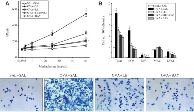
One functional consequence of the inflammatory process that underlies asthma is a AHR [12]. AHR was determined by Penh, and was substantially increased in OVA-challeng- ed mice in response to methacholine (Mch) inhalation as compared with control mice. LE, SB239063, or BAY 11-
Fig. 1.Airway hyperresponsiveness, differential cell counts, and Diff-Quik staining of cells in bronchoalveolar lavage (BAL) fluids follow- ing ovalbumin (OVA) sensitization and treatment with Lentinus edodes ethanol extract. (A) All animals were nebulized with various con- centrations of methacholine as a bronchoconstrictor. Data are shown as the percentage increase in Penh over the baseline, where the baseline Penh of the saline-treated control group is expressed as 100%. (B) The effect of Lentinus edodes ethanol extract, SB239063, or BAY 11- 7085 on OVA-induced differential cell counts in BAL fluids were analyzed. (C) Diff-Quik staining of immune cells in BAL fluids. Magni- fication 200×. EOS, eosinophil; NEU, neutrophil; MAC, macrophage; LYM, lymphocyte. Sampling was performed 48 h after the last OVA challenge in mice. Results from three independent experiments with 7 mice/group are given as mean±SEM. #p⁄0.05 vs. SAL++SAL; *p⁄
0.05 vs. OVA++SAL. Saline-inhaled mice administered saline (SAL++SAL), OVA-inhaled mice administered saline (OVA++SAL), Lentinus edodes ethanol extract (OVA++LE), SB239063 (OVA++SB239063), and BAY 11-7085 (OVA++BAY).
600
400
200
0
SAL++SAL OVA++SAL OVA++LE OVA++SB239063 OVA++BAY
SAL++SAL OVA++SAL OVA++LE OVA++SB239063 OVA++BAY
#
#
#
#
#
#
* *
**
*
* *
* * * * * *
*
* *
**
SAL++SAL OVA++SAL OVA++LE OVA++BAY
10
8
6
4
2
0
012345 10 20 30 40 50
Methacholine (mg/mL)
Total EOS NEU MAC LYM
%Penh Cell no.(105 cells/mL)
A B
C
7085 dramatically prevented AHR to inhaled Mch, as shown in Fig. 1A, suggesting that immune-mediated pathol- ogy in vivo was modified. Next, to examine the effect of LE on chemotaxis, that is, recruitment of inflammatory cells into airway, inflammatory cells were analyzed in BAL fluids of mice 48 h after the last OVA challenge. To inves- tigate the cellular composition of the BAL fluids, the cells were spread on a glass slide and stained with Diff-Quik
solution, and the numbers of each immune cell including eosinophils, neutrophils, macrophage, and lymphocytes were differentially counted under light microscope (Fig. 1B and C). In the saline-treated mice, OVA-challenge resulted in a marked increase of eosinophils and slight increases of neutrophils and lymphocytes when compared with control mice. However, pretreatment of LE, SB 239063, or BAY 11-7085 significantly attenuated OVA-induced recruitment Fig. 2.Histological evaluation of lung inflammation following ovalbumin (OVA) sensitization and treatment with Lentinus edodes ethanol extract. Paraffin-embedded lung sections were stained with hematoxylin-eosin (H&E, A) and periodic acid-Schiff (PAS, C). Bars indicate 50 μm. Data are representative of three independent experiments. (B) Inflammation scores. Total lung inflammation was defined as the average of the peribronchial and perivascular inflammation scores. (D) Quantitation of airway mucus expression. Sampling was performed 48 h after the last OVA challenge in mice. Results from three independent experiments with 7 mice/group are given as mean±SEM. #p⁄
0.05 vs. SAL++SAL; *p⁄0.05 vs. OVA++SAL. Saline-inhaled mice administered saline (SAL++SAL), OVA-inhaled mice administered saline (OVA++SAL), Lentinus edodes ethanol extract (OVA++LE), and BAY 11-7085 (OVA++BAY).
H&E PAS
SAL++SAL OVA++SAL SAL++SAL OVA++SAL
OVA++LE OVA++BAY OVA++LE OVA++BAY
A C
B D
4
3
2
1
0
Peribronchial Perivascular Total
#
# #
#
*
*
*
*
* *
*
* 60
50 40 30 20 10
0
SAL++SAL OVA++SAL OVA++LE OVA++BAY SAL++SAL OVA++SAL OVA++LE OVA++BAY
Inflammation score PAS++Cells/Bronchus(%)
of these cells into airway (p⁄0.05).
2. LE represses OVA-induced airway inflammation in experimental asthma
The observed reduction in chemotaxis into the airway correlated with the histological changes of lung parenchy- ma. Lungs from OVA-challenged mice showed widespread peribronchial and perivascular inflammatory cell infiltrates (Fig. 2A and B). Moreover, the percentage of mucus-pro- ducing goblet cells (indicated by PAS staining) in OVA- inhaled mice was substantially greater than that in control mice (Fig. 2C and D). In contrast, administration of LE led to a significant reduction of inflammatory cell infiltration and goblet cell hyperplasia. These results indicate that LE efficiently attenuates pulmonary inflammation and mucus hypersecretion in OVA-induced asthmatic mice.
3. LE attenuates the release of total and OVA-specific IgE into BAL fluids of OVA-inhaled mice
Total and OVA-specific IgE levels were determined by ELISA in each experimental group. IgE levels in BAL fluids were dramatically elevated in OVA-challenged mice, compared with control mice. However, the administration of LE or BAY 11-7085 to OVA-inhaled mice led to a sig- Fig. 3.Assessment of IgE in bronchoalveolar lavage fluids of oval- bumin (OVA)-sensitized mice treated with Lentinus edodes ethanol extract. The levels of IgE were quantified by ELISAs. Sampling was performed 48 h after the last OVA challenge in mice. Results from three independent experiments with 7 mice/group are given as mean±SEM. #p⁄0.05 vs. SAL++SAL; *p⁄0.05 vs. OVA++SAL.
Saline-inhaled mice administered saline (SAL++SAL), OVA-inhaled mice administered saline (OVA++SAL), Lentinus edodes ethanol extract (OVA++LE), and BAY 11-7085 (OVA++BAY).
10
8
6
4
2
0
#
#
*
*
*
* Total lgE OVA-specific lgE
ng/mL
SAL++SAL OVA++SAL OVA++LE OVA++BAY
Fig. 4.Assessment of pro-inflammatory and Th2 cytokines and adhesion molecules in bronchoalveolar lavage fluids of ovalbumin (OVA)-sensitized mice treated with Lentinus edodes ethanol extract.
The levels of pro-inflammatory (TNF-α and IL-1β) and Th2 cyto- kines (IL-4, IL-5, and IL-13) and adhesion molecules (eotaxin, ICAM-1, and VCAM-1) were quantified by ELISAs. Sampling was performed 48 h after the last OVA challenge in mice. Results from three independent experiments with 7 mice/group are given as mean
±SEM. #p⁄0.05 vs. SAL++SAL; *p⁄0.05 vs. OVA++SAL. Saline- inhaled mice administered saline (SAL++SAL), OVA-inhaled mice administered saline (OVA++SAL), Lentinus edodes ethanol extract (OVA++LE), and BAY 11-7085 (OVA++BAY).
pg/mLpg/mLpg/mL
500
400
300
200
100
0
TNF-α IL-1β
IL-4 IL-5 IL-13
Eotaxin ICAM-1 VCAM-1
#
#
#
#
#
#
#
#
*
*
* *
*
*
*
*
*
*
*
* *
*
*
*
500
400
300
200
100
0
20
16
12
8
4 3 2 1 0
SAL++SAL OVA++SAL OVA++LE OVA++BAY
SAL++SAL OVA++SAL OVA++LE OVA++BAY
SAL++SAL OVA++SAL OVA++LE OVA++BAY
nificant reduction in the total and OVA-specific IgE levels (Fig. 3).
4. LE reduces the levels of cytokines involved in the pathophysiology of asthma in OVA-challenged mice
Allergic airway inflammation is well-recognized to be caused by the release of a series of proinflammatory (TNF- α and IL-1β) and Th2 cytokines (IL-4, IL-5, and IL-13) [16].
To assess the effect of LE on pulmonary inflammation in asthmatic mice, the levels of these cytokines in BAL fluids were measured. ELISA showed that the levels of TNF-α, IL-1β, IL-4, IL-5, and IL-13 in BAL fluids were signifi- cantly increased in OVA-challenged mice compared with the levels in control mice (Fig. 4). The increased levels of these cytokines were significantly decreased by the admin- istration of LE or BAY 11-7085. Moreover, considering
that chemokines and leukocyte-endothelial adhesion mole- cules are critical for the recruitment and migration of leuko- cytes to the sites of inflammation [17], the levels of eotaxin, ICAM-1, and VCAM-1 were measured. Changes in the levels of these cytokines were similar to those seen for the aforementioned cytokines, indicating that the OVA chal- lenge-induced increase in cytokine levels can be reversed by LE or BAY 11-7085.
5. LE suppresses the nuclear translocation of NF-κκB and phosphorylation of IκκB-αα in lung tissues of allergic mice
In view of our data that AHR and eosinophilic inflam- mation in asthmatic mice is blocked by BAY 11-7085, a specific NF-κB inhibitor as well as the knowledge that NF- κB plays a key role in allergic inflammation of the lung by Fig. 5.Effect of Lentinus edodes ethanol extract on ovalbumin (OVA)-induced NF-κB activation. The translocation of p65 to the nucleus (A) as well as IκB-α phosphorylation and degradation in cytoplasm (B) were assessed by Western blot. Sampling was performed 48 h after the last OVA challenge in mice. Density ratio vs. β-actin was measured using a densitometer. Results from three independent experiments with 7 mice/group are given as mean±SEM. #p⁄0.05 vs. SAL+SAL; *p⁄0.05 vs. OVA++SAL. Saline-inhaled mice administered saline (SAL++SAL), OVA-inhaled mice administered saline (OVA++SAL), Lentinus edodes ethanol extract (OVA++LE), and SB239063 (OVA++ SB239063).
SAL
+ +SAL
SAL
+ +SAL
OVA
+ +SAL
OVA
+ +LE
OVA
+ +SB239063 OVA
+ +SAL
OVA
+ +LE
OVA
+ +SB239063
p65 (cytosol)
β-actin
p65 (nuclear)
PARP
1.0 0.8 0.6 0.4 0.2 0.0
1.0 0.8 0.6 0.4 0.2 0.0 p65 (cytosol)
p65 (nuclear)
p-IκB-α IκB-α
#
# #
#
*
*
*
*
*
* *
Relative density * Relative density
p-IκB-α
IκB-α
β-actin
SAL++SAL OVA++SAL OVA++LE OVA++SB239063 SAL++SAL OVA++SAL OVA++LE OVA++SB239063
A B
inducing the transcription of various proinflammatory mediators [8], we hypothesized that LE would attenuate airway inflammatory reactions by suppressing NF-κB acti- vation. To address this issue, we first studied the nuclear translocation of the NF-κB p65 subunit in lung tissues after OVA challenge. There was a decrease and increase of NF- κB p65 levels in the cytosols and nuclei from OVA-chal- lenged lungs, respectively (Fig. 5A). In contrast, cytosolic and nuclear extracts from LE or SB 239063-treated mice showed the suppression of nuclear translocation. Next, the effects of LE on OVA-induced phosphorylation and degra- dation of IκB-α were evaluated to clarify the molecular mechanisms by which LE inhibits NF-κB activity. LE sig- nificantly reduced the OVA-induced phosphorylation and degradation of IκB-α in the cytosol from lung tissues, as did SB 239063 (Fig. 5B). Taken together, these findings indicate that LE represses NF-κB transcriptional activity
possibly by stabilizing IκB-α and impairing the nuclear transport of p65 subunit in lung tissues from OVA-chal- lenged mice.
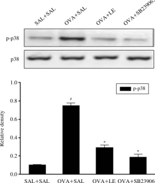
6. LE blocks OVA-induced phosphorylation of p38 MAPK in asthmatic mice
To investigate whether the inhibition of asthmatic respo- nse by LE is mediated through p38 MAPK, phosphoryla- tion of p38 MAPK was examined in OVA-challenged mice pretreated with LE or SB 239063, a p38 MAPK inhibitor.
As shown in Fig. 6, LE or SB 239063 effectively inhibited OVA-induced phosphorylation of p38 MAPK in allergic mice. On the other hand, the expression of p38 was unaf- fected by OVA, LE, or SB 239063.
Discussion
Allergic airway inflammation, characterized by increased infiltration of leukocytes, such as eosinophils, and consid- erable secretion of mucus into the airways, is a major fac- tor in the pathogenesis of asthma. Especially, eosinophil has long been recognized as the principal effector cell and plays pathogenic roles in asthma by its release of cytotoxic granule proteins [18]. Our present findings show that LE prevents eosinophilic infiltration into the airways, as evi- denced by a significant drop in total cell counts and eosi- nophil counts in BAL fluid. Likewise, tissue eosinophilia is also inhibited, as revealed by a marked reduction of inflam- matory cell infiltration in histological examination. Eosino- philic transmigration into the airways is a multistep process that is orchestrated by not only Th2 cytokines such as IL- 4, IL-5, and IL-13 but also proinflammatory cytokines including TNF-α and IL-1β, and coordinated by chemotac- tic cytokine (eotaxin) in combination with adhesion mole- cules, including ICAM-1 and VCAM-1 [19,20]. IL-4 is required for B cell maturation and IgE synthesis, and par- ticipates in the initiation of Th2 inflammatory responses.
IL-5 is pivotal for growth, differentiation, recruitment, and survival of eosinophils. IL-13 can potently induce mucus hypersecretion, eotaxin expression, airway inflammation, and AHR [21,22]. TNF-α and IL-1β also exert similar res- ponses, which include upregulation of eosinophil chemoat- tractants and adhesion molecules, recruitment of eosinophil, increase of cytokine release, and enhancement of AHR [16].
Fig. 6.Effect of Lentinus edodes ethanol extract on ovalbumin (OVA)-induced p38 MAPK activation. Protein expressions of pho- sphorylated (p)-p38 and p38 in lung tissues were evaluated 48 h after the last OVA challenge. Density ratio vs. β-actin was measured using a densitometer. Results from three independent experiments with 7 mice/group are given as mean±SEM. #p⁄0.05 vs. SAL++ SAL; *p⁄0.05 vs. OVA++SAL. Saline-inhaled mice administered saline (SAL++SAL), OVA-inhaled mice administered saline (OVA
+
+SAL), Lentinus edodes ethanol extract (OVA++LE), and SB 239 063 (OVA++SB 239063).
SAL
+ +SAL
OVA
+ +SAL
OVA
+ +LE
OVA
+ +SB239063
p-p38
p38
1.0
0.8
0.6
0.4
0.2
0.0
#
*
*
SAL++SAL OVA++SAL OVA++LE OVA++SB239063
Relative density
p-p38
According to the current study, LE attenuates the increased release of Th2 and proinflammatory cytokines, eotaxin, and adhesion molecules into the airway of OVA-challeng- ed mice. From these findings, we speculate that LE can prevent eosinophilic airway inflammation by diminishing secretion of aforementioned cytokines into lungs.
AHR is a hallmark clinical symptom of asthma, which is defined as the abnormal increase in airflow limitation in response to a provoking stimulus. Although there are less data surrounding the precise mechanisms whereby airway inflammation enhances AHR, it is convincing that various mediators released during allergic inflammation play a crit- ical role in AHR development [23]. For example, it has been established that IL-5 plays a crucial role in AHR by mobilizing and activating eosinophils, leading to the release of proinflammatory products such as major basic protein and cysteinyl-leukotrienes, which are closely associated with AHR [24]. Similarly, IL-4 and IL-13 have been shown to induce AHR in murine asthma models in which cys- teinyl-leukotrienes might be causative agents of AHR [25].
Moreover, AHR could be brought about by a direct effect of TNF-α on airway smooth muscle [26]. As such, the obs- erved reduction of AHR by LE may be related to decrease in Th2 cytokine production, tissue eosinophilia, and TNF- α levels by LE.
The eukaryotic transcription factor NF-κB regulates a wide variety of target genes that encode multiple inflam- matory cytokines, such as TNF-α, IL-1β, IL-4, IL-5, IL-13, ICAM-1, and VCAM-1, all of which are closely implicated in the pathogenesis of asthma, as mentioned above [8]. Our results indicate that LE exerts anti-NF-κB actions in lung tissues of OVA-challenged mice. Moreover, suppression of NF-κB activity by BAY 11-7085, a specific inhibitor of NF-κB, has been shown to induce not only a reduction in the levels of aforementioned cytokines, but the amelio- ration in eosinophilic airway inflammation and AHR in our model, which is in line with the previous report [27].
Taken together, it is proposed that the mechanism under- lying the anti-asthmatic effects of LE may be attributed to inhibition of NF-κB transcriptional activity and to subse- quent reduction of proinflammatory chemical mediators.
As described previously, p38 MAPK has been consid- ered to play a cardinal role in allergic asthma [12]. Suppor- ting this contention, our study has shown that the augmen- tation in asthmatic symptoms as well as the phosphoryla- tion of p38 MAPK after OVA inhalation is significantly
reduced after the administration of SB 239063, a selective p38 MAPK inhibitor. Moreover, SB 239063 prominently represses NF-κB activity in OVA-challenged lungs, sug- gesting that p38 MAPK acts upstream of NF-κB signaling pathway in allergic asthma. Likewise, LE dramatically decreases the activity of p38 MAPK and NF-κB in lung tissues of OVA-challenged mice. Accordingly, these obser- vations encourage the view that treatment of allergic mice with LE leads to the suppression of p38 MAPK and the subsequent disruption of NF-κB activity, reversing patho- physiologic features of asthma.
It is well recognized that mucus hyperproduction in asth- ma results from hypertrophic and metaplastic changes of goblet cells and contributes to clinical symptoms such as airway obstruction and mortality [28]. Although goblet cell hyperplasia is typically more intense in mice with chronic asthma, it could be also identified in a model of acute asth- ma, as indicated in previous and present findings [29]. As for the mechanism of airway mucin hypersecretion, empiri- cal evidence suggests that p38 MAPK as well as Th2 cyto- kines such as IL-4 and IL-13 displays goblet cell hyperpla- sia in the acute asthmatic attack [12,28]. Subsequently, it has been shown that MUC5AC, the major mucin in the goblet cells, is upregulated by inflammatory cytokines such as TNF-α and IL-1β in the ERK and p38 MAPK-depen- dent fashion [30]. Moreover, induction of airway epithelial NF-κB has been reported to cause uncontrolled MUC5AC gene expression and goblet cell hyperplasia in the allergic lung [31]. Taken together, we speculate that LE could sig- nificantly abate marked goblet cell hyperplasia and mucus hypersecretion by modulating not only proinflammatory and Th2 cytokines but the activities of p38 MAPK and NF- κB in OVA-challenged mice.
Ethanol extraction is known to be the first important steps for separation, characterization, and quantification of flavonoids and other phenolic compounds from plant mate- rials. Thus, aqueous ethanol is a popular choice of solvent.
As described in Materials and Methods, the chemical analy- sis has indicated that LE is chiefly made up of polysaccha- rides, which have been demonstrated to exhibit significant biological effects such as anti-inflammatory and anti-oxi- dative activity in previous reports [32,33]. In addition, sev- eral researchers have revealed that lentinan, β-glucan from Lentinus edodes, induces immunomodulatory and immu- nosuppressive behaviors under different conditions through MAPK signaling pathways mediated by ERK1/2 and JNK1
/2 [3,5]. Furthermore, the recent study provides evidence that lentinan could inhibit gut inflammation via modulation of NF-κB activity in the experimental models [34]. In view of these findings and our results, the polysaccharides iso- lated from Lentinus edodes might be active components attributed to the anti-asthmatic activity of LE. Therefore, further work should be directed toward the purification and identification of asthma-relieving constituents from Lentinus edodes.
In conclusion, the present data indicate that LE could ameliorate asthmatic inflammation and AHR by downreg- ulating proinflammatory and Th2 cytokines via inhibiting the p38 MAPK-NF-κB module. These findings suggest that Lentinus edodes is a potential anti-inflammatory agent in asthma treatment.
Acknowledgements
This paper was supported by the National Natural Sci- ence Foundation of China (81260665) and the Project of Research & Innovation of Jilin Youth Leader and Team (20140519013JH).
References
1. Cao X, Liu R, Liu J, Huo Y, Yang W, Zeng M, et al. A novel polysaccharide from Lentinus edodes Mycelia exhibits potential antitumor activity on laryngeal squamous cancer cell line Hep-2. Appl Biochem Biotechnol. 2013; 171:1444- 53.
2. Zembron-Lacny A, Gajewski M, Naczk M, Siatkowski I.
Effect of shiitake (Lentinus edodes) extract on antioxidant and inflammatory response to prolonged eccentric exercise.
J Physiol Pharmacol. 2013; 64:249-54.
3. Xu X, Pan C, Zhang L, Ashida H. Immunomodulatory beta- glucan from Lentinus edodes activates mitogen-activated protein kinases and nuclear factor-kappaB in murine RAW 264.7 macrophages. J Biol Chem. 2011; 286:31194-8.
4. Wasser SP. Current findings, future trends, and unsolved problems in studies of medicinal mushrooms. Applied micro- biology and biotechnology. 2011; 89:1323-32.
5. Xu X, Yasuda M, Nakamura-Tsuruta S, Mizuno M, Ashida H. beta-Glucan from Lentinus edodes inhibits nitric oxide and tumor necrosis factor-alpha production and phosphory- lation of mitogen-activated protein kinases in lipopolysac-
charide-stimulated murine RAW 264.7 macrophages. J Biol Chem. 2012; 287:871-8.
6. Galli SJ, Tsai M, Piliponsky AM. The development of aller- gic inflammation. Nature. 2008; 454:445-54.
7. Medoff BD, Thomas SY, Luster AD. T cell trafficking in allergic asthma: the ins and outs. Annu Rev Immunol. 2008;
26:205-32.
8. Imanifooladi AA, Yazdani S, Nourani MR. The role of nu- clear factor-kappaB in inflammatory lung disease. Inflamm Allergy Drug Targets. 2010; 9:197-205.
9. Gras D, Chanez P, Vachier I, Petit A, Bourdin A. Bronchial epithelium as a target for innovative treatments in asthma.
Pharmacol Ther. 2013; 140:290-305.
10. Choi IW, Kim DK, Ko HM, Lee HK. Administration of antisense phosphorothioate oligonucleotide to the p65 sub- unit of NF-kappaB inhibits established asthmatic reaction in mice. Int Immunopharmacol. 2004; 4:1817-28.
11. Arthur JS, Ley SC. Mitogen-activated protein kinases in innate immunity. Nat Rev Immunol. 2013; 13:679-92.
12. Duan W, Chan JH, McKay K, Crosby JR, Choo HH, Leung BP, et al. Inhaled p38alpha mitogen-activated protein kinase antisense oligonucleotide attenuates asthma in mice. Am J Respir Crit Care Med. 2005; 171:571-8.
13. Tournoy K, Kips J, Schou C, Pauwels R. Airway eosinophi- lia is not a requirement for allergen-induced airway hyper- responsiveness. Clin Exp Allergy. 2000; 30:79-85.
14. Cho JY, Miller M, Baek KJ, Han JW, Nayar J, Lee SY, et al. Inhibition of airway remodeling in IL-5-deficient mice.
J Clin Invest. 2004; 113:551-60.
15. Lee KS, Kim SR, Park HS, Park SJ, Min KH, Lee KY, et al.
A novel thiol compound, N-acetylcysteine amide, attenuates allergic airway disease by regulating activation of NF-kappaB and hypoxia-inducible factor-1alpha. Exp Mol Med. 2007;
39:756-68.
16. Boyce JA, Bochner B, Finkelman FD, Rothenberg ME.
Advances in mechanisms of asthma, allergy, and immuno- logy in 2011. J Allergy Clin Immunol. 2012; 129:335-41.
17. Ulbrich H, Eriksson EE, Lindbom L. Leukocyte and endo- thelial cell adhesion molecules as targets for therapeutic interventions in inflammatory disease. Trends Pharmacol Sci. 2003; 24:640-7.
18. Jacobsen EA, Ochkur SI, Lee NA, Lee JJ. Eosinophils and asthma. Curr Allergy Asthma Rep. 2007; 7:18-26.
19. Lukacs NW. Role of chemokines in the pathogenesis of asthma. Nat Rev Immunol. 2001; 1:108-16.
20. Mattes J, Foster PS. Regulation of eosinophil migration and Th2 cell function by IL-5 and eotaxin. Curr Drug Targets Inflamm Allergy. 2003; 2:169-74.
21. Li L, Xia Y, Nguyen A, Lai YH, Feng L, Mosmann TR, et al. Effects of Th2 cytokines on chemokine expression in
the lung: IL-13 potently induces eotaxin expression by air- way epithelial cells. J Immunol. 1999; 162:2477-87.
22. Kudo M, Ishigatsubo Y, Aoki I. Pathology of asthma. Front Microbiol. 2013; 4:263.
23. Cockcroft DW, Davis BE. Mechanisms of airway hyperre- sponsiveness. J Allergy Clin Immunol. 2006; 118:551-9;
quiz 60-1.
24. Gleich GJ. Mechanisms of eosinophil-associated inflamma- tion. J Allergy Clin Immunol. 2000; 105:651-63.
25. Vargaftig BB, Singer M. Leukotrienes mediate murine bron- chopulmonary hyperreactivity, inflammation, and part of mucosal metaplasia and tissue injury induced by recombi- nant murine interleukin-13. Am J Respir Cell Mol Biol.
2003; 28:410-9.
26. Brightling C, Berry M, Amrani Y. Targeting TNF-alpha: a novel therapeutic approach for asthma. J Allergy Clin Immu- nol. 2008; 121:5-10; quiz 11-2.
27. Kim SR, Lee KS, Park SJ, Min KH, Lee MH, Lee KA, et al.
A novel dithiol amide CB3 attenuates allergic airway disease through negative regulation of p38 mitogen-activated pro- tein kinase. Am J Respir Crit Care Med. 2011; 183:1015- 24.
28. Wills-Karp M, Luyimbazi J, Xu X, Schofield B, Neben TY, Karp CL, et al. Interleukin-13: central mediator of allergic asthma. Science. 1998; 282:2258-61.
29. Kuperman D, Schofield B, Wills-Karp M, Grusby MJ. Sig-
nal transducer and activator of transcription factor 6 (Stat6)- deficient mice are protected from antigen-induced airway hyperresponsiveness and mucus production. J Exp Med.
1998; 187:939-48.
30. Song KS, Lee WJ, Chung KC, Koo JS, Yang EJ, Choi JY, et al. Interleukin-1 beta and tumor necrosis factor-alpha in- duce MUC5AC overexpression through a mechanism involv- ing ERK/p38 mitogen-activated protein kinases-MSK1- CREB activation in human airway epithelial cells. J Biol Chem. 2003; 278:23243-50.
31. Sheller JR, Polosukhin VV, Mitchell D, Cheng DS, Peebles RS, Blackwell TS. Nuclear factor kappa B induction in air- way epithelium increases lung inflammation in allergen- challenged mice. Exp Lung Res. 2009; 35:883-95.
32. He JZ, Ru QM, Dong DD, Sun PL. Chemical characteristics and antioxidant properties of crude water soluble polysac- charides from four common edible mushrooms. Molecules.
2012; 17:4373-87.
33. Yu S, Weaver V, Martin K, Cantorna MT. The effects of whole mushrooms during inflammation. BMC Immunol.
2009; 10:12.
34. Nishitani Y, Zhang L, Yoshida M, Azuma T, Kanazawa K, Hashimoto T, et al. Intestinal anti-inflammatory activity of lentinan: influence on IL-8 and TNFR1 expression in intes- tinal epithelial cells. PLoS One. 2013; 8:e62441.
표고버섯은 알레르기성 천식 마우스 모델에서 NF-κB의 활성을 억제함으로써 기도 과민성과 염증 반응을 저해한다
연광해
1, 최윤호
2,31중국 연변대학교 기초의학원 해부학교실, 2전북대학교 의학전문대학원 해부학교실, 3전북대학교 의과학 연구소
간추림 : 표고버섯은 대표적인 식용버섯으로 면역조절작용을 비롯한 항암, 항염증 및 항바이러스 효과를 지닌 것으로 알려져 있다. 그럼에도 불구하고 표고버섯이 천식에 미치는 효능은 거의 알려지지 않았다. 이에 본 연구 진은 표고버섯의 에탄올 추출물이 마우스에 유발된 천식에서 나타나는 기도 저항과 폐 조직의 염증 반응에 미 치는 영향을 관찰하였다. 표고버섯 추출물을 천식 마우스에게 구강으로 투여할 경우, 기도 저항과 알레르기성 염증반응이 현저하게 감소하는 것을 확인할 수 있었다. 천식 마우스의 폐에서 증가하는 Th2 사이토카인 (IL-4, IL-5 및 IL-13)과 eotaxin, adhesion molecules의 발현이 이 추출물에 의해 유의하게 억제되는 것을 관찰할 수 있 었다. 추가로 표고버섯의 항천식효과에 대한 기전을 규명하고자 표고버섯 추출물이 천식 마우스의 폐조직에서 발현되는 NF-κB에 미치는 영향을 확인하였다. 상기 결과와 마찬가지로 표고버섯 추출물은 NF-κB의 핵 내 이 동을 억제함으로써 이 단백질의 활성을 제어하였다. 이들 결과를 통해 표고버섯 추출물이 천식 마우스에서 NF- κB의 활성을 낮춤으로써 기도 과민 반응 및 염증 반응을 개선하는 것으로 사료된다. 따라서 본 연구결과는 표 고버섯이 천식을 예방하거나 완화시키는 약물로 사용될 수 있는 가능성을 제시한다.
찾아보기 낱말 : 표고버섯, 천식, 알레르기성 염증반응, Th2 사이토카인, NF-κB
교신저자 : 최윤호(전북대학교 의학전문대학원 해부학교실) 전자우편 : [email protected]