Seul-Ki Park1, Jum-Ji Kim1, Sang-Min Sung2& Mi-Young Lee1 1Department of Medical Biotechnology,
SoonChunHyang University, Asan, Chungnam 336-600, Korea 2Susin R & D Institute, Susin Ogapy, Cheonan,
Chungnam 300-881, Korea
Correspondence and requests for materials should be addressed to M. Y. Lee ([email protected])
Accepted 20 November 2009
Abstract
The effects of an extract of Benincasae hispida on allergic inflammation were examined in terms of his-tamine and ββ-hexosaminidase release, serum IgE level and inflammatory cytokine level. The B. hispida extract inhibited the release of histamine and ββ-hex-osaminidase, a degranulation marker, from rat baso-philic leukemia cells (RBL-2H3). When mice were first ovalbumin-challenged and then treated with B. his-pida extract, there was a significant decrease in the IgE level in the mouse serum. The extract treatment reduced the serum IgE level prominently, compared with the ovalbumin-challenged mice. The extract also significantly reduced the TNF-αα and IL-4 levels in the BAL fluid when challenged with antigen. Taken together, the Benincasae hispida extract may be effi-cacious against allergic inflammation.
Keywords: Benincasae hispida, Allergic inflammation, His-tamine release, Inflammatory cytokine, Serum IgE level
Allergic inflammation is an important pathophysio-logical condition and includes asthma, atopic derma-titis, allergic rhinitis, and several ocular allergic dise-ases1. Generally, the pathophysiology of allergic
res-ponses may be divided into two components, the early-and late-phase reactions. The early phase can either subside or progress into a late-phase reaction, which can prolong the symptoms of response substantially and result in tissue damage.
The early-phase reaction is generally referred to as the immediate or type I allergic reaction, caused by
the release of histamine and by activated mast cells following the cross-linking of allergen-specific IgE bound to mast cell FcεRI receptors2. It is well known that FcεRI aggregation triggers sequence of biochem-ical events leading to cell degranulation. Engagement of the receptor leads to the activation of tyrosine kinas-es, activation of phospholipase C (PLC), increased dia-cylglycerol, and mobilization of Ca2++
from internal stores. This is followed by the activation of protein kinase C (PKC), accompanied by an increase in mito-gen-activated protein kinase (MAPK) activity and a Ca2++
influx. In FcεRI signaling, tyrosine phosphoryla-tion of proteins is considered to play an essential role3.
Recent studies have shown that in Syk kinase-deficient mast cells, there is complete abrogation of degranula-tion, elevation of Ca2++
influx, and activation of the ERK and JNK MAP kinase pathways. These observa-tions indicate that Syk kinase is essential for the Fcε RI-mediated degranulation signal transduction4,5.
Activated mast cells can produce a wide variety of inflammatory mediators, such as eicosanoids, proteo-glycans, proteases, and several proinflammatory and chemotactic cytokines, like TNF-α, IL-4, IL-6, IL-13, and TGF-β6. Mediators of allergic inflammation affect
nerve cells, causing itching7, smooth muscle cells,
causing contraction, leading to the airway narrowing in allergic asthma8, and endothelial cells, causing
vasodilation and edema7. Typically, the T-cells
recruit-ed in the early-phase reaction are of the Th2 variety, which belong to a subset of T-cells that produce IL-4 in allergic responses.
Benincasae hispida (Thunb). Cogn, commonly known as white pumpkin, wax gourd, or ash gourd, is cultivated primarily in China, India, and other semi-tropical countries for its edible fruit that has notable medicinal value9. According to an old Korean
medi-cal encyclopedia, the ‘Donguibogam’, which is listed in the “Memory of the World Register” by UNESCO, Benincasae hispida is efficacious against diabetes, edema, dropsy, diseases related to the liver, leucorrhea, and good for the detoxication of minerals, removal of fever, and to strengthen the function of the bladder and intestines10. Moreover, in vitro and in vivo studies
have shown that the seeds of Benincasa hispida have an expectorant11, anti-angiogenic12, anti-oxidative13,
Suppressive Effects of Benincasae hispida on Allergic
gastroprotective14, anti-ulcer15, and anti-inflammatory effects in diabetic vascular complications9. The chem-ical components of the seeds15-17include saponin, urea, citrulline, linoleic acid, oleic acid, and fatty acids, as well as proteins, such as trigonelline, coffearin, and osmotin, as well as phytochemicals, such as triterpenes, sterols, flavonoid C-glycosides, acylated glucose, and benzyl glycoside.
To date, there has been no reported research on the suppressive effects of Benincasae hispida on allergic inflammation. Thus, we examined the inhibitory effects of Benincasae hispida on allergic inflammation by measuring serum IgE level, histamine release, and inflammatory cytokine production.
Reduction of Serum IgE Concentration Elevated levels of IgE are associated with asthma, atopic dermatitis, and allergic rhinitis. In general, un-usual IgE production is regulated primarily by Th2 cells. When Th2 cells are activated, IL-4, IL-5, IL-9, and IL-13 are secreted18-20; among these, 4 and IL-13 play key roles in IgE hyperproduction.
To investigate the effect of B. hispida extract on serum IgE levels, the OVA-specific IgE levels in mice treated with vehicle or B. hispida extract were examin-ed (Figure 1). The IgE levels increasexamin-ed by approxima-tely 4-fold in the OVA-challenged mice, compared with the saline-challenged mice. The oral administra-tion of B. hispida extract during the OVA-challenge period significantly prevented the rise in serum total IgE levels. The B. hispida extract reduced the serum IgE level by 42%, compared with OVA-challenged mice. These results indicate that the B. hispida extract might contain inhibitors of serum IgE elevation. Reduction of Inflammatory Cytokines in BAL Fluid
Cell components of the lower respiratory tract can be obtained from bronchoalveolar lavage (BAL) during respiratory diseases. BAL fluid has been used to assess biochemical and inflammatory changes in the inter-stitial lung tissue. Especially during asthma attacks, the expression of Th2 cytokines in BAL plays an important role in causing allergic inflammation, via enhanced IgE production21. Furthermore, IL-4 induces VCAM-1 (vascular cell adhesion molecule-1) gene expression in the endothelium and gathers eosinophils to the inflammatory lesions, causing increased infiltra-tion, leading to chronic inflammation.
TNF-α is a pivotal proinflammatory cytokine that increases during allergic inflammation. Elevated
TNF-α levels are frequently observed in the BAL fluid of
asthmatic subjects undergoing allergen challenge6. TNF-α expression mediates neutrophil migration and
infiltration; furthermore, it increases particle-induced cytotoxicity and also regulates neutrophil apoptosis in acute inflammation.
Changes in the IL-4 levels in the BAL of ovalbumin-challenged mice are presented in Table 1. The IL-4 level of the ovalbumin-challenged group was about 2-fold higher than that of the PBS group. Interestingly, the increased IL-4 level was lowered significantly, to the PBS-treated level, after B. hispida treatment. The TNF-α level of the ovalbumin-challenged group was about 3-fold higher than the PBS-treated group, and this value decreased by about 68%, compared with the ovalbumin-challenged group after 2% B. hispida treatment (Table 1). These results demonstrated that the B. hispida extract effectively lowered IL-4 and TNF-α in ovalbumin-challenged BAL fluid. B. hispida extract appeared to provide notable ameliorative ben-efit for allergic symptoms, by modulating IL-4 and TNF-α production in BAL fluid.
Histamine and ββ-hexosaminidase Release
Mast cells and basophils play essential roles in the pathogenesis of allergic reactions, such as atopic der-matitis and asthma. The rat basophilic leukemia cell line RBL-2H3, a tumor analog of mast cells, are mu-cosal mast cells that express the immunoglobulin Fc epsilon receptor I (FcεRI). Stimulation of IgE-sensitized RBL2H3 cells with specific antigen triggers a cascade Figure 1. (A) Experimental protocol for the protective effect of Benincasae hispida extract. (B) Inhibition of the elevation in the serum IgE level by treatment with Benincasae hispida extract. *P⁄0.05, significantly different from the OVA sen-sitized value.
B. hispida
0-day 14-day (A)
21-day 22-day 23-day 24-day OVA sensitization OVA intra-nasal challenge Sacrifice
9000 8000 7000 6000 5000 4000 3000 2000 1000 0 * PBS OVA B. hispida Contents of IgE (ng/mL) (B)
of events leading to degranulation, mediator release, activation of mitogen-activated protein kinase (MAPK), tyrosine kinase, and phospholipase C, increased reac-tive oxygen species production, calcium influx, and cytokine production. The secretion of histamine and
β-hexosaminidase from RBL-2H3 is a hallmark of
the allergic reaction resulting from allergen exposure. Histamine is released from intracellular secretory granules, induced by elevation of intracellular Ca2++ concentrations and the activation of protein kinase C22. The role of histamine in allergic inflammation is supported by several types of evidence, including the reproduction of features of allergic inflammation by injected or inhaled histamine, the reduction of allergic inflammation by histamine receptor antagonists, and more recently by the demonstration that mice geneti-cally modified to make less histamine have a dimin-ished capacity to develop allergic inflammation23. β-hexosaminidase is also stored in secretory granules of mast cells and is released concomitantly with hista-mine when mast cells are immunologically activated. Thus, β-hexosaminidase activity in the medium is also used as a marker of mast cell degranulation24.
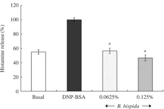
The degranulating effect was measured by assaying histamine release from allergen-sensitized RBL-2H3 cells (Figure 2). About 50% of the histamine was in-duced and released after DNP-BSA challenge. How-ever, upon treatment with 0.0625% B. hispida extract, histamine release was reduced by 44%, compared with DNP-BSA sensitized cells. Treatment with 0.125% of B. hispida extract displayed a 54% inhibitory effect on the histamine release, compared with DNP-BSA sensitized cells.
After stimulation with antigen, cells also release β-hexosaminidase, another marker of mast cell degran-ulation. Thus, to determine whether B. hispida extract could modulate Ag-induced β-hexosaminidase release, IgE stimulated RBL-2H3 cells were treated with B. hispida extract and then challenged with DNP-BSA. The degranulating effect of B. hispida extract, mea-sured by β-hexosaminidase release from
allergen-sensitized RBL-2H3 cells, is depicted in Figure 3. About 30% of the β-hexosaminidase was induced and released after DNP-BSA challenge. Upon treatment with 0.0312% B. hispida extract, however, β-hexosa-minidase release was reduced by 10%, compared with DNP-BSA sensitized cells. Treatment with 0.0625% of B. hispida extract showed a 20% inhibitory effect on the β-hexosaminidase release, compared with DNP-BSA sensitized cells.
These results demonstrate that the allergic inflam-mation-mediating histamine and β-hexosaminidase release were markedly inhibited by treatment with B. hispida extract, suggesting that the extract may be a candidate anti-allergic agent. Tea polyphenols25 and xanthones from G. mangostana26, and guarana seed extract27 and acanthopanax28, have been previously reported to suppress mast cell degranulation.
Table 1. Effect of B. hispida on IL-4 and TNF-α levels in the BAL fluids of mice.
IL-4 TNF-α
(pg/mL) (pg/mL)
PBS 51.9±7.0 75.01±4.1
OVA 110.7±8.1* 229.5±33.0*
B. hispida 52.2±2.6** 74.3±4.1**
The mice were treated with B. hispida and cytokine levels in the BAL fluid were measured as described in the Materials and methods. Re-sults are expressed as mean±SEM (n==6 in each group).
*P⁄0.05 as compared to PBS treated group; **P⁄0.05 as compared
to OVA treated group.
Figure 2. Inhibition of histamine release by treatment with
Benincasae hispida extract in RBL-2H3 cells. *P⁄0.05, sig-nificantly different from the DNP-BSA challenged value.
120 100 80 60 40 20 0 * * Basal DNP-BSA 0.0625% 0.125% B. hispida Histamine release (%)
Figure 3. Inhibition of β-hexosaminidase release by treat-ment with Benincasae hispida extract in RBL-2H3 cells. *P⁄ 0.05, significantly different from the DNP-BSA challenged value. 120 100 80 60 40 20 0 * Basal DNP-BSA 0.0312% 0.0625% B. hispida β -hexosaminidase release (%)
Lipoxygenase Inhibition in vitro
The effect of the aqueous extract of B. hispida on in vitro lipoxygenase activity was examined (Figure 4). Lipoxygenases comprise a family of non-heme iron-containing dioxygenases, representing the key en-zymes in the biosynthesis of leukotrienes that have been postulated to play an important role in the patho-physiology of several inflammatory and allergic dise-ases29. Lipoxygenase activity was reduced by approxi-mately 26 and 38% by treatment with 0.02 and 0.03% B. hispida, respectively. The results indicated that the B. hispida extract had an inhibitory effect on lipoxy-genase activity.
Mast cells produce and release leukotrienes and prostaglandins, as well as histamine. The release of leukotrienes and prostaglandins is caused by the acti-vation of phospholipase A2, an intracellular Ca2
+ + -dependent enzyme, an effect that is followed by the oxidation of fatty acids with lipoxygenase. Several lipoxygenase inhibitors reduce the release of both leukotriene and histamine from mast cells. Tea polyphe-nols have been suggested to suppress leukotriene rele-ase through the inhibition of lipoxygenrele-ase activity30.
Discussion
To treat allergic inflammation, several antagonistic drugs are used to block the action of allergic media-tors, or to prevent the activation of cells and degranula-tion processes. These include antihistamines, steroids, such as cortisone, dexamethasone, and hydrocortisone, epinephrine, and antileukotrienes. In alternative med-icine a number of allergy treatments, particularly from traditional herbal medicine, are known.
In conclusion, our study demonstrated that B. hispi-da extract was capable of alleviating IgE mediating
secretion of histamine and β-hexosaminidase from mast cells. It is also capable of reducing IgE levels in mouse serum and decreasing IL-4 and TNF-α levels in bronchoalveolar lavage fluid. These results show that B. hispida extract has a notable suppressive effect on allergic inflammation. Thus, B. hispida extract may be useful in alleviating a number of allergic inflamma-tions, including atopic dermatitis, asthma, and rhinitis. However, it is not clear whether the inhibitory effect of the extract on allergic inflammation was due to some unidentified component, which displayed an inhibitory effect on the degranulation of mast cells and in preventing increase in serum IgE levels. Thus, it is necessary to further characterize the active com-ponents responsible for the anti-allergic inflammatory action and elucidate the targets of those components, as well as the detailed mechanism by which the B. hispida extract suppresses allergic inflammation.
Materials & Methods
Preparation of Benincasae hispida Extract Benincasae hispida was provided by Susin Ogapy Co. Ltd. (Cheonan, Chungnam, Korea). The whole fruit was freeze-dried, and the dried powder was then dissolved in sterile phosphate-buffered saline (2% w/v). After centrifugation (12,000×g, 5 min), the
supernatant was collected and used in this experiment. Sensitization and Challenge
BALB/c male mice were obtained from Orient Bio Co. Ltd (Seoul, Korea). Mice, 7 weeks old, weighing 18-19 g, were acclimatized for 2 days at 25±2�C and a normal day/night cycle before starting the experi-ment. The mice were sensitized with 0.2 mL of nor-mal saline containing 500μg/mL ovalbumin (OVA; Sigma, St. Louis, MO, USA), adsorbed on 100 mg/mL aluminum hydroxide, intraperitonially (i.p.) on days 0 and 14. Seven days after the final i.p. injection, the mice were given the sample orally on days 21, 22, and 23. At 30 min after oral administration, 100μL OVA (150μg/100 μL) inhalation was performed.
All experimental protocols were approved by the Ins-titutional Animal Care and Use Committee (IACUC) at Soonchunhyang University.
Measurement of OVA-specific IgE
On day 24, each mouse was sacrificed and a blood sample was collected. The sera were separated by cen-trifugation (13,000×g, 10 min) and kept at -70�C until analysis for IgE. OVA-specific IgE levels were measured by enzyme-linked immunosorbent assay (ELISA). The results are expressed as ng/mL of serum.
Figure 4. Inhibition of lipoxygenase activity by treatment
with Benincasae hispida extract. 120 100 80 60 40 20 0 Control 0.02% 0.03% B. hispida
Inhibition of lipoxygenase activity
The plates were coated with diluted anti-mouse IgE overnight at 4�C and then 1 : 250 diluted sera were added to the wells and the plates were incubated for 2 h at room temperature. The bound IgE was detected with biotinylated anti-mouse IgE antibodies and the plates were developed by the addition of horseradish peroxidase and 3,3′,5,5′-tetramethylbenzidine, and measured using a plate reader at 450 nm.
Cytokine Determination by ELISA
Shortly after exsanguination, the trachea was can-nulated and 1 mL of saline was used per lavage and repeated four times for each mouse. About 4 mL of bronchoalveolar lavage (BAL) fluid was centrifuged (12,000×g, 10 min) and the supernatant was kept at -70�C until analysis for cytokines. IL-4 and TNF-α in the BAL fluid were measured using a modified ELISA method, as described in our previous report. MTT Assay for Cell Viability
The MTT assay was used to determine the maxi-mum concentration of the extract that did not affect cell viability, as described previously28,31.
Histamine and ββ-hexosaminidase Release
The rat basophile leukemia cell line RBL-2H3 was maintained in DMEM with 10% fetal bovine serum and 100 unit/mL penicillin/streptomycin at 37�C and 5% CO232. Cells (2×10⁴cells/well) were precultured at 37�C for 24 h in 0.1 mL of medium per well in a 96-well plate. The supernatants were discarded and the cells were incubated at 37�C for 2 h with DMEM containing 2% FBS and anti-DNP IgE. The cells were washed three times with HEPES buffer. After incubat-ing in 0.1 mL of HEPES buffer containincubat-ing Benincasae hispida extract at 37�C for 10 min, the cells were chal-lenged with DNP-BSA (4μg/200 μL) at 37�C for 35 min. The plate was placed at 4�C to stop the reaction. Next, 0.2 mL of 1 N NaOH and 0.1 mL 1% o-phthal-aldehyde (OPT) was added at room temperature, 5 min after the supernatant was removed to a 24-well plate. The reaction was stopped with 0.2 mL/well of 0.1 N HCl. Signals were quantitated using a fluorom-eter (405 nm excitation and 450 nm emission). Results are expressed as a percentage of the total release minus the spontaneous release. Analyses were performed in triplicate.
β-hexosaminidase assays were performed on the
same cell culture conditions as those used for the his-tamine assay. After stimulation by DNP-BSA, the cells were centrifuged (5,000×g, 1 min) and the
super-natants were collected and chilled on ice. Then, 50
μL of each sample was incubated with 50 μL of 1 mM ρ-nitrophenyl-β-acetyl-D-glucosamide (Sigma),
dis-solved in 0.1 M citrate buffer (pH 5), in a 96-well plate at 37�C for 1 h. The reaction was stopped by the addi-tion of 200μL/well of 0.2 M glycine buffer (pH 10.7). The absorbance of the samples was measured using a plate reader at 407 nm.
Lipoxygenase Activity Determination
The assay mixture contained a sufficient amount of soybean lipoxygenase and 0.02% or 0.03% B. hispida extract in 0.1 M borate/NaOH buffer (pH 9.0). Next, 500μM linoleic acid was added after pre-incubation at 30�C for 10 min. The reaction was started by the addi-tion of the enzyme, and increases in UV absorpaddi-tion at 234 nm were measured at 25�C for 1 min. One unit (U) of activity was defined as the amount of enzyme that catalyzed the formation of 1μmol of hydroperoxy linoleate/min under the assay conditions. The ε value used for the calculations was 25,000 mol-1/L cm-1 33. Statistical Analysis
Statistical analyses were performed using SPSS sta-tistical software (SPSS vesion 13.0). Treatment effects were analyzed using one-way ANOVA, followed by Duncan’s multiple range tests. P⁄0.05 was used to
indicate significance.
Acknowledgements
This subject was partly supported by Ministry of Environment as “The Eco-technopia 21 project”.
References
1. Komine, M. Analysis of the mechanism for the devel-opment of allergic skin inflammation and the applica-tion for its treatment: keratinocytes in atopic dermati-tis-their pathogenic involvement. J Pharmicol Sci 110:
260-264 (2009).
2. Hansen, I., Klimek, L., Mösges, R. & Hörmann, K. Mediators of inflammation in the early and the late phase of allergic rhinitis. Curr Opin Allergy Clin Immunol 4:159-163 (2004).
3. Grodzki, A. C., Moon, K. D., Berenstein, E. H. &
Sir-aganian, R. P. FcεRI-induced activation by low
anti-gen concentrations results in nuclear signals in the absence of degranulation. Mol Immunol 46:2539-2547 (2009).
4. Lee, J. H. et al. Curcumin, a constituent of curry, sup-presses IgE-mediated allergic response and mast cell activation at the level of Syk. J Allergy Clin Immunol
121:1225-1231(2008).
5. Sohn, S. H. et al. Genome wide expression profile of Asiasarum sieboldi in LPS-stimulated BV-2 microglial
cells. Mol Cell Toxicol 4:205-210 (2008).
6. Kim, S. H. et al. Gallic acid inhibits histamine release and pro-inflammatory cytokine production in mast cells. Toxicol Sci 91:123-131 (2006).
7. Trocme, S. D. & Sra, K. K. Spectrum of ocular allergy. Curr Opin Allergy Clin Immunol 2:423-427 (2002). 8. Katelaris, C. H. Ocular allergy: implications for the
clinical immunologist. Ann Allergy Asthma Immunol
90:23-27 (2003).
9. Moon, M. K., Kang, D. G., Lee, Y. J., Kim, J. S. & Lee, H. S. Effect of Benincasa hispida cogniaux on high glucose-induced vascular inflammation of human umbilical vein endothelial cells. Vascul Pharmacol
50:116-122 (2009).
10. Choi, H. Y. et al. Donguibogam. Yeogang Press, Seoul, pp. 374-2022 (2001).
11. Kim, Y. J. & Shin, M. G. Mucolytic effects of various parts of Fructus Benincasa extracts in the rat trachea. J Korean Oriental Med 20:165-176 (1999).
12. Lee, K. H., Choi, H. R. & Kim, C. H. Anti-angiogenic effect of the seed extract of Benincasa hispida Cogni-aux. J Ethnopharmacol 97:509-513 (2005).
13. Huang, H. Y., Huang, J. J., Tso, T. K., Tsai, Y. C. & Chang, K. K. Antioxidant and angiotension-convert-ing enzyme inhibition capacities of various parts of Benincase hispida (wax ground). Nahrung 48:230-233 (2004).
14. Manish, A. R. & Sunita, M. J. Gastroprotective effect of Benincasa hispida fruit extract. Indian J Pharma-col 40:271-275 (2008).
15. Grover, J. K., Adiga, G., Vats, V. & Rathi, S. S. Ex-tracts of Benincasa hispida prevent development of experimental ulcers. J Ethnopharmacol 78:159-164 (2001).
16. Kumazawa, Y. et al. Immunopotentiator separated from hot water extract of the seed of Benincasa ceri-fera Savi (Tohgashi). Cancer Immunol Immunother
19:79-84 (1985).
17. Shin, C. T. et al. Purification of an osmotin-like pro-tein from the seeds of Benincasa hispida and cloning of the gene encoding this protein. Plant Sci 160:817-826 (2001).
18. Lee, J. S. et al. Evaluation of the anti-inflammatory and immunomodulatory effects of BSASM using in vitro experiments. Kor J Pharmacogn 34:228-232 (2003).
19. Fish, S. C., Donaldson, D. D., Goldman, S. J., Wil-liams, C. M. & Kasaian, M. T. IgE generation and mast cell effector function in mice deficient in IL-4 and IL-13. J Immunol 174:7716-7724 (2005).
20. Kim, S. C. & Byun, S. H. The effects of oldenlandiae diffusae herbe extract on eosinophil, IgE and IL-4 on experimental asthma induced by ovalbumin. Kor J Herbology 20:35-42 (2005).
21. Zhang, L. et al. Oxidative stress and asthma: proteome analysis of chitinase-like proteins and FIZZ1 in lung tissue and bronchoalveolar lavage fluid. J Proteome Res 8:1631-1638 (2009).
22. Barajas, M., Andrade, A., Hernandez-Hernandez, O., Felix, R. & Arias-Montano, J. A. Hestamine-induced~
Ca2++
entry in human astocytoma U373 MG cells: evi-dence for involvement of store-operated channels. J Neurosci Res 86:3456-3468 (2008).
23. Kozma, G. T. et al. Histamine deficiency in gene-tar-geted mice strongly reduces antigen-induced airway hyper-responsiveness, eosinophilia and allergen-spe-cific IgE. Int Immunol 15:963-973 (2003).
24. Yamada, P., Zarrouk, M., Kawasaki, K. & Isoda, H. Inhibitory effect of various Tunisian olive oils on chem-ical mediator release and cytokine production by baso-philic cells. J Ethnopharmacol 116:279-287 (2008). 25. Tokura, T. et al. Inhibitory effect of
polyphenol-en-riched apple extracts on mast cell degranulation in vitro targeting the binding between IgE and FcεRI.
Biosci Biotechnol Biochem 69:1974-1977 (2005). 26. Itoh, T., Ohguchi, K., Iinuma, M., Nozawa, Y. & Akao,
Y. Inhibitory effect of xanthones isolated from the pericarp of Garcinia mangostana L. on rat basophilic leukemia RBL-2H3 cell degranulation. Bioorg Med Chem 16:4500-4508 (2008).
27. Jippo, T. et al. Inhibitory effects of guarana seed ex-tract on passive cutaneous anaphylaxis and mast cell degranulation. Biosci Biotechnol Biochem 73:2110-2112 (2009).
28. Park, S. K., Lee, C. W. & Lee, M. Y. Inhibitory effect of ore minerals on the allergic inflammation in mouse. J Korean Soc Appl Biol Chem 51:269-275 (2008). 29. Rackova, L., Oblozinsky, M., Kostalova, D., Kettmann,
V. & Bezakova, L. Free radical scavenging activity and lipoxygenase inhibition of Mahonia aquifolium extract and isoquinoline alkaloids. J Inflamm 4:15 (2007).
30. Matsuo, N., Yamada, K., Shoji, K., Mori, M. & Suga-no, M. Effect of tea polyphenols on histamine release from rat basophilic leukemia (RBL-2H3) cells: the structure-inhibitory activity relationship. Allergy 52: 58-64 (1997).
31. Kang, D. K. & Lee, M. Y. Photoprotective effects of minerals from Korean indigenous ores on UVA-irradi-ated human dermal fibroblast. Mol Cell Toxicol 4:150-156 (2008).
32. Oh, M. J. et al. Comparative analysis of gene expres-sion patterns after exposure to nonylphenol in human cell lines. Biochip J 2:261-268 (2008).
33. Ulusu, N. N., Ercil, D., Sakar, M. K. & Tezcan, E. F. Abietic acid inhibits lipoxygenase activity. Phytother Res 16:88-90 (2002).