대한외과학회지:제 65 권 제 2 호
□ 증 례 □
Vol. 65, No. 2, August, 2003
172 서 론
형질세포종은 면역글로불린을 분비하는 B세포의 과잉증 식으로 인해 생긴 종양이다. 골수 이외의 곳에서 발생하는 형질세포종은 보통 다발성 골수종의 종양세포가 골수 이외 의 곳에 침윤되어 이차적으로 발생하는 것이 대부분이며,
원발성으로 골수 이외의 곳에서 발생하는 경우는 매우 드 문 것으로 알려져 있다.(1-3) 원발성 골수 외 형질세포종은 여러 곳에서 발생할 수 있는데 위장관계에 발생하는 원발 성 형질세포종은 원발성 골수 외 형질세포종의 13% 정도를 차지하며, 위장관 중에서는 위에 가장 많이 발생한다.(1-11) 저자 등은 4개월에 걸친 복통과 설사로 인해 시행한 대장 내시경을 이용한 조직생검에서 S상 결장의 궤양성 대장염 으로 진단 받은 환자에게서 시행한 전결장절제술 후의 조직 검사에서 원발성 골수 외 형질세포종으로 진단된 증례의 임 상증상, 방사선학적 소견, 병리 조직학적 소견 및 경과를 문헌 고찰과 함께 보고하는 바이다.
증 례
환 자: 66세, 여자.
주 소: 하복부동통 동반하는 설사.
S상 결장에 발생한 원발성 형질세포종
메리놀병원 1외과, 2해부병리학과
박 용 범1․손 창 목1․김 혜 숙2
Primary Plasmacytoma of the Sigmoid Colon
Yong Beom Bak, M.D.1, Chang Mok Son, M.D.1 and Hye Suk Gim, M.D.2
Plasmacytoma is a lymphoid neoplasm with the histological, immunological, and functional features of an immunoglobulin- secreting B cell proliferation. Intestinal involvement may be a manifestation of a diffuse multiple myeloma or less com- monly, a primary tumor. Gastric plasmacytomas occur more frequently than intestinal plasmacytomas. We experienced a woman who had a primary extramedullary plasmacytoma that originated in the sigmoid colon. She had suffered from abdominal pain and diarrhea for 4 months and underwent a colonoscopy examination. A colonoscopic biopsy revealed it to be ulcerative colitis, and she underwent a total colec- tomy as a result. However, the permanent biopsy finding was a plasmacytoma. We review the clinical manifestations, as well as the radiological and histopathological findings of a plasmacytoma. (J Korean Surg Soc 2003;65:172-175)
Key Words: Plasmacytoma, Sigmoid colon 중심 단어: 형질세포종, S상 결장
ꠏꠏꠏꠏꠏꠏꠏꠏꠏꠏꠏꠏꠏꠏꠏꠏꠏꠏꠏꠏꠏꠏꠏꠏꠏꠏꠏꠏꠏꠏꠏꠏꠏꠏꠏꠏꠏꠏꠏꠏꠏꠏꠏꠏꠏꠏꠏꠏꠏꠏ Departments of 1Surgery, 2Pathology, Maryknoll Hospital, Busan, Korea
책임저자:손창목, 부산광역시 중구 대청동 4가 12번지 ꂕ 600-094, 메리놀병원 외과
Tel: 051-461-2295, Fax: 051-465-8925 E-mail: [email protected]
접수일:2003년 2월 24일, 게재승인일:2003년 4월 17일 2002년 외과 추계학술대회 대장항문 I part 포스터발표(No. 294)되 었음.
Fig. 1. Barium enema demonstrates a luminal narrowing of sig- moid colon (arrow).
박용범 외:S상 결장에 발생한 원발성 형질세포종 173 ꠏꠏꠏꠏꠏꠏꠏꠏꠏꠏꠏꠏꠏꠏꠏꠏꠏꠏꠏꠏꠏꠏꠏꠏꠏꠏꠏꠏꠏꠏꠏꠏꠏꠏꠏꠏꠏꠏꠏꠏꠏꠏꠏꠏꠏꠏꠏꠏꠏꠏꠏꠏꠏꠏꠏꠏꠏꠏꠏꠏꠏꠏꠏꠏꠏꠏꠏꠏꠏꠏꠏꠏꠏꠏꠏꠏꠏꠏꠏꠏꠏꠏꠏꠏꠏꠏꠏꠏꠏꠏꠏꠏꠏꠏꠏꠏꠏꠏꠏꠏꠏꠏꠏꠏꠏꠏꠏꠏꠏꠏꠏꠏꠏꠏꠏ 과거력: 환자는 1990년 영구심박동기를 삽입하였으며,
1993년 승모판 역류로 승모판 치환술을 시행 받고 현재 coumadin 복용 중이다. 1996년 당뇨를 진단 받고 현재 투약 중이다.
가족력: 특이 소견 없음.
현병력: 환자는 4개월 동안의 복통 동반하는 설사와 최근 간헐적인 직장 출혈과 변 굵기 감소를 주소로 내원하였다.
신체검사 소견: 승모판 치환술에 의한 심잡음과 창백한 결막을 제외하고는 복부 및 직장수지검사에서 특이사항은 보이지 않았다.
검사실 검사 소견: 말초혈액 혈색소 9.4 g/dl, 백혈구 4,000/
mm3였으며, Ca2+ 9.2 mEq/L, Phosphate 3.4 mEq/L였다.
단백은 6.5 gm/dl, 알부민 3.9 gm/dl였고, AST 26 U/L, ALT 20 U/L, LDH 443 U/L였으며, 빌리루빈 1.7 mg/dl (직접형 0.4 mg)이었다. PT와 aPTT는 각각 19.0 sec와 53.4 sec였으 며, INR은 2.4이었다. 뇨단백은 음성이었다. CEA 2.5 ng/ml, CA19-9 2.48.6 U/ml였다.
방사선 소견: 바륨 대장 조영술에서 S상 결장의 좁아진 내강이 관찰되며, 전산화단층촬영에서는 S상 결장의 두꺼 워진 장관벽과 결장 주위 지방층으로의 침윤이 관찰된다 (Fig. 1, 2).
내시경 소견: S상 결장의 좁아진 내강과 점막의 발적이 관찰된다. 이곳에서 수회의 생검을 실시하였다(Fig. 3).
내시경 조직검사: 급성 혈관염을 동반한 만성 궤양성
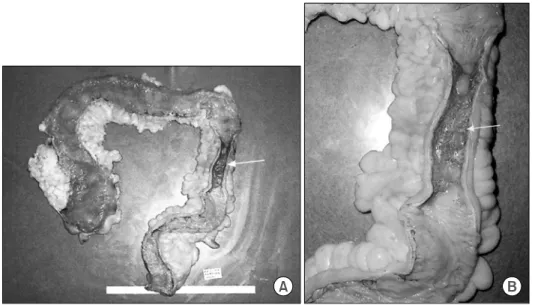
Fig. 4. (A) Cross section of total colon shows a luminal nar- rowing of sigmoid colon (arrow). (B) Cross section of sigmoid colon shows a luminal narrowing and mu- cosal erythema (arrow).
Fig. 2. Abdomen CT scan demonstrates a luminal narrowing and wall thickening of sigmoid colon, and pericolic fat infilt- ration (arrow).
Fig. 3. Colonoscopic finding shows a luminal narrowing and mucosal erythema of the sigmoid colon.
174 대한외과학회지:제 65 권 제 2 호 2003
ꠏꠏꠏꠏꠏꠏꠏꠏꠏꠏꠏꠏꠏꠏꠏꠏꠏꠏꠏꠏꠏꠏꠏꠏꠏꠏꠏꠏꠏꠏꠏꠏꠏꠏꠏꠏꠏꠏꠏꠏꠏꠏꠏꠏꠏꠏꠏꠏꠏꠏꠏꠏꠏꠏꠏꠏꠏꠏꠏꠏꠏꠏꠏꠏꠏꠏꠏꠏꠏꠏꠏꠏꠏꠏꠏꠏꠏꠏꠏꠏꠏꠏꠏꠏꠏꠏꠏꠏꠏꠏꠏꠏꠏꠏꠏꠏꠏꠏꠏꠏꠏꠏꠏꠏꠏꠏꠏꠏꠏꠏꠏꠏꠏꠏꠏ
대장염
시행 수술명: 전결장절제술
검 체: S상 결장의 협착과 종양이 장막으로 침윤된 모습 이 관찰되었다(Fig. 4).
해부병리학적 소견: 응집되어 있는 형질세포들이 관찰되 었다. 조직학적으로는 다발성 골수종과 구별이 되지 않았 다(Fig. 5).
술 후 검사: 다발성 골수종에 의한 이차적인 골수외 형질 세포종과의 감별을 위하여 혈청단백전기영동과 두개촬영 을 시행하였다. 혈청단백전기영동에서는 특이사항이 관찰
되지 않았으며, 두개촬영에서는 다발성 골수종의 특징적인 양상인 반점이 관찰되지 않았다. 이로서 다발성 골수종을 감별진단하고 원발성 골수외 형질세포종으로 진단하였다.
고 찰
골수외 형질세포종은 조직학적, 면역학적 그리고 기능적 으로 면역글로불린을 분비하는 B 세포의 증식을 특징으로 하는 종양이다. 이들은 크게 두 가지로 분류할 수 있는데 하나는 아주 드문 형태로 원발성으로 발생하는 것이고, 다 른 하나는 대부분을 차지하는 것으로 다발성 골수종에 의 해 이차적으로 발생하는 것이다.(1,3,12,13)
골수외 형질세포종의 진단기준은 다음과 같이 정의할 수 있다. 첫째, 파라프로테인요증(paraproteinuria)이 없어야 한 다. 둘째, BenceJones 단백뇨가 없어야 한다. 셋째, 골격구조 가 정상이어야 한다. 마지막으로 골수생검 소견이 정상이 어야 한다.(14)
다발성 골수종이 있으면서 높은 LDH 치, 높은 종양표지 자치, 그리고 복합적인 형질세포형을 보이는 경우 위장관 계를 침범할 확률이 높고 나쁜 예후를 나타낸다.(15) 원발성 골수외 형질세포종은 이차적으로 발생한 것에 비 해 빈혈이나 고칼슘혈증, 신부전의 빈도가 더 낮다.(7-11) 발생 위치는 간, 비장, 임파선, 신장, 피하조직, 뇌 실질 등으 로 다양하다. 그중 위장관계가 차지하는 비율은 13%에 해 당하며 위장관계 중에서는 위가 가장 많은 부분을 차지하 고 있다.(2,4-6,16) 임상양상으로 복부에 발생한 경우 복부 Fig. 5. Microscopically, aggregation of the plasma cells are shown
(arrow). There is no distinguishes from multiple myeloma (H&E, 400).
Fig. 6. Serum protein electrophoresis shows no definite M-protein.
ꠚꠚꠚꠚꠚꠚꠚꠚꠚꠚꠚꠚꠚꠚꠚꠚꠚꠚꠚꠚꠚꠚꠚꠚꠚꠚꠚꠚꠚꠚꠚꠚꠚꠚꠚꠚꠚꠚꠚꠚꠚꠚꠚꠚꠚꠚꠚꠚꠚꠚꠚꠚꠚꠚꠚ
Fraction % % Range g/dl
ꠏꠏꠏꠏꠏꠏꠏꠏꠏꠏꠏꠏꠏꠏꠏꠏꠏꠏꠏꠏꠏꠏꠏꠏꠏꠏꠏꠏꠏꠏꠏꠏꠏꠏꠏꠏꠏꠏꠏꠏꠏꠏꠏꠏꠏꠏꠏꠏꠏꠏꠏꠏꠏꠏꠏ
Albumin 56.7 3.5 5.3 2.38
Alpha 1 6.4 0.1 0.4 0.27
Alpha 2 7.5 0.4 0.9 0.32
Beta 10.1 0.6 1.0 0.42
Gamma 19.3 0.6 1.7 0.81
ꠏꠏꠏꠏꠏꠏꠏꠏꠏꠏꠏꠏꠏꠏꠏꠏꠏꠏꠏꠏꠏꠏꠏꠏꠏꠏꠏꠏꠏꠏꠏꠏꠏꠏꠏꠏꠏꠏꠏꠏꠏꠏꠏꠏꠏꠏꠏꠏꠏꠏꠏꠏꠏꠏꠏ
Total M- 5.8 8.0 4.20
ꠏꠏꠏꠏꠏꠏꠏꠏꠏꠏꠏꠏꠏꠏꠏꠏꠏꠏꠏꠏꠏꠏꠏꠏꠏꠏꠏꠏꠏꠏꠏꠏꠏꠏꠏꠏꠏꠏꠏꠏꠏꠏꠏꠏꠏꠏꠏꠏꠏꠏꠏꠏꠏꠏꠏ
Fig. 7. Skull series shows no mottling.
박용범 외:S상 결장에 발생한 원발성 형질세포종 175 ꠏꠏꠏꠏꠏꠏꠏꠏꠏꠏꠏꠏꠏꠏꠏꠏꠏꠏꠏꠏꠏꠏꠏꠏꠏꠏꠏꠏꠏꠏꠏꠏꠏꠏꠏꠏꠏꠏꠏꠏꠏꠏꠏꠏꠏꠏꠏꠏꠏꠏꠏꠏꠏꠏꠏꠏꠏꠏꠏꠏꠏꠏꠏꠏꠏꠏꠏꠏꠏꠏꠏꠏꠏꠏꠏꠏꠏꠏꠏꠏꠏꠏꠏꠏꠏꠏꠏꠏꠏꠏꠏꠏꠏꠏꠏꠏꠏꠏꠏꠏꠏꠏꠏꠏꠏꠏꠏꠏꠏꠏꠏꠏꠏꠏꠏ 불쾌감, 통증, 구토, 실혈에 의한 빈혈, 장관 폐쇄, 장 천공,
복막염 등의 증상이 나타날 수 있다.(14,16) 술 전 혈액검사 에서 혈청 γ글로불린 증가, M 단백 양성, 요중 light chain 양성 등이 나타날 수 있다.(13,14)
치료로는 이차적으로 발생한 경우 먼저 전신적인 치료가 우선이며 이에 반응을 하지 않거나 증상이 나타날 경우 장 관 절제를 시행한다. 원발성으로 발생한 경우 침범 된 장관 을 부분 절제하는 것을 원칙으로 한다.(17)
결론적으로 골수 외 형질세포종을 가진 환자는 전신질환 의 유무에 대한 검사를 위해 혈청과 소변 단백 검사를 시행 하고, 이 검사에서 M 단백이 양성으로 나타나고 방사선학 적 이상이 있거나 골수검사에서 양성의 소견이 보인다면 다발성 골수종에 의한 이차적인 골수외 형질세포종을 의심 하고 정기적인 혈청, 소변 단백 검사가 필요하다고 할 수 있다.
이번 증례의 경우 지속되는 설사와 혈변을 주소로 하는 환자를 대상으로 술 전 방사선학적 검사(바륨관장술 및 복 부 CT) 결과 S상 결장암을 의심했으나 대장내시경을 통한 조직학적 검사를 통해 궤양성 대장염으로 진단하고, 이의 치료를 위해 대장전절제술을 시행한 후 술 후 조직학적 검 사에서 S상 결장에 발생한 형질세포종으로 진단한 증례에 대한 것이다. 비록 술 전 조직학적 진단이 궤양성대장염이 었지만, 이 질환의 임상양상에 대한 지식과 술 중 병변이 장막을 침범하고 S상 결장에 국한되어 나타나는 양상을 바 탕으로 술 중 동결절편검사를 시행하였다면 대장전절제술 을 피하고 S상 결장 절제술만을 통하여 효과적인 치료를 할 수 있었으리라는 아쉬움을 가져본다.
REFERENCES
1) Gianom D, Famos M, Marugg D, Oberholzer M. Primary extramedullary plasmacytoma of the duodenum. Swiss Surg 1999;5:6-10.
2) Holland AJ, Kubacz GJ, Warren JR. Plasmacytoma of the sigmoid colon associated with a diverticular stricture. J R Coll Surg Edinb 1997;42:47-9.
3) Rappaport H. Tumors of the Hematopoietic System. Fascicle 18, First Series. Atlas of Tumor Pathology. Washington:
Armed Forces Institute of Pathology 1866.
4) Sperling RI, Fromowitz FB, Castellano TJ. Anaplastic solitary extramedullary plasmacytoma of the cecum. Report of a case confirmed by immunoperoxidase staining. Dis Colon Rectum 1987;30:894-8.
5) Ming SC. Tumors of the Esophagus and Stomach, pp. 239-240.
Fascicle 7, Second Series. Atlas of Tumor Pathology, Was- hington:Armed Forces Institute of Pathology 1973.
6) Hampton JM, Gandy JR. Plasmacytoma of the gastrointestinal tract. Ann Surg 1957;145:415-22.
7) Azar HA. Pathology of Multiple Myeloma, pp. 1-84. In:
Multiple Myeloma and Related Disorders. Azar HA, Potte M (Eds.). New York: Harper and Row 1973.
8) Fisher B, Klein DL. Metastasizing plasma cell tumor of the small bowel. Am J Gastroenterol 1975;64:371-5.
9) Gupta DN. Extramedullary plasmacytoma. Indian J Med Sci 1953;7:47-52.
10) Remigio PA, Klaum A. Extramedullary plasmacytoma of stomach. Cancer 1971;27:562-8.
11) Robinson KP. Plasmacytoma and plasma cell polyposis of the colon. Proc R Soc Med 1969;62:818-20.
12) Tomita Y, Ohsawa Hashimoto M, Qiu K, Yang Wi, Park CI, Aozasa K. Plasmacytoma of the gastrointestinal tract in Korea:
higher incidence than in Japan and Epstein-Barr virus association. Oncology 1988;55:27-32.
13) Berger F, Coiffier B, Bonneville C, Scoazec JY, Magaud JP, Bryon PA. Gastrointestinal Lymphomas. Immunohistologic study of 23 cases. Am J Clin Pathol 1987;88:707-12.
14) Fendel EH, Fazio VW. Extramedullary plasmacytoma of the small intestine. Dis Colon Rectum 1981;24:633-5.
15) Sanal SM, Yaylaci M, Mangold KA, Pantazix CG. Exten- siveextramedullary disease in myeloma. An uncommon variant with features of poor prognosis and dedifferentiation. Cancer 1996;77:1298-302.
16) Koike M, Ishiyama T, Hamano Y, Matuda I, Hisatake J, Hino K, et al. Extramedullary plasmacytoma with multiple metas- tasis following a maxillary plasmacytoma. Rinsho Ketsueki 1997;38:336-41.
17) Nielsen SM, Shenken JR, Cawley LP. Primary colonic plas- macytoma. Cancer 1972;30:261-7.