INTRODUCTION
Colonic subepithelial tumors are rare, and therefore, are not encountered as often in daily practice as are adenomas and carcinomas of the colon. Furthermore, they may not cause symptoms before attaining large size. When small in size, co- lonic subepithelial tumors are incidentally detected on a colo- noscopy or cross-sectional images. A wide variety of benign and malignant primary subepithelial tumors may arise from the colonic wall. Some tumors such as lipomas, neuroendo- crine tumors, hemangiomas, and lymphomas originate in the superficial location on the colon wall. Other tumors such as a gastrointestinal stromal tumor (GIST) arise from the deeper
part of the muscularis propria and often demonstrate an exo- enteric growth pattern (1). In this article, we present a variety of radiologic findings from benign and malignant subepithe- lial tumors of the colon, and correlate them with their corre- sponding colonoscopic and pathologic findings.
BENIGN SUBEPITHELIAL TUMORS OF THE COLON
Hemangioma
Colonic hemangiomas are uncommon benign lesions arising from the subepithelial vascular plexus, and are attributable to the embryonic sequestration of mesodermal tissue. The clini-
J Korean Soc Radiol 2011;65(3):275-284
Received June 7, 2011; Accepted July 24, 2011 Corresponding author: Eun Ju Kang, MD
Department of Diagnostic Radiology, Dong-A University College of Medicine, 1 Dongdaesin-dong 3-ga, Seo-gu, Busan 602-715, Korea.
Tel. 82-51-240-5367 Fax. 82-51-253-4931 E-mail: [email protected]
This work was supported by the Dong-A University research fund.
Copyrights © 2011 The Korean Society of Radiology
The colonoscopy has been used to diagnose various colonic lesions. However, this method has its limitations in diagnosing and differentiating subepithelial tumors.
For this reason, the role of cross-sectional radiologic imaging is important for the diagnosis of colonic subepithelial tumors. Moreover, although these tumors are as- sociated with a wide range of radiologic features, they may have unique radiologic features that suggest a specific diagnosis. Hemangiomas typically show transmural colonic wall thickening with phleboliths in intramural or extracolic areas. Colonic lymphangiomas manifest as a multilocular cystic mass at CT and sonography. Co- lonic lipomas are well demonstrated by CT because the masses were present with characteristic fatty density. Schwannomas usually appear as well circumscribed, ho- mogeneous masses with low attenuation at CT. The primary form of colonic lym- phoma has a wide variety of radiologic types, including a polypoid mass, circumfer- ential mural mass, and a cavitary mass. Small gastrointestinal stromal tumors are usually homogeneous, whereas larger tumors tend to have a heterogeneous ap- pearance with central necrosis at contrast-enhanced CT scans. Neuroendocrine tu- mors of the colon are most frequently observed in the rectum and are typically small incidental lesions. Familiarity with these imaging features can help distinguish particular disease entities.
Index terms Colon, Diseases
Colon, Subepithelial Tumors Colon, CT
Unusual Primary Subepithelial Tumors of the Colon: Multimodality Imaging Findings with Endoscopic and Pathologic Correlation
1대장의 드문 원발성 상피하 종양: 영상 소견과 내시경 및 병리 소견과의 비교1
Jong Young Oh, MD
1, Hee Jin Kwon, MD
1, Jin Han Cho, MD
1, Dong Ho Ha, MD
1, Kyung Jin Nam, MD
1, Eun Ju Kang, MD
1, Jong Hoon Lee, MD
2, Suk Kim, MD
3Departments of 1Diagnostic Radiology, 2Internal Medicine, Dong-A University College of Medicine, Busan, Korea
3Department of Diagnostic Radiology, Pusan National University College of Medicine, Busan, Korea
tions (2) (Fig. 1D). In addition, CT scans can ensure the accu- rate diagnosis, which includes inhomogeneously enhancing transmural bowel-wall thickenings containing phleboliths (Fig.
1E). On MR imaging, the colonic wall is markedly thickened with high signal intensity on T2-weighted images. This fea- ture appears to be related to the slow blood flow in hemangio- mas. T1-weighted image shows homogeneous, hypointense colonic wall thickening and MR image after a gadolinium in- jection reveals heterogeneous enhancement of the both co- lonic wall and pericolic area.
Lymphangioma
Lymphangiomas of the colon are rare benign lesions with only 331 cases reported in the world medical literature between 1931 and 2004. They are described as congenital malforma- tions of the lymphatic system and are considered to be lym- phatic hamartomas. Although the peak incidence of colonic lymphangiomas is in the seventh decade, the age distribution cal manifestations usually include acute or recurrent chronic
painless rectal bleeding in a young adult. Colonic hemangio- mas are classified as cavernous and capillary subtype tumors.
Approximately 80% of colonic hemangiomas are cavernous subtype, 50% to 70% of which are located in the rectosigmoid colon. Cavernous hemangiomas are composed of large multi- lobulated thin-walled vascular channels with no capsules, which typically have full-thickness mural involvement, often with infiltration into the surrounding connective tissue (2) (Fig. 1A). At optical colonoscopy, the lesions appear as dilated subepithelial vascular tumors, which are typically soft, rang- ing in color from deep wine to plum, and are associated with mucosal congestion and edema (Fig. 1B). In addition, these tumors collapse easily on insufflation. Moreover, the presence of phleboliths on plain abdominal radiographs is an impor- tant diagnostic clue observed in 26-50% of adult patients (Fig.
1C). Barium enemas expose nonspecific polypoid or multi- lobular annular masses that may collapse with air insuffla-
A
D
B
E
C
Fig. 1. Hemangioma of the descending colon in a 34-year-old woman.
A. Photograph of the resected specimen demonstrates a lobulated subepithelial mass with areas of focal hemorrhage and multiple phleboliths.
B. Colonoscopic image shows dilated subepithelial vascular masses with a deep wine color associated with mucosal congestion and edema.
C. Plain radiograph shows multiple phleboliths (arrows) along the descending colon.
D. Barium study reveals lobulated contour masses (arrows) with multiple phleboliths along the proximal portion of the descending colon.
E. Contrast-enhanced CT scan reveals circumferential mural thickening with multiple phleboliths in the descending colon (arrows).
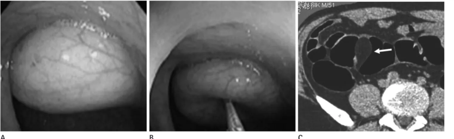
pomas may cause abdominal pain, diarrhea, constipation, hemorrhage, and intussusception (4). Lipomas are composed of well-differentiated adipose tissue originating in the submu- cosa. At optical colonoscopy, the classic characteristics of lipo- mas include a yellowish pale color and soft to the touch when probing with a blunt instrument (Fig. 3A, B). A Barium ene- ma shows a well-defined, smooth intraluminal filling defect, as well as a change in shape under peristalsis (squeeze sign).
These findings are suggestive of lipomas, but do not provide the conclusive evidence for a correct diagnosis. Colonic lipo- mas are well demonstrated by CT because the mass has a typ- ical fat density between -70 and -120 HU (4) (Fig. 3C). On MRI, the typical features of lipomas are a fatty mass with increased signal intensity in the T1-weighted image and intermediately intense to hyperintense in the T2-weighted fast spin-echo im- age relative to muscle. Fat saturation and chemical shift imag- ings are useful imaging techniques for differentiating lipoma from non-fatty masses (4). If a patient is asymptomatic, treat- ment is not necessary; however, endoscopic removal or surgical resection may be required for a large complicated lipoma.
Schwannoma
Schwannomas of the colon are extremely rare with only a small number of cases having been reported in the literature, with average age at presentation of 65 years. Colonic shawan- nomas have been reported, in descending order of frequency, in the cecum, sigmoid, rectosigmoid junction, transverse colon, descending colon, and rectum. Schwannoma usually grow slow- did not demonstrate that the disease is related to aging. Clini-
cal symptoms are not specific and include mild to intense ab- dominal pain, altered bowel habitus, and rectal bleeding. The occurrence of intussusception caused by a colonic lymphan- gioma is extremely rare, with an incidence of only 3 cases out of 79 (4%). Lymphangiomas are masses consisting of endo- thelial cells and supporting connective tissue that are classi- fied into three categories: simple, cavernous, and cystic. More- over, round cells, islands of fat cells and smooth muscle are usually present. The colonoscopic appearance suggests that the presence of a lymphangioma is in the form of a round and smooth, broad based, pinkish color, translucent, tension, and lustrous surface (Fig. 2A). Lymphangiomas show changes in shape caused by peristalsis, compression, and patient posi- tion. These lesions are seen as sharply marginated, oval, or round colonic defects on a barium study. They usually are pli- able, readily changing in shape in response to compression or peristaltic activity during a barium study. CT and sonography properly demonstrate the multilocular cystic mass with inter- nal septa (3) (Fig. 2B, C). Segmental resection seems to be the treatment of choice, except for pedunculated or small lymph- angiomas.
Lipoma
Colonic lipomas are relatively rare and most commonly oc- cur in the nonepithelial submucosal layer of the colon. The most common location for colonic lipomas is the cecum or ascending colon. Most lipomas are asymptomatic, but large li-
A B C
Fig. 2. Lymphangioma of the colon in a 33-year-old woman.
A. Photography from endoscopy reveals a pedunculated subepithelial mass covered with erythematous mucosa in the sigmoid colon.
B. Contrast-enhanced CT scan shows a well-defined low attenuated cystic mass in the sigmoid colon (arrow).
C. Transabdominal sonogram shows an anechoic cystic mass in the sigmoid colon (arrow).
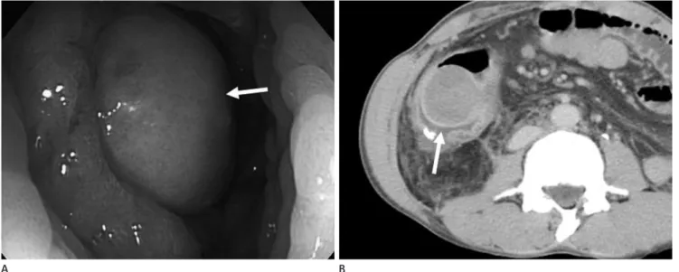
this tumor has imaging characteristics similar to those of co- lonic GISTs. A Barium study demonstrates the presence of an intraluminal or subepithelial mass, possibly with ulceration of the overlying mucosa. Further, schwannoma manifest as a non- specific soft tissue attenuated mass at CT (Fig. 4). However, the lack of hemorrhage, necrosis, and degeneration at CT was believed to be helpful in distinguishing schwannomas from GISTs, where these findings typically occur (5).
Inflammatory Fibroid Polyp
Inflammatory fibroid polyp (IFP) is an uncommon benign polypoid lesion of the gastrointestinal tract. Most cases of IFP are located in the stomach and small bowel, but a colonic oc- currence is rare. Most colonic IFPs tend to be found in the right colon. The clinical manifestation may rely on the gross mor- phology of the lesion and its location in the gastrointestinal tract. The major clinical symptoms include abdominal pain, bloody stools, weight loss, diarrhea, and anemia. A large IFP may cause intestinal obstruction or intussusception. Macro- scopically, an IFP is a smooth sessile or pedunculated polyp.
Microscopically, it is composed of loose connective tissue with abundant inflammatory cells including plasma cells and eosinophils. Immunohistochemically, the stromal cells of IFPs are often CD34-immunoreactive, but do not stain for KIT. En- doscopically, IFPs appear to have a smooth sessile or peduncu- lated configuration with most cases having erythematous or ulcerative mucosa (Fig. 5A). A Barium enema reveals the pe- dunculated or sessile mass, but with no distinct radiologic ly, so most patients are asymptomatic at the time of diagnosis.
Large schwannomas may cause latent or acute bleeding, bow- el obstruction, constipation, and abdominal pain. Macroscop- ically, schwannomas appear as a spherical, solid, and well-en- capsulated tumor that is gray in color. Mucosal ulceration is often visible, but necrosis and hemorrhage are not typical fea- tures. Histopathologically, schwannomas are composed of elon- gated bipolar spindle cells with zonally variable cellularity and a focally prominent nuclear palisading pattern. Immunohisto- chemistrically, schwannomas are strongly positive for S-100 and are constantly negative for CD117 (KIT), which differs from GISTs. The combination of a positive S-100 and negative KIT establishes the diagnosis of schwannomas. Radiographically,
A B C
Fig. 3. Lipoma of the sigmoid colon in a 51-year-old man.
A. Colonoscopic image shows a yellowish subepithelial mass.
B. The mass reveals a cushion sign at compression with a blunt instrument.
C. Axial CT scan shows a well-defined fatty mass in the sigmoid colon (arrow).
Fig. 4. Schwannoma of the cecum in a 44-year-old man. Contrast- enhanced CT scan shows a homogeneously enhancing, exoenteric growing mass (arrow) in the posterior wall of the cecum.
nal pain and weight loss. Obstruction is rare because it does not elicit a desmoplastic response, and subepithelial lymphoid infiltration weakens the muscularis propria of the wall (7).
Nearly all occurrences of primary lymphoma of the large bowel are non-Hodgkin B-cell lymphomas. Imaging manifestations of colonic lymphomas included polypoid masses, which oc- curred most frequently near the ileocecal valve (Fig. 6); 2) cir- cumferential infiltration (Fig. 7); 3) a cavitary mass excavat- ing into the mesentery; 4) endoexoenteric tumors; 5) mucosal nodularity; and 6) fold thickening. Polypoid lesions may be predisposed to experience intussusception, have homogeneous intermediate signal intensity on T1-weighted MR images, and heterogeneous high signal intensity on T2-weighted images.
Mild to moderate enhancement is seen after the intravenous administration of gadolinium-based contrast material (7).
Treatment usually consists of surgical resection followed by adjuvant chemotherapy.
Gastrointestinal Stromal Tumor
GISTs of the colon are extremely rare, consisting of only about 0.1% of colon and rectal tumors. The most common site of colonic GISTs is the anorectum. GI bleeding, constipa- tion, rectal or pelvic pain, obstruction, and a mass on examina- tion are the most common initial symptoms. GISTs are typical- ly well circumscribed masses without a true capsule. Moreover, they exhibit a propensity for exophytic growth because they features. Contrast-enhanced CT scan shows low attenuated,
homogeneous intraluminal or intramural mass, but there is no unique CT finding that will aid in the diagnosis of an IFP (6) (Fig. 5B). For the treatment of symptomatic IFPs, surgical resection has been performed in most cases. However, in the case of small pedunculated lesions, treatment can be adminis- tered in the form of an endoscopic polypectomy.
MALIGNANT SUBEPITHELIAL TUMORS OF THE COLON
Primary Colorectal Lymphoma
Primary lymphoma of the large bowel accounts for 0.4% of all tumors of the colon, and colorectal lymphomas constitute 6-12% of gastrointestinal lymphomas. Dawson’s diagnostic criteria for primary intestinal lymphomas require the absence of lymphadenopathy, normal chest radiography, normal com- plete blood count, predominance of the bowel lesion at lapa- rotomy, and the absence of tumor in the liver and spleen (7).
Most cases consisted of males in the 5th to 7th decades. Im- munosuppression and inflammatory bowel disease are known to be associated with the development of primary colorectal lymphomas. The most common location for the occurrence of primary colorectal lymphoma is the cecum, followed by the rectum. The clinical manifestations are often nonspecific;
however, the two most common symptoms include abdomi-
A B
Fig. 5. Inflammatory fibroid polyp of the ascending colon in a 35-year-old man.
A. Photograph from endoscopy reveals a subepithelial mass with a smooth mucosal surface (arrow).
B. Contrast-enhanced CT shows a well-defined, homogeneously low attenuated mass with intact overlying mucosa (arrow) in the ascending colon.
of mitoses (8). These guidelines are now being updated after the publication of very large studies by Miettinen and colleagues who found that small bowel or colonic GISTs show a much higher rate of aggressive behavior than gastric GISTs. Accord- ing to updated guideline, tumors greater than 2 cm with greater than 5 mitoses per 50 HPF or a size greater than 10 cm with any mitotic rate, indicates a high risk for malignancy in colonic GISTs. Colorectal GISTs may have a broad based appearance that is subtle and nonspecific at colonoscopy because they tend to exhibit exoenteric growth and only rarely demonstrate a prominent intraluminal component (9). Most lesions are shown usually involve the outer muscular layer (8). Focal areas of
hemorrhage, cystic degeneration, and necrosis may occur, particularly in large lesions (Fig. 8A). GISTs can be histologi- cally classified by their predominant cell morphology, consist- ing of either a spindle cell or epithelioid (8). Immunohisto- chemically, GISTs are positive for KIT (CD117), and as a result, a definite diagnosis of GISTs now relies upon a tumor’s posi- tive expression of KIT. The most commonly cited characteris- tics indicative of a malignancy include tumor size and mitotic activity (8). The GIST Workshop created a consensus recom- mendation of risk assessment based on tumor size and number C
A
D B
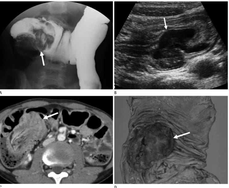
Fig. 6. Polypoid primary lymphoma of the cecum in an 18-year-old man.
A. Barium study reveals a lobulated filling defect with a “coiled spring” appearance in the hepatic flexure of the colon (arrow).
B. Transabdominal sonogram shows a well-defined hypoechoic mass (arrow) with intussusception of the ascending colon.
C. Corresponding contrast-enhanced CT scan shows the inhomogeneously enhancing mass combined with intussusception within the ascending colon.
D. Photograph of the resected specimen shows a well-demarcated fungating mass with focal ulceration in the cecum.
hanced CT imaging (Fig. 8B) and it usually has not undergone a lymphadenopathy. MRI findings of GISTs show low-interme- diate signal intensity on T1-weighted images and heteroge- neous high signal intensity on T2-weighted images (Fig. 8C, D).
as extraluminal or intramural masses at CT. Smaller tumors are usually homogeneous, whereas larger tumors tend to have a heterogeneous appearance with necrosis or hemorrhage.
Prominent enhancement of a tumor is typical on contrast- en-
A B C
Fig. 7. Circumferential infiltrative primary lymphoma of the rectum in a 29-year-old man.
A. Pre-contrast CT scan shows low attenuated circumferential mural thickening of the rectum.
B. Contrast-enhanced CT scan reveals poor contrast enhancement of the lesion.
C. Barium study demonstrates segmental luminal narrowing with lobulated contour defect (arrows) in the rectum.
A
D
B
E
C
Fig. 8. Gastrointestinal stromal tumor of the rectum in a 56-year-old man.
A. Photograph of the resected specimen shows intramural mass with intact overlying mucosa in the rectal wall.
B. Contrast-enhanced CT scan shows an exoenteric growing mass with inhomogeneous enhancement in the anterior wall of the rectum.
C. T1-weighted axial image.
D. T2-weighted sagittal image.
E. Gd-enhanced fat suppressed T1-weighted image demonstrate a well-defined exoenteric subepithelial mass with heterogeneous enhancement (arrows).
logically, serotonin-producing EC-cell neuroendocrine tu- mors of the cecum mimic cecal adenocarcinomas in that they are frequently large, polypoid, or ulcerating masses. L-cell rectal carcinoids are usually solitary, small, subepithelial nod- ules or have focal areas of plaque-like thickening. Neuroen- docrine tumors of the proximal colon are polypoid intralumi- nal masses that are indistinguishable from adenocarcinoma at CT. Rectal carcinoids are commonly seen as small mural or polypoid masses at CT (10) (Fig. 9B, C).
Other Primary Malignant Subepithelial Tumors of the Colon
Malignant subepithelial tumors of the colon, other than GISTs, include leiomyosarcomas and malignant schwanno- mas. Leiomyosarcomas are extremely rare and are often large and ulcerative by the time they are detected (1) (Fig. 10). They arise from smooth muscle cells of the colonic wall and sup- pose less than 1% of the malignant colorectal tumors. Present- ing symptoms and endoscopic findings may be non-specific.
On immunohistochemical study, the c-KIT determination is negative unlike the GISTs, whereas they are positive for actin, vimentin and desmin. They are aggressive tumors with a high local recurrence rate as well as having significant hematoge- neous spread, the liver being the most affected organ.
CONCLUSION
Subepithelial tumors of the colon represent a wide range of benign and malignant tumors, some of which have similar ra- Furthermore, GISTs show intense, heterogeneous enhance-
ment (9) (Fig. 8E). Surgical resection is the treatment of choice for colonic GISTs. However, recurring and metastatic tumors of the peritoneum or liver can effectively be treated with ima- tinib mesylate (8).
Neuroendocrine Tumor
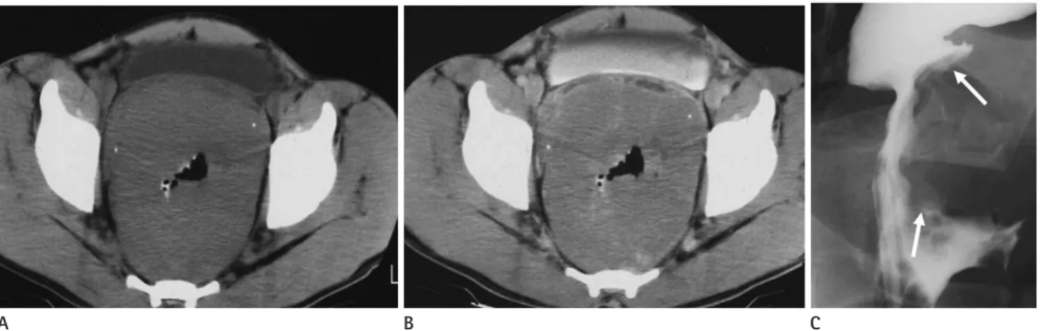
Neuroendocrine tumors of the colon are uncommon, with most cases arising in the rectum and fewer cases occurring in the cecum. Abdominal pain and weight loss are typical symp- toms for neuroendocrine tumors of the proximal colon, where- as more than 50% of patients with rectal neuroendocrine tu- mors are asymptomatic and the lesions are discovered at routine rectal examination or screening colonoscopy (Fig. 9A). Patho-
A B C
Fig. 9. Neuroendocrine tumor of the rectum in a 55-year-old woman.
A. Colonoscopic image reveals a small subepithelial mass (arrow) with intact overlying mucosa in the lower rectum.
Axial (B) and coronal (C) reformatted CT images show a small well-enhancing subepithelial mass (arrows) in the lower rectum.
Fig. 10. Leiomyosarcoma of the rectum in a 64-year-old man. On contrast-enhanced CT scan, a well-circumscribed, moderately enhanc- ing mural mass with focal necrosis is noted in the rectum.
findings of asymptomatic intra-abdominal gastrointesti- nal system lipomas. J Comput Assist Tomogr 2008;32:841- 847
5. Levy AD, Quiles AM, Miettinen M, Sobin LH. Gastrointesti- nal schwannomas: CT features with clinicopathologic cor- relation. AJR Am J Roentgenol 2005;184:797-802
6. Harned RK, Buck JL, Shekitka KM. Inflammatory fibroid polyps of the gastrointestinal tract: radiologic evaluation.
Radiology 1992;182:863-866
7. Ghai S, Pattison J, Ghai S, O’Malley ME, Khalili K, Stephens M. Primary gastrointestinal lymphoma: spectrum of imag- ing findings with pathologic correlation. Radiographics 2007;27:1371-1388
8. Levy AD, Remotti HE, Thompson WM, Sobin LH, Miettinen M. Gastrointestinal stromal tumors: radiologic features with pathologic correlation. Radiographics 2003;23:283- 304, 456; quiz 532
9. Levy AD, Remotti HE, Thompson WM, Sobin LH, Miettinen M. Anorectal gastrointestinal stromal tumors: CT and MR imaging features with clinical and pathologic correlation.
AJR Am J Roentgenol 2003;180:1607-1612
10. Levy AD, Sobin LH. From the archives of the AFIP: Gastro- intestinal carcinoids: imaging features with clinicopatho- logic comparison. Radiographics 2007;27:237-257 diologic features that make differentiation difficult. However,
despite overlaps in the radiologic findings, some tumors have characteristic radiologic features that may suggest a specific diagnosis. Therefore, it is important that radiologists are fa- miliar with tumors that have unique radiologic findings. Last- ly, awareness of the various radiologic findings in colonic sub- epithelial tumors can help ensure a correct diagnosis and proper management of the condition.
REFERENCES
1. Pickhardt PJ, Kim DH, Menias CO, Gopal DV, Arluk GM, Heise CP. Evaluation of submucosal lesions of the large in- testine: part 1. Neoplasms. Radiographics 2007;27:1681- 1692
2. Hsu RM, Horton KM, Fishman EK. Diffuse cavernous hem- angiomatosis of the colon: findings on three-dimensional CT colonography. AJR Am J Roentgenol 2002;179:1042- 1044
3. Wan YL, Lee TY, Hung CF, Ng KK. Ultrasound and CT find- ings of a cecal lymphangioma presenting with intussus- ception. Eur J Radiol 1998;27:77-79
4. Genchellac H, Demir MK, Ozdemir H, Unlu E, Temizoz O.
Computed tomographic and magnetic resonance imaging
대장의 드문 원발성 상피하 종양: 영상 소견과 내시경 및 병리 소견과의 비교1
오종영
1· 권희진
1· 조진한
1· 하동호
1· 남경진
1· 강은주
1· 이종훈
2· 김 석
3대장 내시경은 다양한 대장 질환의 진단에 사용되고 있으나 상피하 종양의 진단과 감별에는 제한적이다. 이러한 이유로 대장 상피하 종양의 진단에 영상의학적 단면 영상들이 중요한 역할을 할 수 있다. 상피하 종양들은 다양한 영상 소견을 보 여 감별진단이 어려운 경우도 있지만 일부 종양은 특징적인 영상 소견을 보여 영상 소견만으로도 진단이 가능하다. 혈관종 은 CT에서 두꺼워진 대장 벽 혹은 대장 주위로 특징적인 정맥돌이 보일 경우 진단이 가능하며, 림프종은 초음파나 CT에 서 대장 벽 내에 격막을 가진 낭성 종괴 형태로 나타난다. 지방종은 CT상 전형적인 지방 음영을 보일 경우 진단이 가능하 며, 신경초종은 경계가 좋고 균질한 저음영의 조영 증강을 보이는 종괴로 나타난다. 대장의 일차성 림프종은 다양한 영상 소견을 보이는데 융기형 종괴, 원주형 종괴 및 공동형의 종괴로 나타난다. 위장관 간질종양은 크기가 작을 경우 CT상 균 질한 조영 증강을 보이나 크기가 큰 경우에는 내부 괴사 등으로 인해 비 균질한 조영 증강 소견을 보인다. 대장의 신경내 분비종양은 대개 크기가 작아 우연히 발견되는 경우가 많으며 직장에서 흔히 발생한다. 이러한 영상 소견들에 대한 숙지 는 대장의 상피하 종양의 감별진단에 도움이 되리라 생각한다.
동아대학교 의과대학 1영상의학과학교실, 2내과학교실, 3부산대학교 의과대학 영상의학과학교실