submit.radiology.or.kr
대한영상의학회지 2012;67(2):117-120117 서론
아밀로이드증은 아밀로이드 단백에서 기원한 아밀로이드 섬 유의 침착으로 조직기능과 장기에 손상을 일으키는 질환이다.
다양한 장기에 전신적 혹은 국소적으로 생길 수 있으며, 그 중 방광의 국소적 아밀로이드증은 남성과 여성 간의 비율이 동일 하고 40대와 50대 연령에 주로 호발한다(1). 또한 주로 전신 성으로 발현하나 약 10~20% 가량에서 국소적 형태로 관찰된 다. 그 중 방광의 국소적 아밀로이드증의 경우 임상적, 영상학 적으로 요로상피세포암과 유사한 양상을 보인다(2).
방광의 아밀로이드증은 약 100예가 조금 넘는 수의 보고가 있다(3). 그러나 대부분 병리학적, 임상적 양상을 기초로 한 보 고이며, 영상학적 소견을 포함한 예는 드물다.
이에 저자들은 방광의 요로상피세포암과 유사한 CT 소견을 보인 국소적 원발성 아밀로이드증 1예를 경험하여 영상소견, 문헌고찰과 함께 보고하고자 한다.
증례 보고
65세 남자 환자가 내원 2주 전부터 시작된 지속적인 육안적
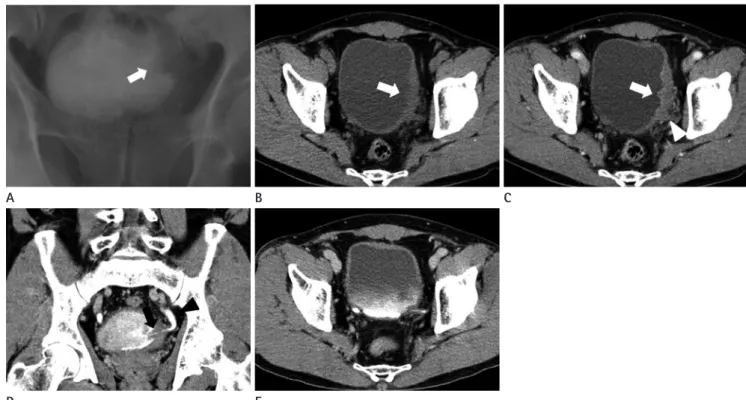
혈뇨를 주소로 타 병원에서 시행한 경정맥요로조영술에서 방 광 좌측 벽의 불규칙 음영이 발견되어 추가적인 검사 및 치료를 위해 본원으로 전원되었다(Fig. 1A).
환자는 과거력상 암이나 외상, 고혈압, 당뇨 등의 병력은 없 었으며, 이학적 검사와 일반적인 혈액학적 검사에서도 이상소 견은 없었다. 또한 내원하여 시행한 소변 검사에서 육안적 혈뇨 를 보였다. 내원 후 시행한 조영 전 CT에서는 방광 좌측 벽에 불규칙한 변연을 보이는 국소적인 방광벽의 비후가 관찰되며, 내부에 석회화는 관찰되지 않았다(Fig. 1B). 조영 후 신조영기 CT 영상에서 병변부의 비후된 방광벽은 변연이 불규칙하며, 주변의 정상적인 방광벽과 비슷한 정도로 조영증강되었고, 방 광 주변 지방음영의 증가가 있어 방광 주위 침범이 의심되었다 (Fig. 1C). 조영 후 배설기 영상에서 좌측 요관방광이음부 (ureterovesical junction)와 원위부 요도벽 비후와 내강의 감소 가 관찰되었다. 또한 그보다 근위부의 좌측 요도에는 경도의 요도확장이 동반되었다(Fig. 1D). 이외의 복강 내 장기와 양측 폐 하부에는 특이소견이 없었다. 이와 같은 소견을 종합하여 방 광 주위 침범을 동반한 방광의 미만성 요로상피세포암을 우선 적으로 진단하였다.
경요도방광종양절제술을 시행하여 방광 좌측 벽의 미만성 고
Case Report
pISSN 1738-2637
J Korean Soc Radiol 2012;67(2):117-120
Received March 26, 2012; Accepted June 23, 2012 Corresponding author: See Hyung Kim, MD Department of Radiology, Keimyung University College of Medicine, Dongsan Hospital, 56 Dalseong-ro, Jung-gu, Daegu 700-712, Korea.
Tel. 82-53-250-7767 Fax. 82-53-250-7766 E-mail: [email protected]
Copyrights © 2012 The Korean Society of Radiology
Primary localized amyloidosis is a rare disease that impairs the organs and tissue function, caused by extracellular deposition of amyloid protein. Although amyloid proteins can be deposited in localized or systemic organs, the bladder is a rare site. CT showed diffuse infiltrative mass in the left urinary bladder wall, and the left uretero- vesical junction with enhancement and perivesical invasion, which mimics urothelial carcinoma. However, pathological diagnosis was localized primary amyloidosis. We re- port here on the CT findings of a rare case of primary localized amyloidosis of the uri- nary bladder with brief review of the literature.
Index terms
Amyloidosis Urinary Bladder Computed TomographyCT Findings of Primary Localized Amyloidosis of the Urinary Bladder: A Case Report
1방광에 국소적으로 호발한 원발성 아밀로이드증의 CT 소견: 증례 보고
1Young Hwan Kim, MD
1, See Hyung Kim, MD
1, Sang Pyo Kim, MD
2, Mi Jeong Kim, MD
1Departments of 1Radiology, 2Pathology, Keimyung University College of Medicine, Dongsan Hospital, Daegu, Korea
방광에 국소적으로 호발한 원발성 아밀로이드증의 CT 소견
submit.radiology.or.kr
대한영상의학회지 2012;67(2):117-120
118
methyl sulfoxide therapy) 3개월 후 시행한 추적 CT 영상에서 좌측 방광벽과 요관방광이음부의 비후는 모두 호전되었다 (Fig. 1E).
고찰
아밀로이드증은 선행 요인에 따라 원발성 및 속발성으로 분 형종괴를 확인하였고, 그 일부를 절제하였다. 현미경적 소견에
서 Brunn’s nest 주위로 경도의 림프구성 침범과 호산성 물질이 관찰되었으며(Fig. 2A), 이 호산성 물질은 콩고레드 염색(Con- go-red stain) 후 편광현미경상에서 이중굴절 양성의 연녹색 (apple-green birefringence)으로 보여 아밀로이드증으로 진단하 였다(Fig. 2B).
수술 후 방광 내 다이메틸설폭시화물 치료(intravesical di- D
A B
A
E
B C
Fig. 1. Intravenous pyelogram and CT. Intravenous pyelogram (A) reveals diffuse ill-defined filling defect and wall irregularity (arrow) at the left wall of the urinary bladder. Non-enhanced axial CT (B) scan shows irregular focal wall thickening (arrow) without calcification in the left wall of the urinary bladder. On the contrast enhanced axial CT (C) scan during nephrographic phase, the wall thickening shows weak enhancement (ar- row), there reveals wall irregularity with perivesical irregular density (arrowhead). Coronal image during the excretory phase (D) shows wall thickening of the left ureterovesical junction (black arrow). Further, there shows a mild ipsilateral hydroureter (black arrowhead). After 3 months later, contrast enhanced CT (E) scan shows disappearing of the focal wall thickening in the urinary bladder and the left uterovesical junction.
Fig. 2. Microscopic features. Microscopic examination (A) shows an amorphous eosinophilic materials (arrows) and mild lymphocytic infiltrates around the Brunn’s nests (Hematoxilin and Eosin, × 200). Congo-red stain (B) shows strong apple-green birefringences (arrows) under the polar- izing microscopy (Hematoxilin and Eosin, × 200).
김영환 외
submit.radiology.or.kr
대한영상의학회지 2012;67(2):117-120119
외에 미만성 벽 비후를 보이는 출혈성 방광염, 미만성 비후나 종괴의 형태를 나타내는 방광의 림프종 등을 생각할 수 있다.
본 증례에서는 CT에서 전반적으로 주변 조직과 비교하여 약하 게 조영증강되어 종양의 가능성이 낮아 보였으나, 방광 좌측 벽과 좌측 요관방광이음부가 국소적으로 비후되어 있었고 방 광주변 조직을 침범하고 있어 침습적 악성 종양과의 감별이 어 려웠다. 이러한 영상소견의 아밀로이드증의 경우 영상학적 감 별에 어려움이 있으나, 치료와 예후에 있어 임상적으로 중요하 므로 영상의학적 감별진단의 하나로 생각해 볼 수 있어 증례로 보고하고자 한다.
참고문헌
1. Wilkinson M, Fanning DM, Flood H. Primary bladder amy- loidosis. BMJ Case Rep 2011:2011
2. Kawashima A, Alleman WG, Takahashi N, Kim B, King BF Jr, LeRoy AJ. Imaging evaluation of amyloidosis of the uri- nary tract and retroperitoneum. Radiographics 2011;31:
1569-1582
3. Ng CS, Wan S, Yim AP, Vale J. Idiopathic localised bladder amyloidosis: rare cause of haematuria. Int Urol Nephrol 2002;34:55-58
4. Merlini G, Westermark P. The systemic amyloidoses: clearer understanding of the molecular mechanisms offers hope for more effective therapies. J Intern Med 2004;255:159- 178
5. Urban BA, Fishman EK, Goldman SM, Scott WW Jr, Jones B, Humphrey RL, et al. CT evaluation of amyloidosis: spec- trum of disease. Radiographics 1993;13:1295-1308 류되고, 아밀로이드가 분포된 조직의 범위에 따라 전신성과 국
소성으로 분류할 수 있다. 또한 침착된 아밀로이드의 구성 단백 에 따라 amyloid immunoglobulin light chain amyloidosis (이 하 AL), serum amyloid A amyloidosis (이하 AA), transthyre- tin amyloidosis 등의 아밀로이드증으로 분류한다. 흔한 아밀로 이드증의 형태는 형질세포질환(plasma cell dyscrasia)으로 인 한 단일클론 면역글로불린 경쇄단백(monoclonal immuno- globulin light-chain protein)의 침착(AL)과 류마티스 관절염 (rheumatoid arthritis), 강직성 척수염(ankylosing spondyli- tis), 건선성 관절염(psoriatic arthritis)과 같은 급성, 만성 염증 동안 생성된 급성기 단백(acute-phase protein)의 침착(AA)이 있다(4).
방광의 국소성 아밀로이드증의 임상양상은 무통성 혈뇨로, 임상적으로 악성 종양과 구분되는 특징은 없다. 아밀로이드 침 착은 황색반(yellow plaque)부터 다수의 궤양성 용종과 방광벽 의 불규칙한 미만성 비후에 이르기까지 다양하기 때문에 진단 에 조직학적 검사는 필수적이다(1).
방광 아밀로이드증의 CT 소견으로 국소적, 혹은 미만성 벽 비후, 종괴로 인한 방광 내 조영제의 충만 결손 등이 나타날 수 있어 영상학적으로 방광의 악성 종양이나 염증성 병변과 감별 하기 어렵다(5). 방광 아밀로이드증은 특징적으로 점막하 혹은 근육층 내 석회화를 보일 수 있어, CT에서 발견된 석회화가 유 일하게 아밀로이드증의 가능성을 시사하는 소견이 되기도 한 다(2). MRI에서는 T2-강조영상에서 저신호 강도를 보여 T2-강조영상에서 고신호 강도를 보이는 요로상피세포암, 염 증성 병변과의 감별에 유용하나, 결합조직형성전이(desmo- plastic metastasis)나 림프종성 침범 또한 비슷한 소견을 보인 다. 아밀로이드증은 특징적으로 약한 조영증강을 보이나, 조영 증강의 정도는 다양할 수 있다(2). 감별진단으로 악성 종양 이
방광에 국소적으로 호발한 원발성 아밀로이드증의 CT 소견
submit.radiology.or.kr
대한영상의학회지 2012;67(2):117-120
120
방광에 국소적으로 호발한 원발성 아밀로이드증의 CT 소견:
증례 보고
1김영환
1· 김시형
1· 김상표
2· 김미정
1아밀로이드증은 장기나 조직의 세포외 공간에 아밀로이드 단백의 침착으로 조직기능의 손상을 일으키는 질병이다. 아밀 로이드 원섬유들은 국소적 혹은 전신 장기에 침착할 수 있으며, 드물게 방광의 국소적 침착도 일으킬 수 있다. CT에서 방 광의 좌측 벽과 요관방광이음부에 국소적인 방광벽의 비후와 조영증강 및 방광주위 조직의 침윤이 관찰되어 요로상피세 포암과 유사한 소견을 보였으나, 병리학적 소견에서 아밀로이드증으로 진단되었다. 이에 저자들은 국소적 원발성 아밀로 이드증 1예의 CT 소견을 간단한 문헌고찰과 함께 보고하고자 한다.
계명대학교 의과대학 동산의료원 1영상의학과, 2병리과