Korean J Gastroenterol Vol. 58 No. 5, 284-287 http://dx.doi.org/10.4166/kjg.2011.58.5.284
CASE REPORT
Korean J Gastroenterol, Vol. 58 No. 5, November 2011 www.kjg.or.kr
결장경 시행 중 발견된 장 게실염에 의한 방광-S결장루
정용욱, 유정현, 이정수, 장병익, 김경옥, 정상훈
1영남대학교 의과대학 내과학교실, 외과학교실1
Sigmoidovesical Fistula Caused by Diverticulitis Detected with Sigmoidoscopy
Yong Wook Jung, Jung Hyun Yoo, Jung Soo Lee, Byung Ik Jang, Kyeong Ok Kim and Sang Hun Jung1 Departments of Internal Medicine and Surgery1, Yeungnam University College of Medicine, Daegu, Korea
Enterovesical fistular is an abnormal communication between the intestine and the bladder. It represents a rare complication of intestinal diverticulitis, colorectal malignancy, bladder cancer, inflammatory bowel disease, radiotherapy, and trauma. The most common etiology is diverticular disease. A 70-year-old man came to our hospital due to frequent urinary tract infection, dysuria, pneumaturia and fecaluria. Sigmoidoscopy revealed a large diverticulum with impacted stool at the sigmoid colon.
When the scope was inserted into the site, the patient complained of severe urgency and pneumaturia. CT scan was performed.
1.5 cm sized fistular tract between the sigmoid colon and bladder was noted. According to the endoscopy and CT finding, the diagnosis of colovesical fistula was made. The patient underwent surgical intervention. At laparotomy, there were multiple diverticula and fistular tract was noted. (Korean J Gastroenterol 2011;58:284-287)
Key Words: Enterovesical fistula; Diverticulitis; Sigmoidoscopy
Received September 3, 2010. Revised October 18, 2010. Accepted November 10, 2010.
CC This is an open access article distributed under the terms of the Creative Commons Attribution Non-Commercial License (http://creativecommons.org/licenses/
by-nc/3.0) which permits unrestricted non-commercial use, distribution, and reproduction in any medium, provided the original work is properly cited.
교신저자: 장병익, 705-717, 대구시 남구 대명동 317-1 영남대학교 의과대학 내과학교실
Correspondence to: Byung Ik Jang, Department of Internal Medicine, Yeungnam University College of Medicine, 371-1, Daemyeong-dong, Nam-gu, Daegu 705-717, Korea. Tel: +82-53-620-3830, Fax: +82-53-654-8386, E-mail: [email protected]
Financial support: None. Conflict of interest: None.
서 론
방광-장루(enterovesical fistula)는 장관과 방광 사이의 비 정상적인 교통을 말하며, 서구에 비하여 동양에서는 매우 드 문 질환으로 장 게실염, 대장암, 방광암, 염증상 장질환 등이 그 원인으로 알려져 있다.1 그 중 게실염이 50-70%로 가장 흔한 원인으로 알려져 있고, 명확하게 밝혀져 있지는 않지만 게실염을 가진 환자에게서 방광-장루의 빈도는 1-4% 정도로 보고된다.2-8 저자들은 지속적인 요로 감염, 기뇨(pneumatu- ria), 분뇨(fecaluria)를 주소로 내원한 환자에서 시행한 결장 경에서 S결장에 분변이 박혀있는 게실이 관찰되고, 검사 중 심한 요의를 호소하면서 기뇨가 관찰되어 방광-S결장루(sig- moidovesical fistula)를 의심하고, 추가적으로 시행한 컴퓨 터 전산화단층촬영에서 방광과 S결장 사이에 누공을 확인하 여 수술적 치료를 한 1예를 경험하였기에 보고하는 바이다.
증 례
70세 남자가 2달 전부터 심해진 빈뇨, 배뇨통과 간헐적인 기뇨를 주소로 비뇨기과로 내원하였다. 환자는 혼탁한 분변이 섞인 소변을 보았으며, 수년 전부터 배뇨곤란과 빈뇨 등의 증 상과 간헐적인 하복부 통증이 있었고, 반복적인 요로 감염과 전립선 비대증으로 비뇨기과에서 치료 중이었다. 과거력에서 4년 전부터 심방 세동, 확장성 심근병증, 전립선 비대증으로 약물 치료 중이었고, 지속적인 요로 감염으로 입원을 반복하 였다. 사회병력에서 주 2-3회의 음주 외에 특이사항은 없었 다.
내원 당시 혈압은 100/60 mmHg, 맥박수 70회/분, 호흡수 20회/분, 체온은 37oC였고, 신체 검사에서는 직장 수지 검사 에서 전립선이 중등도로 커져있는 것 외에 열감이나 압통은 없었다.
Jung YW, et al. Vesicosigmoidal Fistular
285
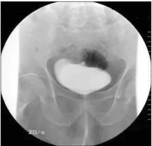
Vol. 58 No. 5, November 2011 Fig. 1. Cystogram finding. About 300 cc of contrast media was filled
in the urinary bladder and no contrast leakage was seen. The margin
of the urinary bladder was relatively regular. Fig. 2. Sigmoidoscopic finding. At the sigmoid colon-25 cm from anal verge, a large diverticulum with impacted stool was noted.
Fig. 3. CT scan finding. (A) There was about 1.5 cm sized fistular tract between the sigmoid colon and posterior wall of the urinary bladder (white arrow). Perilesional bladder wall thickening and fat infiltrations were also noted. (B) Multiple diverti- cula were seen in the sigmoid colon (white arrows).
일반혈액검사에서 백혈구 13,370/mm3, 혈색소 11.2 g/dL, 혈소판 235,000/mm3이었다. 혈액생화학검사에서 혈액요소 질소 33.05 mg/dL, 크레아티닌 2.88 mg/dL, 총 단백 5.38 g/dL, 알부민 3.76 g/dL, 총빌리루빈 0.81 mg/dL, 직접빌리 루빈 0.15 mg/dL, AST/ALT 35/27 U/L이었다. 요 검사에서 현미경 고배율 시야당 적혈구 10-20개, 백혈구 30-40개가 관 찰되었고, 요 배양검사에서 Escherichia coli (extended- spectrum β-lactamases+)가 10만 집락(colony) 이상 배양 되었다. 경직장 초음파 검사에서 전립선은 24 cc로 비대 소견 을 보였고 그 밖의 특이한 석회화 음영이나 저반향 음영은 나타나지 않았다. 방광 조영술에서는 조영제 누출 등의 특이 소견은 관찰되지 않았다(Fig. 1).
비뇨기과에서 의뢰되어 시행한 결장경 검사에서 항문연에 서 25 cm에 분변이 매복된 게실이 관찰되었고(Fig. 2), 검사 중 환자가 심한 요의와 하복부 불편감을 호소하여 검사를 더 이상 진행하지 못하였으며, 검사 종료 후 분뇨와 기뇨가 관찰
되어 방광-장루를 의심할 수 있었다. 컴퓨터 전산화단층촬영 에서 방광 후벽이 두꺼워져 있고, 방광 후벽과 S결장 사이에 1.5 cm 크기의 누공이 형성되어 있었으며(Fig. 3A), S결장에 다발성의 게실이 관찰되어(Fig. 3B), 결장 게실을 통한 방광과 의 누공 형성을 진단할 수 있었다. 환자는 외과로 전과 후 방 광-S결장 누공 진단 하에 개복수술을 시행하였다. 수술 시야 에서 중하부 S결장에 다발성 게실들이 관찰되었고, 하부 S결 장이 방광과 유착되어 있었다. 유착 박리 후 S결장과 방광과 의 누공을 육안으로 확인하고 전방절제술 시행 후 결장 직장 문합을 시행하였고, 방광은 일차봉합하였다. 절제된 표본에서 10개 이상의 게실들이 있었으며 이 중 하나의 게실에서 생긴 누공임을 확인할 수 있었다(Fig. 4). 환자는 현재 수술 후 경과 관찰 중이다.
고 찰
방광-장루(enterovesical fistular)는 장관과 방광 사이의 비정상적인 교통을 말하며 1888년에 Cripps9이 처음으로 보 고하였다. 병태 생리로는 천공된 게실이 직접적으로 방광에 연결되거나, 게실 주위의 농양이 방광으로 침윤하여 발생한다
286
정용욱 등. 결장경 시행 중 발견된 장 게실염에 의한 방광-S결장루The Korean Journal of Gastroenterology Fig. 4. Gross finding of resected specimen. The mucosal surface of
the sigmoid colon showed multiple diverticula (black arrow) and fistular opening (white arrow).
고 알려져 있다.1,4,11 이번 증례에서는 환자가 과거부터 간헐 적인 하복부 통증을 호소하였고, 결장경에서 분변이 채워진 게실이 관찰되었으며, 컴퓨터 전산화단층촬영에서 결장에 다 발성의 게실이 관찰된 것으로 미루어, 과거부터 반복된 경미 한 증상의 S결장의 게실염으로 인해 결장과 방광 사이에 누공 이 형성되었을 가능성이 크다.
방광 장루는 주로 60-70대에서 발생하는 경향을 보이고, 남자에게서 주로 호발하는데, 그 이유는 여자에서는 자궁 및 자궁 부속기관에 의하여 방광과 장이 격리되어 누공 형성을 막아주기 때문으로 생각된다.1,10,11
환자의 75% 이상에서 호소하는 방광-장루의 가장 흔한 증 상은 반복적인 요로 감염, 기뇨, 분뇨 등이다.2,3,12 그 외에 배 뇨통, 빈뇨 등의 방광 자극 증상과 설사, 복통, 구토 및 체중감 소 등의 소화기계 증상 또한 흔한 증상이다.13 이번 증례에서 는 반복적인 요로 감염과 기뇨, 분뇨 등의 특징적인 증상을 나타냈으나 설사, 복통 등의 소화기계 증상은 나타나지 않았 다.
방광-장루의 진단에 있어서 컴퓨터 전산화단층촬영은 가장 좋은 진단법으로 민감도가 60-100%에 이르며, 특징적인 방광 내 가스음영과 국소적인 방광과 장벽의 비대 및 주위 조직과 의 유착 등을 확인할 수 있어서 진단을 도와 준다. 바륨 관장 검사 또한 게실증과 누공 등의 발견에 효과적인 검사로 알려 져 있으나, 민감도는 20-65% 정도로 다양하게 보고된다.3,5,11 방광경, 방광조영술, 배뇨요로조영술, 자기공명단층촬영 등도 유용하다. 그러나 대장 내시경검사는 게실증, 종양 등을 진단 하는 데 있어서는 가치가 있지만 누공과 게실의 구분이 어렵 고, 민감도가 6% 정도로 효과적인 검사는 아니라고 알려져 있다.14 Melchior 등11에 의하면 1982년부터 2007년까지 S결 장 게실염을 가진 환자 중 방광-장루가 발생한 47명에서 대장 내시경으로 4명이 진단되어 8.5%의 낮은 진단율을 보고하였 다. 이번 증례에서는 결장경 검사 시행시 S결장에서 분변이 채워진 게실이 관찰되었고, 검사를 위해 송기 시 환자가 심한
요의를 느끼고, 검사 후 배뇨 시 기뇨가 관찰되어 방광-장루를 강하게 의심할 수 있었고, 추가적으로 시행한 컴퓨터 전산화 단층촬영에서 S결장과 방광 사이에 누공을 확인하여 진단이 가능하였다.
방광-장루의 치료는 원인질환, 환자 상태, 병변의 위치, 합 병 질환의 유무에 따라 다르지만 자연적으로 막히는 경우가 거의 없기 때문에 일반적으로 수술적 치료를 요한다. 이 증례 에서도 S결장 전방 절제술 시행 후 결장직장문합을 시행하였 고, 방광은 일차봉합하였다.
방광-장루는 드문 질환이고 대장 내시경으로 진단이 어렵 다고 알려져 있지만, 저자들은 결장 내시경을 통해 방광-S장 루를 의심하고, 추가 검사로 확진 후 수술할 수 있었던 방광-S 장루를 경험하였기에 문헌 고찰과 함께 보고하는 바이다.
REFERENCES
1. Young-Fadok TM, Roberts PL, Spencer MP, Wolff BG. Colonic di- verticular disease. Curr Probl Surg 2000;37:457-514.
2. Kavanagh D, Neary P, Dodd JD, Sheahan KM, O'Donoghue D, Hyland JM. Diagnosis and treatment of enterovesical fistulae.
Colorectal Dis 2005;7:286-291.
3. Daniels IR, Bekdash B, Scott HJ, Marks CG, Donaldson DR.
Diagnostic lessons learnt from a series of enterovesical fistulae.
Colorectal Dis 2002;4:459-462.
4. Pollard SG, Macfarlane R, Greatorex R, Everett WG, Hartfall WG.
Colovesical fistula. Ann R Coll Surg Engl 1987;69:163-165.
5. Najjar SF, Jamal MK, Savas JF, Miller TA. The spectrum of colo- vesical fistula and diagnostic paradigm. Am J Surg 2004;188:
617-621.
6. Solkar MH, Forshaw MJ, Sankararajah D, Stewart M, Parker MC.
Colovesical fistula-is a surgical approach always justified?
Colorectal Dis 2005;7:467-471.
7. Shatila AH, Ackerman NB. Diagnosis and management of colo- vesical fistulas. Surg Gynecol Obstet 1976;143:71-74.
8. Bahadursingh AM, Virgo KS, Kaminski DL, Longo WE. Spectrum of disease and outcome of complicated diverticular disease. Am J Surg 2003;186:696-701.
9. Cripps H. Gow: passage of gas and faeces through the urethra:
colostomy, recovery, remarks. Lancet 1888;2:619-620.
10. Garcea G, Majid I, Sutton CD, Pattenden CJ, Thomas WM.
Diagnosis and management of colovesical fistulae; six-year ex- perience of 90 consecutive cases. Colorectal Dis 2006;8:
347-352.
11. Melchior S, Cudovic D, Jones J, Thomas C, Gillitzer R, Thüroff J.
Diagnosis and surgical management of colovesical fistulas due to sigmoid diverticulitis. J Urol 2009;182:978-982.
12. Ferguson GG, Lee EW, Hunt SR, Ridley CH, Brandes SB.
Management of the bladder during surgical treatment of enter- ovesical fistulas from benign bowel disease. J Am Coll Surg 2008;207:569-572.
13. Hsieh JH, Chen WS, Jiang JK, Lin TC, Lin JK, Hsu H. Enterovesical
Jung YW, et al. Vesicosigmoidal Fistular
287
Vol. 58 No. 5, November 2011 fistula: 10 years experience. Zhonghua Yi Xue Za Zhi (Taipei)
1997;59:283-288.
14. Kwon EO, Armenakas NA, Scharf SC, Panagopoulos G, Fracchia
JA. The poppy seed test for colovesical fistula: big bang, little bucks! J Urol 2008;179:1425-1427.