Anisakiasis is a parasitic disease of the human body and it is caused by infestation with anisakis larvae.
These larvae exist in raw or undercooked fish such as
mackerel, cod, pollack, herring, whiting, bonito and squid (1). Although the anisakis larvae can intrude into any part of the human body, the majority of cases are the gastric and intestinal forms (2). Especially, intestinal aniskiasis is the rarer form (3).
The known radiological findings of intestinal anisakia- sis were mainly based on gastrointestinal contrast stud- ies or ultrasonography (1, 4-6). To the best of our knowl- edge, the CT findings of intestinal anisakiasis are not
CT Findings of Small Bowel Anisakiasis:
Analysis of Four Cases
1Wee Kyoung Kim, M.D., Soon-Young Song, M.D., On Koo Cho, M.D., Byung-Hee Koh, M.D., Yongsoo Kim, M.D.2, Woo Kyoung Jung, M.D.2, Min Yeong Kim, M.D.2
1Department of Radiology, College of Medicine, Hanyang University
2Department of Radiology, College of Medicine, Hanyang University Guri Hospital
Received September 7, 2010 ; Accepted October 14, 2010
Address reprint requests to : Soon-Young Song, M.D., Department of Radiology, College of Medicine, Hanyang University Hospital, 17, Haendang-dong, Seongdong-gu, Seoul 133-792, Korea.
Tel. 82-2-2290-9164 Fax. 82-2-2293-2111 E-mail: [email protected]
Purpose: We wanted to describe the CT findings of small bowel anisakiasis with the pathologic correlation.
Materials and Methods: Four patients with surgically and pathologically proven small bowel anisakiasis were included in this retrospective study. They were three men and one woman and their ages ranged from 28 to 43 years (mean age: 38 years). We evalu- ated their clinical, CT and histological findings.
Results: All the patients had a history of ingesting raw fish within 24 hours from the time of symptom onset. They complained of abdominal pain (n=4), nausea (n=4), vomiting (n=2) and diarrhea (n=1). Physical examination revealed tenderness (n=4), rebound tenderness (n=4) and increased bowel sounds (n=3). Leukocytosis was noted in all the patients on the laboratory examination. None of the patients showed eosinophilia. The CT findings were segmental small bowel wall thickening with pre- served layering (n=4), focal segmental luminal narrowing with proximal dilatation (n=4), peritoneal thickening (n=3), mesenteric or omental infiltration (n=4) and vary- ing degrees of ascites (n=4). On the histopathologic examination, they revealed an in- filtration of eosinophils (n=4) in all layers of the bowel wall with severe edema. The larvae were found on surgico-pathologic examination in all the cases.
Conclusion: The CT findings may be helpful to make the specific diagnosis of small bowel anisakiasis in a patient with the clinical findings of an acute abdomen and a his- tory of eating raw fish.
Index words :Anisakiasis
Tomography, X-Ray Computed Intestinal Diseases, Parasitic infection
(5, 7-11) and one original research study on this (12). We experienced four cases of surgically proven intestinal ansakiasis. We analyzed their CT findings and clinical features with the pathologic correlation.
Material and Methods
From November 2004 to February 2008, we experi- enced four cases of intestinal anisakiasis. Our institu- tional review board approved this retrospective study and informed consent was waived.
These cases were proven surgically and pathologically via laparotomy. The patients were three men and one woman; their age range was 28 to 43 years old (mean age: 38 years). All of them were evaluated by abdominal CT. Through a review of the medical records, we evalu- ated their clinical manifestations and the CT findings, and this was all correlated with the histological findings.
The CT images were obtained using a 16-MDCT scan- ner (Somatom Sensation-16; Simens, Erlangen, Germany). Scanning was performed from the top of the liver to the symphysis pubis with 120 kVp, 140 mA, 16
×1.5-mm collimation, a rotation speed of 0.5 seconds and a pitch of 0.75. From the raw data, the transverse section datasets were reconstructed with 5-mm thick- ness and the coronal sections were 3-mm thick.
Intravenous nonionic contrast material (2 mL/kg;
Ultravist 370; Schering, Berlin, Germany) was adminis- tered at a rate of 3 mL/s. Bolus-tracking software was used to trigger scanning 10 seconds after aortic enhance- ment reached a threshold of 150-HUs. In all patients, the three phased CT images were obtained, including the pre-, arterial- and portal phases.
The preoperative laboratory parameters that were ex- amined were the complete blood cell count, including the eosinophil count, several indices through urine
quired about each patient’s clinical information such as the kinds of food they had eaten within the 3 previous days, the medical history, the operation history and the family history through a review of the medical records.
All the patients underwent segmental resection of the small bowel.
Results
Clinical Features
All the patients had a history of ingesting raw fish with- in 24 hours before their symptoms. They complained of various gastrointestinal symptoms, including acute ab- dominal pain (n=4), nausea (n=4), vomiting (n=2) and diarrhea (n=1). On physical examination, they showed tenderness (n=4), rebound tenderness (n=4) and in- creased bowel sounds (n=3). That is, all the cases clini- cally showed the features of an acute surgical abdomen.
Three of them showed leukocytosis (n=3). Eosinophilia was not found in any patients and the other laboratory indicies were within the normal range (Table 1).
CT Findings
The affected segments of the small bowel were the je- junum in two patients and the ileum in two. All the pa- tients mostly showed common CT features; segmental small bowel wall thickening (range: 7-17 cm in length [mean: 13 cm]) (Table 2), preserved layering of the in- testinal wall in the affected segments with mucosal en- hancement, and irregular and segmental luminal nar- rowing in the involved segments with proximal dilata- tion. In addition, there was peritoneal thickening (n=3), mesenteric infiltration (n=4) and various degree of as- cites (n=4) (Table 2) (Fig. 1).
Table 1. Clinical Features of the Small Bowel Anisakiasis Patients Case Gender/ Raw Fish
Symptoms Signs Laboratory Findings
Number Age Ingestion White Blood Cell Count (/mm3) Eosinophil Count
1 M/28 <12hr Cramping abdominal pain, Td (+)/RTd (+) 15000 1.6%
Nuasea, Vomiting
2 M/43 <24hr Colicky abdominal pain, Td (+)/RTd (+),
Nausea, Vomiting increased bowel sounds 13700 1.0%
3 F/40 <24hr Intermittent abdominal pain, Td (+)/RTd (+),
Diarrhea, Nausea Increased bowel sounds 09000 4.0%
4 M/40 <12hr Colicky abdominal pain, Td (+)/RTd (+)
Nausea Increased bowel sounds 14800 1.9%
Note.─Td = tenderness, RTd = rebound tenderness
A B
C D
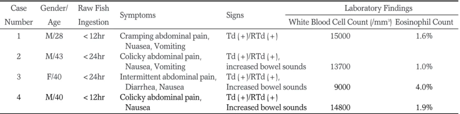
Fig. 1. A 43-year-old male patient with small bowel anisakiasis (Case 2). The CT findings (A and B): The axial scan of abdominal CT at the level of the iliac crest (A) shows circumferential wall thickening or edema of a long segment of the jejunum with peritoneal thickening and mesenteric infiltration. Proximal luminal dilatation from the transition zone is also noted (arrow). There is ascites at the right gutter. The axial scan at the level of the kidney (B) shows dilated proximal bowel loops that mimic obstructive ileus (ar- rows). The pathologic features (C and D): Photomicroscopy (C) show a helminthic larva (arrows) penetrating the bowel mucosa (H
& E, ×40). There is a dense infiltration of eosinophils with other inflammatory cells in all the layers of the small bowel (H & E, × 20) (D).
Table 2. CT Findings of the Small Bowel Anisakiasis Patients Case Involved Site Segmental Luminal
Number and Length Narrowing Pattern of Wall Thickening Ascites
Target appearance, Circumferentially preserved layering,
1 Jejunum, 15 cm + Mucosal enhancement +
Mesenteric infiltration
Target appearance, Circumferentially preserved layering,
2 Jejunum, 13 cm + Mucosal enhancement +
Peritoneal thickening, Mesenteric infiltration
Target appearance, Circumferentially preserved layering,
3 Ileum, 7 cm + Mucosal enhancement +
Peritoneal thickening, Mesenteric infiltration
Target appearance, Circumferentially preserved layering,
4 Ileum, 17 cm + Mucosal enhancement +
Peritoneal thickening, Mesenteric infiltration
Four patients underwent surgical resection of the small bowel. The pathologic examination showed the infiltra- tion of eosinophils and other inflammatory cells, includ- ing neutrophils, in all the layers of the bowel along with severe transmural edema (Fig. 1D). In all the cases, there were larvae penetrating the intestinal mucosa (Fig. 1C).
Discussion
Gastrointestinal anisakiasis is a rare parasitic disease in human beings and it is caused by penetration of a nema- tode larva. Its incidence has considerably increased in the last few years in many countries (13). The major in- flicted species are Anisakis simplex and Pseudoterranova decipiens and the final hosts are marine mammals such as whale and dolphin (6, 14). It is known as these larvae can involve any part of the human body, including the colon and the extraintestinal organs such as the peri- toneum, lungs, lymph nodes, ovaries, liver, pancreas and spleen. However, its major forms are the gastric and in- testinal types (2, 7). Gastrointestinal anisakiasis is classi- fied into the luminal and invasive forms, and most of the gastric and intestinal types belong to the invasive form, which causes clinical problems (8).
The clinical presentations of gastrointestinal anisakia- sis in the previous reports were acute abdominal pain of varying severity, nausea, vomiting, urticaria, chills and diarrhea within a few hours after ingestion of raw fish (1, 2, 7, 8, 14, 15). All our cases also had similar clinical manifestations and a history of eating raw fish.
Leukocytosis is a common laboratory finding of gas- trointestinal anisakiasis. Peripheral eosinophilia is fre- quently presented in cases of the gastric form of anisaki- asis. However, peripheral eosionophilia is known to be an uncommon finding in cases of intestinal anisakiasis (1, 4, 8). Intestinal anisakiasis can be classified as two types; one is the mild initial infection type and the other is the fulminant reinfection type. The fulminant reinfec- tion type shows featured of allergic reactions by the Arthus-type and these reactions can be clinically prob- lematic. Therefore, when sensitized patients are rein- fected by anisakis larvae, enteric anaisakiasis reveals fo- cal enteritis accompanied with an infiltration of inflam- matory cells, and mainly eosinophils, around the sites penetrated by larvae (7, 16). The clinico-pathologic man- ifestations of our cases could be explained by this mech- anism.
The intestinal type is a rarer form of gastrointestinal
few studies have reported on the radiologic features of intestinal anisakiasis (5). To recapitulate the brief radio- logic findings that had been described in several studies, edema of the intestinal wall and luminal narrowing along a long segment of intestine with an adjacent peri- toneal fluid collection are the fundamental features re- gardless of what modalities were used (1, 4, 5, 7-10, 17, 18). The CT findings of intestinal anisakiasis have been described in some studies (5, 7-12). Previous studies have revealed a relatively long segment of circumferen- tial and symmetric thickening of the bowel wall, lumi- nal narrowing and diffuse mucosal enhancement with the target sign suggesting ‘edema’ in the involved seg- ment. Our cases also showed similar findings. Based on the results of the previous studies and our study, small bowel anisakiasis shows constant CT findings.
These radiologic findings of intestinal anisakiasis re- flect pathologic changes such as marked edema or con- gestion by a diffuse eosinophilic infiltration, and granu- loma formation (4). Sometimes, well-preserved helminthic larvae that have penetrated the bowel wall, which was surrounded by a thick cuff of acute inflam- matory cells with numerous eosinophils, may be micro- scopically seen to be present in the involved small bow- el (2, 6, 8).
Gastric or colonic anisakiasis can be diagnosed by en- doscopy and that’s why rapid diagnosis and treatment are possible (1, 11, 14, 17). According to the previous re- ports and our study, the clinical symptoms and signs of intestinal anisakiasis mimicked an acute abdomen re- quiring surgery and its diagnosis is difficult (5, 7-10, 15).
However, intestinal anisakiasis can be highly suggested if a patient has a history of raw fish ingestion with the characteristic radiologic findings. Because most patients with anisakiasis have a self-limiting course or they need only medical treatment (13), the differential diagnosis from various conditions that cause true intestinal ob- struction or strangulation may be important to avoid un- necessary laparotomy.
Although our study has a small sample size and this can be regarded as a limitation, all the patients showed common clinical and CT findings, which could be a clue for making the correct diagnosis. In conclusion, intesti- nal anisakiasis should be considered when there are fa- vorable CT findings in a patient with a history of recent- ly ingesting raw or undercooked fish.
References
1. Shirahama M, Koga T, Ishibashi H, Uchida S, Ohta Y, Shimoda Y.
Intestinal anisakiasis: US in diagnosis. Radiology 1992;185:789-793 2. Takei H, Powell SZ. Intenstinal anisakidosis (anisakiosis). Ann
Diagn Pathol 2007;11:350-352
3. Ishikura H, Kikuchi K, Nagasawa K, Ooiwa T, Takamiya H, Sato N, et al. Anisakidae and anisakidosis. Prog Clin Paraisitol 1993;3:
43-102
4. Ido K, Yuasa H, Ide M, Kimura K, Toshimitsu K, Suzuki T.
Sonographic diagnosis of small intestinal anisakiasis. J Clin Ultrasound 1998;26:125-130
5. Matsuo S, Azuma T, Susumu S, Yamaguchi S, Obata S, Hayashi T.
Small bowel anisakiosis: a report of two cases. World J Gastroenterol 2006;12:4106-4108
6. Kim LS, Lee YH, Kim S, Park HR, Cho SY. A case of anisakiasis causing intestinal obstruction. Korean J Parasitol 1991;29:93-96 7. Sasaki T, Fukumori D, Matsumoto H, Ohmori H, Yamamoto F.
Small bowel obstruction caused by anisakiasis of the small intens- tine: report of a case. Surg Today 2003;33:123-125
8. Yoon SW, Yu JS, Park MS, Shim JY, Kim HJ, Kim KW. CT find- ings of surgically verified acute invasive small bowel anisakiasis resulting in small bowel obstruction. Yonsei Med J 2004;45:739-742 9. Ishida M, Harada A, Egawa S, Watabe S, Ebina N, Unno M. Three
successive cases of enteric anisakiasis. Dig Surg 2007;24:228-231
10. Masui N, Fujima N, Hasegawa T, Kigawa S, Kagei N, Nagashima K, et al. Small bowel strangulation caused by parasitic peritoneal strand. Pathol Int 2006;56:345-349
11. Hong SS, Kim JH, Park ST, Chan YW, Kim HJ, Kwon KH, et al.
Duodenal anisakiasis presenting as bowel obstruction and fistula formation: a case report. J Korean Soc Radiol 2009;60:419-422 12. Chung TW, Kang HK, Jeong YY, Jeong GW, Seo JJ, Kim YH, et al.
Radiographic findings of gastrointestinal anisakiasis: clinical and pathologic correlation. J Korean Radiol Soc 2000;43:209-213 13. Montalto M, Miele L, Marcheggiano A, Santoro L, Curigliano V,
Vastola M, et al. Anisakis infestation: a case of acute abdomen mimicking Crohn’s disease and eosinophilic gastroenteritis. Dig Liver Dis 2005;37:62-64
14. Nakata H, Takeda K, Nakayama T. Radiological diagnosis of acute gastric anisakiasis. Radiology 1980;135:49-53
15. Pellegrini M, Occhini R, Tordini G, Vindigni C, Russo S, Marzocca G. Acute abdomen due to small bowel anisakiasis. Dig Liver Dis 2005;37:65-67
16. Asaishi K, Nishino C, Ebata T, Totsuka M, Hayasaka H, Suzuki T.
Studies on the etiologic mechanism of anisakiasis. Gastroenterol Jpn 1980;15:120-127
17. Matsumoto T, Iida M, Kimura Y, Tanaka K, Kitada T, Fujishima M. Anisakiasis of the colon: radiologic and endoscopic features in six patients. Radiology 1992;183:97-99
18. Matsui T, Iida M, Murakami M, Kimura Y, Fujishima M, Yao Y, et al. Intestinal anisakiasis: clinical and radiologic features. Radiology 1985;157:299-302
대한영상의학회지 2011;64:167-171
소장 아니사키스증의 CT 소견:
네 증례를 통한 분석11한양대학교 의과대학 영상의학과
2한양대학교 의과대학 구리병원 영상의학과
김위경∙송순영∙조온구∙고병희∙김용수2∙정우경2∙김민영2
목적: 소장 아니사키스증의 CT 소견을 기술하고 병리학적 소견과 연계성을 알아보고자 하였다.
대상과 방법: 수술 후 병리소견으로 확진된 4예의 소장 아니사키스증을 대상으로 하였다. 환자는 3명의 남자 환자와 1명의 여자 환자를 포함하였고 평균 38세로 28-43세의 연령범위에 분포한 환자군을 대상으로 CT 소견과 임상 소견 및 조직학적 소견을 평가하였다.
결과: 모든 환자가 증상이 시작된 시점으로부터 24시간 내에 날생선 섭취력이 있었고 모든 예에서 복부 통증과 오심 을 호소하였으며, 구토는 2예에서, 설사는 1예에서 각각 나타났다. 이학적 검사에서 복부 압통과 반동 압통이 4명에 서, 장음의 증가는 3명에서 있었다. 검사 소견으로는 백혈구 증가증이 모든 예에서 있었으나 호산구 증가증은 없었 다. CT 소견은 4명의 환자 모두에서 장층이 유지되었고 분절성의 장벽 비후와 국소 분절의 협부가 있었으며, 협부 상부의 소장은 내강 확장 소견을 보였다. 복막 두께의 증가는 3증례에서, 장간막 또는 장막 침윤은 모든 예에서 있었 고, 동반된 다양한 양의 복수가 모든 예에서 있었다. 유충은 모든 증례에서 확인되었으며, 병리적으로 모든 층의 호 산구 침윤과 장벽의 부종 소견이 모든 예의 소장 조직에서 관찰되었다.
결론: 급성 복증과 유사한 임상 증상과 날생선 섭취의 과거력이 있는 환자들에서 CT 소견은 소장 아니사키스증의 특정 진단에 도움이 될 것이다.