CASE REPORT
복강내 농양과 연관되어 S자 결장으로 연결된 원발성 대동맥 장관루
이원호, 정철민, 조은희, 류동렬, 최대희, 김재환
강원대학교 의학전문대학원 내과학교실
Primary Aortoenteric Fistula to the Sigmoid Colon in Association with Intra-abdominal Abscess
Wonho Lee, Chul Min Jung, Eun-Hee Cho, Dong Ryeol Ryu, Daehee Choi and Jaihwan Kim
Department of Internal Medicine, Kangwon National University College of Medicine, Chuncheon, Korea
Primary aortoenteric fistula (PAEF) is a rare but catastrophic cause of massive gastrointestinal bleeding. Diagnosis of PAEF is difficult to make and is frequently delayed without strong clinical suspicion. Timely surgical intervention is essential for patient’s survival. We report on a case of an 86-year-old woman with no history of abdominal surgery, who presented with abdominal pain. Initially, computed tomography scan showed an intra-abdominal abscess, located anterior to the aortic bifurcation.
However, she was discharged without treatment because of spontaneous improvement on a follow-up computed tomography scan, which showed a newly developed right common iliac artery aneurysm. One week later, she was readmitted due to recurrent abdominal pain. On the second day of admission, sudden onset of gastrointestinal bleeding occurred for the first time. After several endoscopic examinations, an aortoenteric fistula bleeding site was found in the sigmoid colon, and aortography showed progression of a right common iliac artery aneurysm. We finally concluded that intra-abdominal abscess induced an infected aortic aneurysm and enteric fistula to the sigmoid colon. This case demonstrated an extremely rare type of PAEF to the sigmoid colon caused by an infected abdominal aortic aneurysm, which has rarely been reported. (Korean J Gastroenterol 2014;63:239-243)
Key Words: Primary aortoenteric fistula; Abdominal aortic aneurysm; Abdominal abscess; Sigmoid colon; Gastrointestinal hemor- rhage
Received June 3, 2013. Revised August 30, 2013. Accepted September 5, 2013.
CC This is an open access article distributed under the terms of the Creative Commons Attribution Non-Commercial License (http://creativecommons.org/licenses/
by-nc/3.0) which permits unrestricted non-commercial use, distribution, and reproduction in any medium, provided the original work is properly cited.
교신저자: 김재환, 200-722, 춘천시 백령로 156, 강원대학교병원 소화기내과
Correspondence to: Jaihwan Kim, Division of Gastroenterology, Department of Internal Medicine, Kangwon National University Hospital, 156 Baengnyeong-ro, Chuncheon 200-722, Korea. Tel: +82-33-258-9225, Fax: +82-33-258-2404, E-mail: [email protected]
Financial support: This study is supported by a 2013 Kangwon National University Hospital Grant. Conflict of interest: None.
INTRODUCTION
Aortoenteric fistula is defined as a pathological direct com- munication between the aorta and any portion of the bowel.
It is classified as the primary and secondary aortoenteric fistulas. Primary aortoenteric fistula (PAEF) should be dis- tinguished from secondary aortoenteric fistula, which occurs as a complication after aortic repair with vascular prosthesis.
The incidence of PAEF is approximately 0.04-0.07% and the secondary one is more frequent in the general population.1
Since 1817, only approximately 300 cases of PAEF have been reported in the English literature. The majority of cases are found in the third and fourth portions of the duodenum, how- ever, the sigmoid colon is rarely affected.2 The most common cause of PAEF is atherosclerosis; however, septic aortitis, tu- berculosis infection, tumor, radiotherapy, and foreign bodies have rarely been reported.3 Spontaneous rupture of an aortic aneurysm through an aortoenteric fistula may lead to mas- sive gastrointestinal bleeding with lethality. Early diagnosis and prompt surgical intervention are crucial for patient survival.
240 이원호 등. S자 결장으로 연결된 원발성 대동맥 장관루
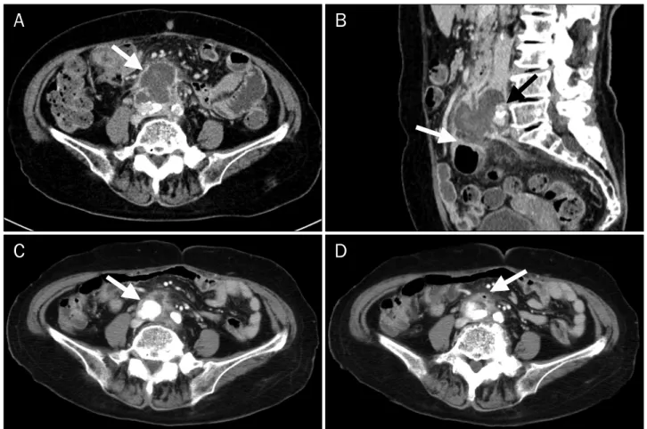
Fig. 1. Contrast-enhanced CT scans. (A) Low density lesion measuring approximately 5.2 cm in size (arrow) with peripheral enhancement was observed anterior to the aortic bifurcation without aortic aneurysm on an initial CT scan. (B) Low density lesion invaded the common-iliac artery (black arrow) and distal sigmoid colon (white arrow) on an initial CT scan. (C, D) Ten days later, follow-up CT scan showed a newly developed right common iliac artery aneurysm with air (arrows).
In this case, we report on PAEF to the sigmoid colon caused by an infected abdominal aortic aneurysm, which will be help- ful in increasing awareness of this rare disease.
CASE REPORT
An 86-year-old woman visited a urologist in our center, with abdominal pain, which had begun one week ago. She was a non-smoker. Her other medical history included diabetes mellitus for approximately 10 years and a left ureteral stone treated as spontaneous passing two years ago. With a suspi- cion that her symptoms were due to a recurrent ureteral stone, non-enhanced abdominal CT was performed. No stone was found; however, a soft tissue mass was observed at the anterior portion of the aortic bifurcation. The urologist had doubts about the mass and was suspicious that it was a mes- enteric abscess, therefore, he referred her to a surgeon for follow-up with contrast-enhanced CT. Five days later, the CT
scan showed a low density lesion measuring 5.2 cm in size with peripheral enhancement, which was reported as an ab- scess located anterior to the aortic bifurcation, between the upper to the distal sigmoid colon and posterior to the superior mesenteric root (Fig. 1A, B). When she visited the outpatient clinic of the department of surgery in our center, she com- plained of persistent abdominal pain and difficulty in defecation. On the physical exam, the abdomen was soft without tenderness, rebound tenderness, or a palpable mass.
Due to suspicion of sigmoid colon cancer, she underwent a colonoscopic exam, which revealed only a small polyp on the transverse colon, which showed a pathological result of inflammation. Serologic tumor markers were within the nor- mal range. Fifteen days after the initial contrast-enhanced CT, follow up CT imaging was performed due to the absence of colon cancer. The lesion on the retroperitoneal cavity showed a marked decrease in size; however, an adjacent focal aneur- ysmal dilatation (about 2 cm) of the right common iliac artery,
Fig. 2. (A) Initial colonoscopy showed a nodular lesion with central dimpling at the sigmoid colon and biopsy was performed here. (B) Follow-up colono- scopy showed bloody oozing at the previous lesion after biopsy. (C, D) Electrocauterization and clipping were performed at the bleeding site. After the endoscopic treatments, there was no further bleeding.
which had not been seen before, was found on the enhancing phase (Fig. 1C, D). She was discharged a couple of days later due to improvement of her symptoms and radiological findings. The day after discharge, abdominal pain recurred and continued for one week. Because of poor oral intake due to pain, she visited the emergency department with hypo- glycemic-related mental changes. When the patient visited the emergency department, the white blood cells count was 6,400/mm3 and CRP was 2.062 mg/dL. There was no fever at that time. She was readmitted to the department of endo- crinology under the suspicion of adrenal insufficiency. Fever was first observed on the second hospital day after admis- sion to the department of endocrinology. A rapid adrenocorti- cotropic hormone test was performed and prednisone 10 mg was administered. On the second hospital day, hema- tochezia (approximately 150 g) occurred; however, she was hemodynamically stable with a hemoglobin level of 9.4 g/dL.
Under colonoscopic examination, there was no active bleed- ing focus, except for a single nodular lesion with central dim-
pling 20 cm above the anal verge (Fig. 2A). Biopsy performed on this lesion showed inflammation. In addition, antibiotics (third generation cephalosporin) were administered after blood cultures were taken because of fever and chills during the bleeding event.
On the fifth hospital day, a small amount of hematochezia occurred; however, her vital signs were stable. Follow-up he- moglobin was 8.9 g/dL, and a blood transfusion was given.
Due to a mild fever without other symptoms, a blood culture was performed. At night, she reported three more episodes of hematochezia, involving a small amount each time, with- out hemodynamic instability. Because the initial blood cul- ture results identified Bacteroides fragilis, intravenous met- ronidazole was added.
The next day, there was no further bleeding and the hemo- globin was 8.5 g/dL. Gastroscopic findings were normal to the third portion of the duodenum. However, follow up colono- scopy showed fresh blood at the beginning of the anus and bloody oozing at the site of the previous biopsy (Fig. 2B).
242 이원호 등. S자 결장으로 연결된 원발성 대동맥 장관루
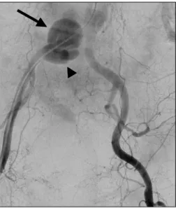
Fig. 3. After massive bleeding, emergency aortography was performed. It showed a known aneurysm measuring approximately 2 cm size (arrowhead) and a newly developed aortic aneurysm measuring approximately 5 cm in size (arrow) at the right common iliac artery.
Electrocauterization and clipping were performed for treat- ment of a suspiciously pulsatile vessel (Fig. 2C, D). However, massive bleeding with syncope occurred during the night.
The patient was hemodynamically unstable and the level of hemoglobin was 6.5 g/dL. Due to failure of endoscopic treat- ment, angiographic embolization was attempted and aneur- ysmal progression of approximately 5 cm was observed at the right common iliac artery (Fig. 3). However, the patient experi- enced sudden cardiac arrest during the evaluation. As a re- sult of unresponsiveness to the resuscitative maneuvers, she expired soon after angiography. Two days after her death, the results of the follow-up blood culture were reported, showing growth of Bacteroides fragilis, Enterococcus raffino- sus, Enterococcus faecalis, and Bacteroides thetaiotaomicron.
DISCUSSION
A PAEF originating from spontaneous erosion of the aorta into the gastrointestinal tract was first described by Sir Astley Cooper in 1822.4 The most common site of PAEF is the duode- num (54%), particularly the third and fourth portions due to its fixed retroperitoneal position and proximity to the aorta.2 PAEF to the sigmoid colon is rarely reported, and this is the first case report of PAEF to the sigmoid colon caused by an infected aortic aneurysm.
The etiology of PAEF is mainly an atherosclerotic aneurysm and only 4% of cases are septic aortitis. Other causes include radiotherapy, pancreatic carcinoma, metastases, divertic- ulitis, appendicitis, ulcers, gallstones, and foreign body.3 In the current case, we assumed that there was a micro- perforation of the sigmoid colon and that it had caused the intra-abdominal abscess, according to the results of the blood cultures, which showed growth of Enterobacteriaceae.
The decreased size of the abscess could be explained by drainage of the abscess through the fistula into the sigmoid colon. Serial CT scans showed a growing abdominal aortic aneurysm in an abscess over a two-week period. Based on such rapid radiological changes, results of the blood culture and the patient’s fever, the authors thought that the PAEF was induced from the mycotic process, which produced an in- fected aortitis and ulceration of the sigmoid colon due to the adjacent abscess. As other studies reported that an interval between the first herald bleed and massive exsanguinations ranged from hours to months,5,6 a rapid course within one month in this case could be further evidence for the process.
The clinical manifestations of PAEF vary, and include inter- mittent back pain, fever, sepsis, weight loss, and syncope.
The classical triad of abdominal PAEF is upper gastro- intestinal bleeding (94%), abdominal pain (32%), and a pul- satile abdominal mass (25%).7,8 However, the classical triad is only observed in 11-38.5% of patients with an abdominal aortoenteric fistula.9 In this case, despite the presence of ab- dominal pain and lower gastrointestinal bleeding, an ab- dominal pulsatile mass was not palpable during the initial physical examination. On the second hospital day, there was a sentinel hemorrhage, which was self-limited and massive bleeding occurred four days later.
Diagnosis of PAEF is difficult without a high index of suspicion. The most useful tool for diagnosis of PAEF is a con- trast-enhanced CT scan, which has a detection rate of 61%.10 CT is an optimal method because of its benefits, including its noninvasiveness, rapid scanning, high resolution, and good image quality. Considering that the suggestive findings for PAEF include visualization of the contrast medium within the bowel, air within the calcified wall of the aneurysm with ad- herent bowel loops, and destruction of the fat plane between the aneurysm and gastrointestinal tract,11 in this case, air within the aneurysm with an adherent sigmoid colon was compatible with PAEF. In regard to endoscopy, the preferred
initial method for PAEF, the detection rate is only 25-38% be- cause visualization of the third and fourth duodenum is very difficult.12 Therefore, endoscopy cannot exclude the possi- bility of PAEF despite negative findings. Although this case was PAEF to the sigmoid colon and not to the duodenum, it was also difficult to find the lesion as a fistula opening.
Similar to endoscopy, aortography showed a limited value of approximately 26% for diagnosis of PAEF.2 Actually, in this case, aortography showed not just the fistula but also the in- creased size of the right common iliac aneurysm.
The treatment for PAEF is surgical intervention and anti- biotics. The overall mortality from PAEF is 61-100%; however, the mortality rate from the surgery is 30-40%.2 The standard method for PAEF is an open surgical technique with anatomic in situ repair with an aortic graft or extra anatomical bypass graft in cases of gross infection.13 Endovascular repair may be an alternative for patients with hemodynamic instability or who are poor surgical candidates.14 In addition, antibiotic treatment should be administered for at least one week fol- lowing a negative culture and should be continued for 4-6 weeks if cultures prove positive.3 In this case, the patient could not undergo surgical treatment due to late detection.
PAEF is a rare cause of gastrointestinal bleeding, which could be fatal if the diagnosis is delayed. Prompt diagnosis and surgical treatment are essential for this disease. The au- thors hope that this case will increase recognition of such an extremely rare type of PAEF to the sigmoid colon, caused by an infected abdominal aortic aneurysm.
REFERENCES
1. Voorhoeve R, Moll FL, de Letter JA, Bast TJ, Wester JP, Slee PH.
Primary aortoenteric fistula: report of eight new cases and re- view of the literature. Ann Vasc Surg 1996;10:40-48.
2. Saers SJ, Scheltinga MR. Primary aortoenteric fistula. Br J Surg 2005;92:143-152.
3. Lemos DW, Raffetto JD, Moore TC, Menzoian JO. Primary aorto- duodenal fistula: a case report and review of the literature. J Vasc Surg 2003;37:686-689.
4. Cooper A. The lectures of sir Astley Cooper on the principles and practice of surgery. 5th ed. Philadelphia: Haswell, Barrington and Haswell, 1839.
5. Sweeney MS, Gadacz TR. Primary aortoduodenal fistula: mani- festation, diagnosis, and treatment. Surgery 1984;96:492-497.
6. Steffes BC, O'Leary JP. Primary aortoduodenal fistula: a case re- port and review of the literature. Am Surg 1980;46:121-129.
7. Perler BA, Ernst CB. Infected aneurysms. In: Veith FJ, Hobson RW, Williams RA, Wilson SE, eds. Vascular surgery: principles and practice. New York: McGraw-Hill, 1994:589-608.
8. Korkut AK, Arpinar E, Yasar T, Guney D. Primary aortoduodenal fistula complicated by abdominal aortic aneurysm. J Cardiovasc Surg (Torino) 2000;41:113-115.
9. Song Y, Liu Q, Shen H, Jia X, Zhang H, Qiao L. Diagnosis and man- agement of primary aortoenteric fistulas--experience learned from eighteen patients. Surgery 2008;143:43-50.
10. Hill SL, Knott LH, Alexander RH. Recurrent aortoduodenal fistu- la: a lesson in management. Am Surg 1982;48:137-140.
11. Lee JT, Saroyan RM, Belzberg G, Pianim NA, Bongard FS. Primary aortoenteric fistula: computed tomographic diagnosis of an atypical presentation. Ann Vasc Surg 2001;15:251-254.
12. Delgado J, Jotkowitz AB, Delgado B, Makarov V, Mizrahi S, Szendro G. Primary aortoduodenal fistula: Pitfalls and success in the endoscopic diagnosis. Eur J Intern Med 2005;16:363- 365.
13. Jayarajan S, Napolitano LM, Rectenwald JE, Upchurch GR Jr.
Primary aortoenteric fistula and endovascular repair. Vasc Endovascular Surg 2009;43:592-596.
14. Baril DT, Palchik E, Carroccio A, et al. Experience with endovas- cular abdominal aortic aneurysm repair in nonagenarians. J Endovasc Ther 2006;13:330-337.