기관을 침범하는 비종양성 병변은 전신 질환의 일환이나 감 염, 외상 후 또는 특발성으로 발생한다. 크게 국소적으로 침범 하는 병변과 미만성 병변으로 나눌 수 있으며 국소적 병변은 대체로 기관이나 주기관지 내경을 감소시키고 미만성 병변은 크게 내경을 감소시키는 것과 증가시키는 것으로 나눌 수 있 다. 이러한 질환을 가진 환자들은 기침, 호흡곤란, 천명과 같은 비특이적인 증상으로 내원하므로 천식으로 오진되어 진단이 늦 어지는 경우가 많다. 반면 영상 소견은 특징적이어서 그 차체 로 진단이 가능하거나 진단에 결정적인 단서를 제공할 수 있 다. 이에 저자는 기관을 침범한 다양한 비종양성 병변의 영상 소견과 문헌 고찰을 통해 질병의 이해 및 진단에 도움을 주고 자 한다.
기관의 정상 해부학
기관은 윤상연골 하방에서부터 시작하여 기관용골(carina)까 지 연결되는 약 10-12 cm 길이의 연골과 근섬유로 구성된 관 으로 흉곽 외부와 내부에 걸쳐 있다. 벽은 점막, 점막하부, 연 골, 근육과 외막으로 구성되어 있다 (Fig. 1). 말발굽모양의 유 리 연골이 전벽과 측벽을 이루고 후벽은 얇은 평활근으로 이 루어져 있다.
CT에서 기관은 1-3 mm 두께의 띠로 보이며 안쪽으로는 내강, 바깥쪽으로는 종격동 지방이나 폐와 접하고 있다. 기관 후벽에는 연골이 없어 호흡상태에 따라 다양한 모양을 보일 수 있다. 흡기 시에는 원이나 타원형으로 보이며 호기 시에는 후 벽이 함입되어 말발굽 모양을 보인다 (Fig. 2). 호기 시에 전 후경 뿐 아니라 횡경도 다소 감소되며 단면적이 정상인에서도
흡기의 60%까지 감소될 수 있다 (1). 연골은 CT에서 약간 희 게 보이며 기관과 기관지 연골 석회화는 40세 이상의 환자에 서 흔히 볼 수 있다 (2). 방사선 소견은 두드러지지만 임상적 의의는 없으며 병적인 경우와 감별을 요한다 (Fig. 3).
기관 내경을 증가시키는 질환
기관 내경이 증가되는 질환은 감소되는 경우보다 훨씬 드물 다. 그중 대표적이고 드물게 볼 수 있는 것이 기관기관지 거대 증이다.
기관기관지 거대증(Tracheobronchomegaly, Mounier-Kuhn Syndrome)
기관기관지 거대증은 기관과 중심기관지 확장, 반복되는 폐 감염을 특징으로 하는 질환이다. 원인은 병리조직학적으로 기
기관과 주기관지를 침범하는 비종양성 병변의 영상소견
1전 경 녀・강 덕 식・배 경 수
다양한 비종양성 질환이 기관과 주기관지를 침범한다. 이런 질환을 가진 환자들은 임상경과 가 길고 기침, 천명, 호흡곤란과 같은 비특이적인 증상을 보이며 천식으로 오진되기 쉽다. 반 면 영상 소견은 비교적 특징적이어서 그 자체로 또는 임상 양상을 고려하여 정확한 진단이 가 능한 경우가 많다. 그러므로 이들의 영상 소견을 숙지하고 있는 것은 진단을 위해 필수적이다.
저자들은 본 화보에서 기관과 주기관지를 침범하는 비종양성 질환의 다양한 영상 소견을 보 여주어 이해 및 진단에 도움을 주고자 한다.
1경북대학교병원 진단방사선과
이 논문은 2002년 1월 7일 접수하여 2002년 6월 3일에 채택되었음. Fig. 1. A schematic drawing of normal trachea.
관의 근육층, 탄력섬유층의 위축과 근신경총 결손이 있고 Ehlers-Danlos 증후군이나 피부이완증과 같은 결체조직질환 과 동반된 보고가 있어 선천성이라고 보는 견해가 지배적이다 (2). 그러나 대부분의 환자에서 다른 결체조직 이상이 없고 20 대 이후에 주로 발견되어서 인공호흡기와 산소 치료에 의한 기 압손상, 흡연이나 감염 등의 만성적 자극에 의해 후천적으로 발생한다고 보는 견해도 있다 (3).
방사선 소견은 기관과 기관지 확장, 약해진 기관 연골 고리 사이로 점막과 점막하 조직이 돌출되어 생긴 게실 형성을 특 징으로 한다 (Fig. 4). 단순 흉부 사진이나 기관지조영 검사에 서 기관은 대동맥궁 2 cm 상방에서 직경 3 cm, 우측 주관지 는 2.4 cm, 좌측 주기관지는 2.3 cm을 넘는 경우 진단할 수 있다 (3).
기관 내경을 미만성으로 감소시키는 질환
기관기관지 골연골 증식증, 재발성 다발성 연골염, 아밀로 이드증, 베게너 육아종, 기관기관지 결핵, 기관연골연화, saber-sheath 기관 등이 있다.
기관기관지 골연골증식증(Tracheobronchopathia Osteochondroplastica)
이 질환은 기관과 기관지의 점막하층에 골이나 연골로 구성 된 다발성의 결절 형성을 특징으로 하는 질환이다. 결절들이 기관강 내로 돌출하여 내경을 감소시킨다. 원인은 선천성, 감 염이나 만성 자극, 대사이상, 퇴행성 변화 등 여러 가지 가설 이 제기되었지만 가장 유력한 것으로는 연골막으로부터 증식 하여 생긴다는 것이며 주로 고령의 환자에서 자주 발견되므로 일종의 퇴행성 변화로 보고 있다 (4). 대부분 무증상이나 기 침, 객혈, 발열, 폐렴 등을 보일 수 있다.
CT에서는 기관의 전, 측벽의 석회화된 결절이 기관강 내로 돌출한 특징적인 모양으로 진단 할 수 있다. 아밀로이드증과 달리 후벽은 침범하지 않으며 정상적인 기관 연골 석회화나 재 발성 다발성 연골염 보다 훨씬 더 불규칙적인 모양을 보인다 (Fig. 5). 기관연골 연화는 잘 동반되지 않는다.
재발성 다발성 연골염 (Relapsing Polychondritis)
이 질환은 귀, 코, 관절, 기관, 기관지 등의 연골을 침범하는 드문 전신성 질환이다. 원인은 acid mucopolysaccharide 대사 이상이나 결체조직을 침범하는 자가면역성 질환으로 보고 있 다 (5). 기도 침범은 50% 이상의 환자에서 볼 수 있으며 기관 을 침범하는 경우 염증은 연골과 연골막에 국한 된다. 병리적 으로는 연골막을 둘러싸는 염증이며 연골의 호염기성 염색과
Fig. 3. A 67-year-old woman with dense calcification of tra- cheal cartilage.
High-resolution CT scan shows calcified cartilage. Posterior tracheal membrane appears thin and uncalcified.
A B
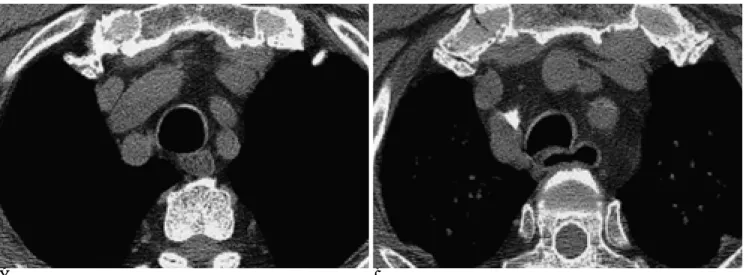
Fig. 2. Cross sectional shape of normal trachea in healthy person.
A. Inspiratory high-resolution CT scan shows slightly dense, horseshoe-shaped tracheal cartilage supporting anterior and lateral tracheal walls. Mucosa and submucosa internal to cartilage are not seen on CT scan. Posterior tracheal membrane is relatively thin.
B. Expiration results in anterior bowing of posterior tracheal membrane with a decrease in sagittal diameter.
lacunar 구조가 소실된다 (6). 기관 내경 감소는 기관 벽에 염 증을 동반한 부종이나 섬유화 또는 연골 허탈에 기인 한다 (5).
기도 침범과 반복적 폐렴이 사망의 주요 원인이 된다. CT는 빠르고 안전한 검사 방법이며 전벽과 연골에 국한된 기관벽 비 후와 기관 내경이 감소된 소견을 볼 수 있다. 연골 석회화를 동반하는 경우 정상 연골 석회화 보다 훨씬 두껍게 보인다 (Fig.
6).
아밀로이드증(Amyloidosis)
아밀로이드증은 드문 전신성 질환으로 세포밖에 불용성 단 백 침착을 특징으로 한다. 10-20% 정도는 국한성으로 발생 할 수 있고 전신성인 경우는 원발성이거나 다발성 골수종, 류 마티스양관절염, 기관지 확장증, 가족성 지중해열, 결핵과 같은 만성 감염에 이차적으로 발생 한다 (7). 호흡기계는 약 50%
에서 침범하며 기관기관지형이 가장 흔하다 (7). 주 증상은 객 혈, 천명, 쉰목소리, 기침 등이다. CT에서는 결절형(아밀로이
A B C
Fig. 4. Tracheobronchomegaly in a 51-year-old man who presented with recurrent pneumonia.
A. Chest PA shows marked dilatation of the trachea with outbulging appearance (arrowheads).
B. Tracheogram with barium reveals tracheal luminal dilatation and diverticula (arrows).
C. CT scan demonstrates multiple saccular outpouching (diverticula) of posterior tracheal wall (black arrows). T=trachea, aster- isk(*)=esophagus.
A B
Fig. 5. A 42-year-old woman with tracheobronchopathia osteochondroplastica.
A. CT scan demonstrates calcified nodules protruding into tracheal lumen. Note sparing of the membranous posterior wall. Both main bronchus are also involved (not shown here).
B. Specimen histology (hematoxylin-eosin stain, ×200) shows mature bone formation within submucosal layer (arrows).
드종)이나 미만성 기관-기관지벽 비후와 내경 협착으로 나타 난다 (Fig. 7).
베게너육아종증(Wegener’s Granulomatosis)
베게너 육아종은 신장이나 다른 장기와 더불어 상, 하기도를 침범하는 육아종성 혈관염으로 점막과 점막하 염증과 궤양 형 성을 특징으로 한다. 미만성 기관 침범은 드물고 대개 질병의 후기에 볼 수 있다 (6). 갑작스러운 호흡곤란을 유발할 수 있 으므로 기관 침범은 치료 경과에 중대한 영향을 미친다. 기관
의 상부, 특히 성문하부를 침범하는 것이 특징적이며 원위부 기관과 양쪽 주기관지를 침범하기도 한다 (2). 기관 연골 파괴 가 동반될 수 있다. CT 소견은 후두연골과 기관벽의 평탄한 비후와 기관내경 감소, 만성 염증에 의한 기관 연골의 석회화 이다 (Fig. 8).
기관과 기관지 결핵(Tracheobronchial Tuberculosis)
기관과 기관지 결핵은 폐결핵 환자의 10-20% 정도에서 동 반 된다 (8). 폐실질의 공동성 병변으로부터 방출된 균이 직접
A
B
C D
Fig. 6. A 48-year-old man with relapsing polychondritis. He had recurrent episodes of arthritis and physical examination revealed destruction of auricular cartilage.
A. Chest PA shows diffuse wall thickening and deformity of the trachea (arrowheads).
B, C. CT scan of the trachea and main bronchi shows thickening and calcification of the cartilage. Note lung cancer in right upper lobe.
D. Shaded-surface-display three dimensional image demonstrates diffuse luminal narrowing and deformity.
A
C
B
Fig. 7. Tracheobronchial amyloidosis in a 63-year-old man. He had blood-tinged sputum and dyspnea for four months.
A, B. CT demonstrates diffuse bronchial wall thickening with concentric calcification. A submucosal nodule is noted in pos- terolateral wall of left upper lobe bronchus (arrow in A). Smooth wall thickening of distal trachea is also noted (not shown here).
C. Under polarized light, apple-green birefringence of amyloid is noted within biopsy specimen (Congo red stain, ×200).
A
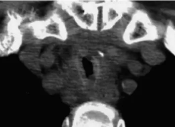
B Fig. 8. Tracheal involvement of Wegener’s granulomatosis in a 19-year-old girl.
Diagnosis was established from histologic examination of tissues obtained from the trachea, kidney and skin.
A. CT scan at the level of subglottic trachea reveals circumferential thickening of the wall.
B. Coronal reformatted image shows diffuse thickening of the tracheal wall.
착상되어 생기거나 기관지 주위의 림프 경로를 따라 파급되거 나, 종격동 림프절염으로부터 또는 혈행성으로 파급되어 생긴 다. CT 소견은 병변의 활성도에 따라 다르다. 활동성인 경우 는 기관벽의 불규칙한 비후와 내경감소, 염증성 혈류 증가에 의한 조영 증강 소견을 볼 수 있고 흔히 주위에 임파절 종대 를 동반한다 (8) (Fig. 9). 반면 치유된 기관 기관지 결핵의 경 우 내벽이 평활하며 기관이나 기관지벽 비후가 심하지 않다 (Fig. 10). 활동성인 경우는 악성 종양과 감별이 힘들 수 있으 나 3 cm 이상 긴 분절의 원위부 기관을 침범하며 기관지 침
범을 동반한다는 점이 감별점이다 (8). 치유된 섬유화 협착은 대개 좌측 주기관지에서 볼 수 있다.
Saber-sheath 기관 (Saber-sheath trachea)
만성폐쇄성 폐질환이 있는 환자에서 흉곽내 기관의 횡경이 전후경의 2/3 또는 그 이하인 saber-sheath 모양을 보일 수 있다 (Fig. 11). 단면적을 줄이기 위해 기관 후벽이 함입되는 것보다 측벽의 약해진 연골이 내측으로 심하게 허탈되어 생기 는 것이다 (9). 원인은 만성 폐쇄성 폐질환에서 흉곽내 압력의
A
B Fig. 9. Active caseating tracheobronchial tuberculosis in a 22-year-old woman.
Bronchoscopic biopsy revealed chronic granulomatous inflammation with caseation necrosis.
A. CT scan at the level of thoracic inlet shows irregular wall thickening and lumi- nal narrowing of the trachea.
B. Three dimensional CT scan demonstrates a long segmental involvement of the distal trachea. Right main bronchus is also stenotic (white arrow).
A B
Fig. 10. Fibrotic stage of tracheobronchial tuberculosis in a 23-year-old woman. She had a history of prior antituberculous therapy.
Acid-fast-bacillus staining and culture of bronchial aspirate were negative.
A, B. CT scan shows mild wall thickening of the distal trachea (arrow) and diffuse luminal narrowing of the left main bronchus (ar- rowheads). The left lung is collapsed.
정도와 형태에 따른 이차적인 변형으로 추정된다. 흉곽 바깥쪽 의 기관은 정상이며 초기에는 흉곽입구의 기관에서 이런 변화 를 볼 수 있다. 호기시에 찍은 CT에서 더욱 내측으로 허탈되 는 모양을 볼 수 있으며 기관연골 석회화를 동반할 수 있다.
기관연골연화(Tracheomalacia)
기관연골 연화는 기관벽과 연골이 약해져서 호기나 기침과 같이 흉곽 내압을 증가시키는 상황에서 쉽게 허탈되는 상태를 말한다. 원발성으로 발생하기도하지만 대부분 기관내 삽관, 외 상, 염증, 만성 폐쇄성 폐질환 등과 연관하여 이차적으로 발생 한다. 증상은 호흡 천명, 호기의 제한, 반복적 상기도 감염, 청 색증, 호흡곤란 등이다. 투시검사로 연골 연화부위의 역동적 변
화를 알 수 있으나 기도협착 정도와 범위가 과소평가 될 수 있 으며 단면을 보여 주지 못하는 단점이 있다. 전자선 단층촬영 과 같은 초고속 CT를 이용하면 숨쉬는 동안의 기관벽의 동적 변화를 볼 수 있고 호기 시에 기관 단면이 흡기에 비해 50- 70% 이상 감소되면 진단할 수 있다 (6) (Fig. 12).
국소적으로 기관을 침범하는 질환 기관협착(Tracheal Stenosis)
기관협착은 주로 기관절개술, 기관 삽관, 외상 등에 의해 이 차적으로 발생한다. 기관절개나 삽관에 의한 경우 절개부나 기 관삽관부와 풍선 확장부에 협착이 잘 생긴다 (8). 원인은 기관
Fig. 11. A 72-year-old man with history of chronic obstructive pulmonary disease.
Translateral narrowing of trachea below thoracic inlet, with- out thickening of tracheal wall, is typical of saber-sheath tra- chea.
A B
Fig. 12. A 36-year-old man with tracheomalacia after prolonged intubation.
CT scan obtained after forced expiration (A) reveals marked collapse and abnormal decrease in cross sectional area compared with inspiration (B) (in this case 72%).
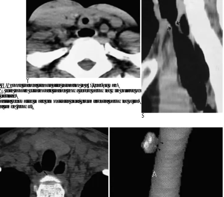
Fig. 13. Acute tracheal stenosis due to granulation tissue in a 29-year-old man. He was intubated for three weeks due to head trauma. Bronchoscopic biopsy revealed granulation tis- sue.
CT scan shows narrowing of tracheal lumen by soft tissue in- crease inward to tracheal cartilage and thickening of posterior membrane. The cartilage appears normal in shape.
점막의 실핏줄에 가해진 압력에 의한 허혈과 괴사, 이에 따른 반흔형성이다. 때로는 기관삽관부과 풍선확장부 사이의 염증에 의해 연골이 파괴되고 이차적으로 연골연화가 생겨 호기 시에 협착이 초래 된다. 또 관의 말단부가 기관벽에 직접적인 손상 을 주어 생긴 육아종으로 인해 내경 협착이 초래되기도 한다.
협착된 부위의 길이는 대개 1.5-2.5 cm 정도로 짧다. CT는 협착된 부위를 잘 보여줄 뿐 아니라 협착이 육아종이나 반흔 에 의한 것인지 연골 허탈에 의한 것인지를 구분하여 치료 방 침을 결정하는데 도움을 준다 (2, 9) (Fig. 13, 14). CT는 협
착정도는 과대평가하며 협착 길이는 과소평가하는 경향이 있 다 (6). 나선형 CT를 이용하여 재조합한 영상이나 삼차원 영 상을 얻으면 경증의 협착 부위를 잘 파악할 수 있고 협착정도 와 길이를 비교적 정확하게 알 수 있다.
기관 게실(Tracheal Diverticulum)
기관 게실은 비교적 드물며 성인에서는 만성 폐쇄성 폐질환 이나 만성적인 기침과 같이 기관 내압이 증가하는 상황에서 기 관벽의 약한 부위로 점막이 탈출되어 생긴다 (10). 이는 saber-
A
B Fig. 14. Chronic tracheal stenosis after intubation in a 37-year-old man.
A. CT scan through extrathoracic trachea shows severe narrowing with collapse of the cartilage.
B. Sagittal reformatted image demonstrates focal, circumferential narrowing of tra- cheal lumen (arrows).
A B
Fig. 15. Tracheal diverticulum in a 22-year-old man. He has suffered from prolonged, severe cough due to bronchial compression by thymic lymphoma.
A. Axial CT scan at the level of thoracic inlet shows right paratracheal air cyst with communicating channel (arrow) to tracheal lu- men.
B. Anterior view of shaded-surface-display 3D image of the trachea demonstrates diverticulum and its comunication with the tra- cheal lumen (arrowheads).
sheath 기관과 마찬가지로 폐쇄성 폐질환의 징후가 된다. 거의 대부분 흉곽 입구에서 우측 후벽에 생기며 하나의 기낭 또는 다낭성으로 보일 수 있다. 기관과 따로 떨어져 보일 수도 있고 세절편 CT 영상에서 기관 내강과 연결 통로를 보이기도 한다 (Fig. 15).
참 고 문 헌
1. Stern EJ, Graham CM, Webb WR, Gamsu G. Normal trachea dur- ing forced expiration: dynamic CT measurements. Radiology 1993;
187:27-31
2. Webb EM, Elicker BM, Webb WR. Using CT to diagnose nonneo- plastic tracheal abnormalities: appearance of the tracheal wall. AJR Am J Roentgenol 2000;174:1315-1321
3. Shin MS, Jackson RM, Ho KJ. Tracheobronchomegaly (Mounier- Kuhn syndrome): CT diagnosis. AJR Am J Roentgenol 1988;150:
777-779
4. Onisuka H, Hirose N, Watanabe K, et al. Computed tomography of tracheopathia osteoplastica AJR Am J Roentgenol 1983;140:268- 270
5. Mendelson DS, Som PM, Crane R, Cohen BA, Spiera H. Relapsing polychondritis studied by computed tomography. Radiology 1985;
157:489-490
6. Kwong JS, Muller, NL, Miller RR. Diseases of the trachea and main-stem bronchi: correlation of CT with pathologic findings.
RadioGraphics 1992;12:645-657
7. Urban BA, Fishman EK, Goldman SM, et al. CT evaluation of amy- loidosis: spectrum of disease. RadioGraphics 1993;13:1295-1308 8. Moon WK, Im JG, Yeon KM, Han MC. Tuberculosis of the central
airways: CT findings of active and fibrotic disease. AJR Am J Roentgenol 1997;169:649-653
9. Gamsu G, Webb WR. Computed tomography of the trachea: nor- mal and abnormal. AJR Am J Roentgenol 1982;139:321-326 10. Goo JM, Im JG, Ahn JM, et al. Paratracheal air cysts in thoracic in-
let: clinical and radiologic significance. AJR Am J Roentgenol 1999;
173:65-70
J Korean Radiol Soc 2002;47:269-277
Address reprint requests to : Kyungnyeo Jeon, M.D., Department of Diagnostic Radiology, Kyungpook National University Hospital, 50, Samduk-dong 2ga, Chung-gu, Taegu 700-412, Korea.
Tel. 82-53-420-5390 Fax. 82-53-422-2677 E-mail: [email protected]
Imaging Features of Nontumorous Conditions Involving the Trachea and Main-stem Bronchi
1Kyungnyeo Jeon, M.D., Duk-Sik Kang, M.D., Kyungsoo Bae, M.D.
1Department of Diagnostic Radiology, Kyungpook National University Hospital
A number of nontumorous diseases may affect the trachea and main-stem bronchi, and their nonspecific symptoms may include coughing, dyspnea, wheezing and stridor. The clinical course is often long-term and a misdiagnosis of bronchial asthma is common. The imaging findings of these nontumorous conditions are, however, relatively characteristic, and diagnosis either without or in conjunction with clinical information is often possible. For specific diagnosis, recognition of their imaging features is therefore of prime importance. In this pictorial essay, we illustrate the imaging features of various nontumorous conditions involving the trachea and main-stem bronchi.
Index words :Bronchi, diseases Trachea, CT Trachea, diseases
Trachea, stenosis or obstruction