Korean J Gastroenterol Vol. 68 No. 1, 45-48 http://dx.doi.org/10.4166/kjg.2016.68.1.45 pISSN 1598-9992 eISSN 2233-6869
CASE REPORT
Korean J Gastroenterol, Vol. 68 No. 1, July 2016 www.kjg.or.kr
내시경 절제술로 제거한 구불결장의 상피하종양 형태의 고립성 신경섬유종
이원직, 박성민, 김병욱, 김준성, 지정선, 최 황
가톨릭대학교 인천성모병원 소화기내과
Solitary Neurofibroma of the Sigmoid Colon Presenting as a Subepithelial Tumor Successfully Removed by Endoscopic Resection
Won Jik Lee, Sung Min Park, Byung-Wook Kim, Joon Sung Kim, Jeong-Seon Ji, and Hwang Choi
Department of Internal Medicine, Incheon St. Mary’s Hospital, College of Medicine, The Catholic University of Korea, Incheon, Korea
Neurofibromas are benign, slow-growing nerve sheath tumors of the peripheral nervous system, arising from Schwann cells, and classically associated with neurofibromatosis type 1 (Nf1, von Recklinghausen’s disease). They occur rarely in the gastro- intestinal tract as isolated neoplasms, outside the classical clinical feature of neurofibromatosis. We herein present an isolated colonic neurofibroma without any systemic signs of neurofibromatosis. A 59-year-old female came to our hospital for constipation.
On physical examination, general appearance showed no definite skin lesions. A subepithelial tumor measuring 0.8 cm was detected at the distal descending colon on colonoscopy. The lesion was removed completely by endoscopic resection. Microscopic examination showed proliferation of spindle cells in the mucosa and infiltration of inflammatory cells. Immunohistochemical staining was positive for S-100 protein. The above morphological and immunohistochemical characteristics were consistent with a diagnosis of a solitary neurofibroma of the sigmoid colon. (Korean J Gastroenterol 2016;68:45-48)
Key Words: Neurofibroma; Sigmoid colon; Endoscopic resection
Received December 6, 2015. Revised April 27, 2016. Accepted April 27, 2016.
CC This is an open access article distributed under the terms of the Creative Commons Attribution Non-Commercial License (http://creativecommons.org/licenses/
by-nc/4.0) which permits unrestricted non-commercial use, distribution, and reproduction in any medium, provided the original work is properly cited.
Copyright © 2016. Korean Society of Gastroenterology.
교신저자: 김병욱, 21431, 인천시 부평구 동수로 56, 가톨릭대학교 인천성모병원 소화기내과
Correspondence to: Byung-Wook Kim, Department of Internal Medicine, The Catholic University of Korea, Incheon St. Mary’s Hospital, 56 Dongsu-ro, Bupyeong-gu, Incheon 2131, Korea. Tel: +82-32-280-5052, Fax: +82-32-280-5987, E-mail: [email protected]
Financial support: None. Conflict of interest: None.
서 론
신경섬유종(neurofibroma)은 말초신경계의 신경초(nerve sheath)에서 발생하는 양성 종양으로, 주로 신경섬유종증 1 형(neurofibromatosis type 1)과 동반되어 나타난다.1,2 신경 섬유종증 환자 중 위 장관을 침범하는 경우는 25% 정도로 보고되었고, 주로 30대-50대에 복통, 출혈, 설사, 장 염전, 장 폐쇄, 천공 등의 증상으로 나타난다.3,4 그러나 신경섬유종증 이 없이 위장관에 단발성 병변으로 나타나는 경우는 거의 없 다. 대부분 양성이나, 신경섬유종증의 1-5%는 악성화 가능성 이 있으며, 신경섬유종증과 동반되지 않은 경우엔 더 낮은 것
으로 알려져 있다.5 위장관에서의 발생 빈도는 공장, 위, 회장, 십이지장, 결장의 순서로 알려져 있고, 내시경 검사에서 다양 한 형태로 나타날 수 있는데, 무경성 용종의 형태가 가장 흔하 다.6 면역조직화학 검사에서 데스민(desmin), 평활근 액틴 (smooth muscle actin)에 음성이고 S-100 단백에 양성일 때 확진할 수 있다.7,8 저자들은 전신적 신경섬유종증의 특징을 동반하지 않은 환자에서 구불결장에 상피하종양 형태의 고립 성 신경섬유종을 대장내시경하 절제술로 성공적으로 치료한 예를 경험하였기에 문헌 고찰과 함께 보고한다.
46
이원직 등. 내시경 절제술로 제거한 구불결장의 고립성 신경섬유종The Korean Journal of Gastroenterology
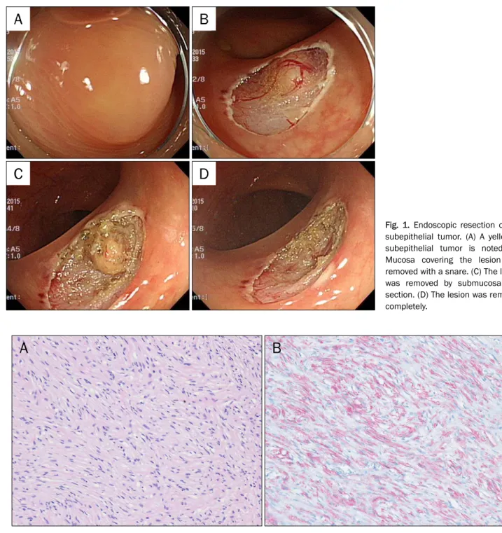
Fig. 1. Endoscopic resection of the subepithelial tumor. (A) A yellowish subepithelial tumor is noted. (B) Mucosa covering the lesion was removed with a snare. (C) The lesion was removed by submucosal dis- section. (D) The lesion was removed completely.
Fig. 2. Histopathologic finding of the resected tissue. (A) Bundles of spindle cells with elongated and wavy nuclei are noted (H&E, ×200). (B) Immunohistochemical staining for S-100 protein (×200). The spindle cells are stained in red, positive for S-100 protein.
증 례
59세 여자가 수개월간의 변비를 주소로 대장내시경 검사를 시행 받았다. 과거력에서 특이 병력은 없었고, 가족력에서도 특별한 이상은 없었다. 신체검사에서 피부에 특별한 반점이나 결절 소견, 겨드랑이 혹은 서혜부의 주근깨와 특징적 골병변 등은 없었다. 복부 소견에서 장음은 정상이었고, 복통이나 촉 지되는 종괴는 없었다. 말초혈액 검사에서 특별한 이상 소견
은 발견되지 않았고 대변 잠혈 검사도 음성이었다. 대장내시 경 검사에서 구불결장에 0.8 cm 크기의 노란색 점막하종양이 관찰되었다. 육안적으로 상피하층에서 기원한 것으로 생각되 어 우선 점막을 올가미로 제거하였고, 점막 하층의 병변은 생 리적 식염수와 에피네프린을 섞은 용액을 주입한 후 dual knife (KD-650U; Olympus, Tokyo, Japan)를 이용하여 핵제 거술로 완전 제거하였다(Fig. 1).
제거된 병변은 0.8×0.8 cm로, 조직학적 검사에서 분화도
Lee WJ, et al. Solitary Neurofibroma of Sigmoid Colon Removed by Endoscopic Resection
47
Vol. 68 No. 1, July 2016
가 높지 않고 유사분열이 관찰되지 않았다. 다수의 방추상세 포 증식과 주위 염증세포 침윤이 관찰되었으며(Fig. 2A), 절 제면 음성으로 완전 제거가 확인되었다. 면역조직화학 검사에 서 데스민, 크로모그라닌(chromogranin), 시냅토피신(synap- tophysin) 음성이었으나 S-100 단백 양성으로(Fig. 2B), 구불 결장의 단발성 신경섬유종으로 최종 진단하였다. 환자는 12 개월 후 추적 대장내시경 검사를 시행하였으나 특별한 이상 소견이 없는 상태로 현재 경과 관찰 중이다.
고 찰
신경섬유종증은 신경피부 장애를 보이는 상염색체 우성 유 전질환의 한 범주로 신경섬유종증 1과 2로 표현된다.1 신경섬 유종증 1형은 염색체 17q11.2에 존재하는 암억제 유전자인 NF1 유전자의 돌연변이가 원인이 되며, 진단 기준으로 “cafe- au-lait” 피부 반점(6개 이상, 사춘기가 지난 후 1.5 cm 이상)과 신경섬유종, 액와부나 서혜부의 얼룩, 홍체의 색소 침착 및 과 오종(Lisch nodule, 2개 이상), 골 병변(가관절증), NF1 유전자 돌연변이의 가족력, 그리고 시신경 교종 등을 포함한다.4,9
신경섬유종증 1형 환자의 25%에서 소화관을 침범하며 그 부 위는 공장, 위, 회장, 십이지장, 결장의 순서로 흔히 발생하고, 대부분 위나 소장에서 발생한다.5 소화관을 침범한 경우 고립 성, 다발성, 얼기 모양 등 세 가지 형태로 나타날 수 있고, 내시 경 검사에서 흔히 바닥이 넓은 무경성으로 나타나지만 유경성 용종 형태로도 관찰될 수 있다.10 결장의 신경섬유종증은 흔하 지 않으며 나타나더라도 주로 신경섬유종증 1형의 결장 침범이 며, 주로 30대에서 50대 사이에 관찰된다.4 결장의 신경섬유종 은 임상적으로 복통이나 종괴 촉지, 설사, 지방변, 출혈에서부 터 보다 심각한 합병증인 장중첩, 천공, 거대결장 등으로 나타 날 수 있으며, 무증상으로 우연히 진단되는 경우도 있다.3,4
면역조직화학 검사에서 S-100 단백 양성이면서 데스민, 평 활근 액틴 음성이고 신경섬유가 증식하여 모여 있을 경우 진 단이 가능하다.7,8 대부분 양성 경과를 보이고, 1-5%에서 악성 변화가 가능하지만 장기간 관찰한 연구는 없다.5 고립성 신경 섬유종의 경우 넓은 범주에서 위장관 기질 종양에 포함하여 크기 및 유사분열의 수에 따라서 악성, 양성을 구분하기도 한 다.11 치료는 대장 병변의 경우에는 악성 종양으로의 변화가 드물기 때문에 보존적 수술이나 다발성일 경우라도 제한된 장 절제술만을 시행한다.12,13 이전에 전신적인 신경섬유종증 없 이 결장에 고립성 신경섬유종이 발생한 국내 사례로 용종 형 태로 나타난 단 1예의 보고가 있었으나,14 상피하종양 형태로 보고된 증례는 없었다.
이번 증례의 경우 신경초총(schwannoma)과 감별이 필요 한데, 신경초총에서 보이는 특징적인 Verocay body가 관찰
되지 않았고, 혈관 확장 변환(antiectatic change)도 관찰되 지 않았으며, S-100의 면역조직화학염색에서 주변과의 경계 가 모호한 미만형 염색상을 보여 신경섬유종으로 진단하였다.
대장의 고립성 신경섬유종을 보고한 이전의 5개 증례를 분 석해보면,10,14-17 한국인, 백인, 흑인 모두에서 발생하였고, 남 녀 모두에서 30대-60대 사이에 발생하였다. 위치는 구불결장 부터 횡행결장까지 다양하였고 크기는 대부분 1 cm 미만이었 으나, 1예는 4 cm였다. 대부분 무증상이었고, 크기가 4 cm였 던 증례는 출혈로 내원하였다. 이전의 증례들은 용종 형태로 대장내시경 검사 및 조직생검으로 진단만 시행하였으나, 이번 증례는 상피하종양 형태였으므로 내시경 절제술로 병변을 완 전히 제거하였다. 악성의 가능성이 낮으므로 경과 관찰을 하 는 것도 한 방법이 될 수 있으나, 내시경 절제술로 비교적 간 단하게 제거하는 것은 이러한 병변에 유용한 치료법이 될 수 있을 것으로 생각한다. 이전의 증례처럼 출혈 등 합병증이 발 생하거나 크기가 1 cm 이상으로 큰 경우, 환자의 정신적 스트 레스, 조직학적 진단이 꼭 필요한 경우 등은 내시경 절제술이 필요할 것으로 생각한다. 저자들은 전신적 신경섬유종증 없이 결장에 상피하종양 형태의 고립성 신경섬유종이 발견되어 내 시경 절제술로 성공적으로 제거한 증례를 경험하여 문헌 고찰 과 함께 보고한다.
REFERENCES
1. Ferner RE, Gutmann DH. International consensus statement on malignant peripheral nerve sheath tumors in neurofibro- matosis. Cancer Res 2002;62:1573-1577.
2. Korf BR. Neurofibromatosis. Handb Clin Neurol 2013;111:
333-340.
3. Feinstat T, Tesluk H, Schuffler MD, et al. Megacolon and neuro- fibromatosis: a neuronal intestinal dysplasia. Case report and review of the literature. Gastroenterology 1984;86:1573-1579.
4. Reynolds RM, Browning GG, Nawroz I, Campbell IW. Von Recklinghausen's neurofibromatosis: neurofibromatosis type 1.
Lancet 2003;361:1552-1554.
5. Riddle ND, Gorden L, Rojiani MV, Hakam A, Rojiani AM. CD44 and p53 immunoexpression patterns in NF1 neoplasms - indicators of malignancy and infiltration. Int J Clin Exp Pathol 2010;3:
515-521.
6. Hochberg FH, Dasilva AB, Galdabini J, Richardson EP Jr.
Gastrointestinal involvement in von Recklinghausen's neuro- fibromatosis. Neurology 1974;24:1144-1151.
7. Fisher C. Immunohistochemistry in diagnosis of soft tissue tumours. Histopathology 2011;58:1001-1012.
8. Turner MS, Goldsmith JD. Best practices in diagnostic im- munohistochemistry: spindle cell neoplasms of the gastro- intestinal tract. Arch Pathol Lab Med 2009;133:1370-1374.
9. Cichowski K, Jacks T. NF1 tumor suppressor gene function: nar- rowing the GAP. Cell 2001;104:593-604.
48
이원직 등. 내시경 절제술로 제거한 구불결장의 고립성 신경섬유종The Korean Journal of Gastroenterology 10. Bononi M, De Cesare A, Stella MC, et al. Isolated intestinal neuro-
fibromatosis of colon. Single case report and review of the literature. Dig Liver Dis 2000;32:737-742.
11. Amin MB, Ma CK, Linden MD, Kubus JJ, Zarbo RJ. Prognostic val- ue of proliferating cell nuclear antigen index in gastric stromal tumors. Correlation with mitotic count and clinical outcome. Am J Clin Pathol 1993;100:428-432.
12. Franklin A, Uff JS, Spencer J. Anal neurofibrosarcoma treated with fast neutron irradiation. Br Med J 1978;1:959-960.
13. Raszkowski HJ, Hufner RF. Neurofibromatosis of the colon: a unique manifestation of von Recklinghausen's disease. Cancer 1971;27:134-142.
14. Kim KO, Jang BI, Moon HJ, et al. A solitary colonic neurofibroma
in a patient without neurofibromatosis. Korean J Gastrointest Endosc 2008;36:44-47.
15. Hindy P, Parvin R, Hanna K, Andrawes S, Gress F, Goodman A. An isolated neurofibromal polyp of the colon. Case Rep Gastroenterol 2012;6:58-62.
16. Wiesen A, Davidoff S, Sideridis K, Greenberg R, Bank S, Falkowski O. Neurofibroma in the colon. J Clin Gastroenterol 2006;40:85-86.
17. Panteris V, Vassilakaki T, Vaitsis N, Elemenoglou I, Mylonakou I, Karamanolis DG. Solitary colonic neurofibroma in a patient with transient segmental colitis: case report. World J Gastroenterol 2005;11:5573-5576.