INTRODUCTION
The family Cottidae, in the suborder Cottoidei, contains 256 species and 56 genera worldwide, with 37 species and 19 genera in Korea(NIBR, 2011; Froese and Pauly, 2016). The species of Cottidae are characterized by a large head, large eyes placed high on the head, a sep- arated dorsal fin, no spines on the anal fin, pelvic fins with one spine, 2~5 fin rays, and no swim bladder(Kim et al., 2005; Nelson, 2006). The genus Artediellus con- tains 15 species throughout the world, distributed in the Pacific Ocean, North Atlantic Ocean, and Arctic Ocean (Nelson, 1986; Froese and Pauly, 2016). Eight species of the genus Artediellus occur in Japan: A. camchaticus Gilbert and Burke, 1912, A. aporosus Soldatov, 1921, A.
ochotensis Gilbert and Burke, 1912, A. neyelovi Muto, Yabe and Amaoka, 1994, A. fuscimentus Nelson, 1986, A. schmidti Soldatov, 1915, A. dydymovi Soldatov, 1915, and A. minor(Watanabe, 1958)(Nakabo and Kai, 2013), but no occurrence of the genus Artediellus has been re-
ported in Korea(NIBR, 2011). The genus Artediellus is characterized by two or four preopercular spines, a dor- sally curved first preopercular spine, pelvic fins with one spine and three rays, no developed anal papilla, a sin- gle row of lateral line pores, and a smooth naked body, except for lateral line scales(Jordan and Starks, 1904;
Schmidt, 1927; Nelson, 1986; Muto et al., 1994). In this study, we confirmed that specimens collected from Donghae and Samcheok, Korea, were A. fuscimentus us- ing morphological and molecular methods, and provide a morphological description of these specimens as a new record based on these specimens(Fig. 1).
MATERIALS AND METHODS
Sixteen specimens of A. fuscimentus, of the family Cot- tidae, were collected from the coastal waters off Donghae and Samcheok, Korea, in December 2010 and February 2015, respectively. The specimens were fixed in 10%
formalin and then preserved in 70% ethanol. Counts and measurements were made according to the methods of Hubbs and Lagler(2004) and Nelson(1986), and body
—207 — http://www.fishkorea.or.kr
* Corresponding author: Jin Koo Kim Tel: 82-51-629-5927, Fax: 82-51-629-5931, E-mail: [email protected]
Department of Marine Biology, Pukyong National University, Yongso-ro 45, Nam-gu, Busan 48513, Republic of Korea
1Coastal Water Fisheries Resources Research Division, National Institute of Fisheries Science, Busan 46083, Republic of Korea
ABSTRACT Sixteen specimens of Artediellus fuscimentus, belonging to the family Cottidae, were collected from the coastal waters off Samcheok and Donghae, Korea, in December 2010 and February 2015, respectively. The specimens are characterized by four preopercular spines, 12~13 second dorsal fin rays, 10~12 anal fin rays, a first preopercular spine with no minute spine on the inner side, and pectoral fin extending past the origin of the anal fin. We also analyzed 600 base pairs of the mitochondrial DNA cytochrome b sequence to confirm the taxonomic status of the specimens. The sequences of our specimens corresponded completely to those of Japanese A. fuscimentus, but differed from those of A. atlanticus(d=0.057~0.061). We propose the new Korean names “Eom-ji- hoet-dae-sok” for the genus Artediellus and “Eom-ji-hoet-dae” for the species A. fuscimentus.
Key words: Artediellus fuscimentus, new record, Cottidae, East Sea, Korea
parts were measured to the nearest 0.1mm with Vernier calipers. The terminology for describing the head cirri and the cephalic sensory system was according to Nel- son(1986). All fin rays and the numbers of vertebrae were counted from radiographs(Sehwa Medical System SMS-CM, Korea). The specimens of A. fuscimentus were deposited at the Ichthyological Laboratory, Pukyong National University(PKU), Korea. Genomic DNA was
extracted from the muscle tissues with 10% Chelex 100 Resin(Bio-Rad, Hercules, CA). A polymerase chain re- action(PCR) was used to amplify the mitochondrial cy- tochrome b gene using primers, GluDG(5′-TGACTTGA ARAACCAYCGTTG-3′) and CB3(5′-GGCAAATA GGAARTATCATTC-3′)(Palumbi et al., 2002). The con- dition of PCR, purification of PCR products and sequenc- ing were followed by the method of Kim et al.(2006),
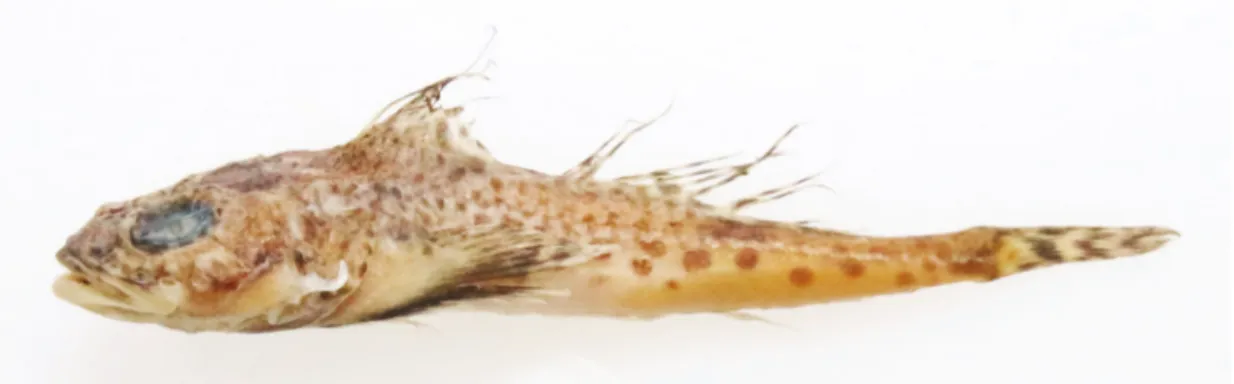
Fig. 1. Artediellus fuscimentus Nelson, 1986, PKU 53729, 74.3mm standard length, collected Samcheok, Gangwon-do.
Table 1. Comparison of counts and measurements of Artediellus fuscimentus
Present study Nelson(1986)
Nakabo & Kai(2013) Holotype Holotype+Paratypes
No. specimens 16 1 27 -
Standard length(mm) 52.4~74.3 52.7 38.1~61.5 -
Counts
Dorsal fin rays VII~VIII-12~13 VII-13 VII~VIII-12~14 VII~VIII-12~14
Anal fin rays 10~12 11 10~12 10~12
Pectoral fin rays 19~21 19 19~22 19~22
Vertebrae 28~30 30 28~30 -
Lateral line pores 27~29 28 26~29 26~29
Measurements In % standard length
Head length 33.4~37.4(35.4) 34.9 33.0~40.7(36.4)
Snout length 7.6~9.5(8.6) 8.2 7.4~9.7(8.5)
Orbit diameter 9.5~10.5(10.1) 12.1 10.3~13.4(11.3)
Interorbital width 1.6~2.4(2.0) 2.1 1.6~2.1(1.9)
Upper jaw length 13.7~17.0(14.8) 16.7 14.0~16.7(15.2)
Body depth 15.0~20.7(17.3) 20.5 17.5~22.1(19.7)
Predorsal fin length 30.0~35.6(32.7) 30.4 28.5~36.0(32.7)
Prepelvic fin length 24.2~29.3(26.8) 26.9 24.8~32.2(29.2)
Pelvic fin length 14.1~18.7(16.7) - 15.2~21.4(17.5)
Length of 1st dorsal base 15.2~21.3(17.9) 19.9 15.3~22.8(19.0)
Length of 2nd dorsal base 27.5~33.3(30.1) 31.8 28.7~33.1(31.1)
Length of anal base 26.7~30.2(28.5) 29.0 27.0~30.6(29.6)
Pelvic-anal space 22.8~29.6(26.8) 25.6 23.5~30.5(26.6)
Dorsal-caudal space 13.5~17.7(15.9) 17.0 13.3~18.0(15.8)
Anal-caudal space 15.4~18.5(17.4) 18.1 15.0~18.1(16.6)
Caudal peduncle depth 5.2~6.6(5.8) 7.0 5.6~7.1(6.3)
Parenthesis indicates average value.
Genus Artediellus Jordan, 1885 (New Korean genus name: Eom-ji-hoet-dae-sok) Artediellus Jordan, 1885: 898(type species: Cottus unci-
natus Reinhardt, 1835=Artediellus uncinatus).
Evermanniana Taranetz, 1935: 91(type species: Blenn- icottus clarki Evermann and Gill, 1907).
Description. Two or four preopercular spines, first preopercular spine curved dorsally; pelvic fins with one spine and three rays; no anal papilla developed; single row of lateral line pores; body naked and smooth, except for lateral line scales(Jordan and Starks, 1904; Schmidt, 1927; Nelson, 1986; Muto et al., 1994).
Artediellus fuscimentus Nelson, 1986 (New Korean name: Eom-ji-hoet-dae)
(Table 1, Fig. 1)
Artediellus fuscimentus Nelson, 1986: 41(type locality:
Southern Sea of Japan); Nakabo and Kai in Nakabo, 2013: 1178.
Materials examined. PKU 5062, 15 specimens, 52.4~
65.1mm SL, Donghae, Gangwon-do, Korea, 4 Dec 2010;
PKU 53729, one specimen, 74.3mm SL, Samcheok, Gangwon-do, Korea, 1 Feb 2015.
Description. All counts and measurements are listed in Table 1. Head depressed and large. Snout short and blunt.
Mouth terminal; anterior tips of upper and lower jaws almost equal; small conical teeth on both jaws in three rows; posterior margin of the maxilla round, reaching to the middle of the eye. Two pairs of nostrils, located in front of the orbit; nostrils small and circular. Eyes oval and close to the dorsal margin of the head. Four preoper- cular spines; first preopercular spine larger than other spines, strongly curved dorsally; first preopercular spine has no minute spine on the inner side. The posterior tip of the opercular region extends to the second spine of the dorsal fin. Two dorsal fins; dorsal fin base long(Table 1).
Pectoral fin large, extending past the origin of the anal fin. Pelvic fins located below the pectoral fins; posterior tips of pelvic fin rays do not reach the anus. Origin of the
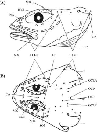
anal fin vertically below the posterior tip of first dorsal fin base. Anus located in front of the origin of the anal fin. Caudal fin truncated. Body naked and smooth, except for lateral line scales. Lateral line single, beginning at the upper tip of the gill opening and extending to the base of the caudal fin. Cirri on head(Fig. 2): maxillary, nuchal, eye, supraocular, and opercular cirri present. Cirri are ribbon-like in shape, except on the eye. Supraocular cirri longer than other cirri. Tip of nuchal cirri branched. Eye cirri present as granules on upper surfaces of eyeballs.
Pores on the cephalic sensory system(Fig. 2): second to fifth supraorbital pores present; first supraorbital pore absent. First to eighth infraorbital pores present. Anterior and posterior coronal pores present. First to sixth postor- bital pores present. Lateral anterior central, posterior me- dial, lateral posterior central, and posterior lateral pores present. Area of preopercular mandibular pores damaged.
Other pores absent.
Coloration. After fixation in formalin, dorsal half of (B)
MX IO 1-8 CP
CA
SO2 SO3
SO4 SO5
T 1-6
OCLA OCP OLP OCLP
Fig. 2. Cephalic semisensory system of Artediellus fuscimentus. A.
Lateral views. B. Dorsal views. CA, anterior coronal pore; CP, pos- terior coronal pore; EYE, eye cirri; IO, infraorbital pores; MX, max- illary cirrus; NA, nuchal cirri; OCLA, anterior central pores; OCLP, lateral posterior central pores; OCP posterior medial pore; OLP, posterior lateral pores; OP, opercular cirrus; SO, supraorbital pores;
SOC, supraorbital cirri; T, postorbital pores.
body brown, ventral half of body light yellow. Many dark brown spots on back and sides. Branchiostegal mem- branes black or light yellow.
Distribution. East Sea, Korea(present study), Japan (Nakabo and Kai, 2013).
Remarks. The present specimens were collected from the coastal waters of Donghae and Samcheok, Korea, and identified as A. fuscimentus, based on the following features: four preopercular spines, 12~13 second dorsal fin rays, 10~12 anal fin rays, first preopercular spine with no minute spine on the inner side, and the pectoral fin extending past the origin of the anal fin. Most counts and measurements in our specimens corresponded well to the original description by Nelson(1986), although our specimens differed slightly in the orbit diameter(Table 1). These subtle differences in some measurements seem to be geographic variations within the species in samples from the coast of the East Sea and the Southern Sea of Japan. We analyzed 600 base pairs of the mitochondrial cytochrome b sequence. The sequences of our specimens (KX368985) corresponded completely to those of Japa- nese A. fuscimentus, but differed considerably from those of A. atlanticus(genetic distance, d=0.057~0.061)(Fig.
3). Artediellus fuscimentus differs from A. camchaticus, A. aporosus, A. ochotensis, and A. neyelovi in having four preopercular spines(Nelson, 1986; Muto et al., 1994;
Nakabo and Kai, 2013), and from A. schmidti and A. dy- dymovi in that its first preopercular spine has no minute spine on the inner side(Nelson, 1986; Nakabo and Kai, 2013). Artediellus fuscimentus can be distinguished from A. minor by the number of dorsal fin rays(12~14 in A.
fuscimentus vs 11~12 in A. minor), the number of anal fin rays(10~12 vs 9~10, respectively), and the length of the pectoral fin(extending past origin of anal fin in A.
fuscimentus but not reaching the origin of the anal fin in A.
minor)(Nelson, 1986; Nakabo and Kai, 2013). We pro-
pose the new Korean genus name “Eom-ji-hoet-dae-sok”
for Artediellus and the new Korean name “Eom-ji-hoet- dae” for A. fuscimentus.
ACKNOWLEDGMENTS
We are grateful to anonymous reviewers for valuable advice and suggestions for improvement of the paper.
This research was supported by the project on Institute of Marine Bio-resources of Marine-Bio Technology Pro- gramme under the Ministry of Oceans and Fisheries, Ko- rea.
REFERENCES
Froese, R. and D. Pauly. 2016. FishBase. World Wide Web electron- ic publication. www.fishbase.org(01/2016).
Gilbert, C.H. and C.V. Burke. 1912. Fishes from Bering Sea and Kamchatka. Bull. U.S. Bur. Fish., 30: 31-96.
Hall, T.A. 1999. BioEdit: A user-friendly biological sequence align- ment editor and analysis program for Windows 95/98/NT.
Nucleic Acids Symp., 41: 95-98.
Hubbs, C.L. and K.F. Lagler. 2004. Fishes of the Great Lakes Re- gion. Revised ed. Michigan University Press, Ann Arbor, MI, 276pp.
Jordan, D.S. 1885. A catalogue of the fishes known to inhabit the waters of North America, north of the Tropic of Cancer, with notes on species discovered in 1883 and 1884. U. S.
Fish. Comm., 13: 789-973.
Jordan, D.S. and E.C. Starks. 1904. A review of the Cottidae or scul- pins found in the waters of Japan. Proc. U.S. Nat. Mus., 27:
231-335.
Kim, I.S., Y. Choi, C.L. Lee, Y.J. Lee, B.J. Kim and J.H. Kim. 2005.
Illustrated book of Korean fishes. Kyohak Publishing Co.
Ltd., Seoul, 615pp.(in Korean)
Fig. 3. Neighbor joining tree showing the relationships among two species of the genus Artediellus including A. fuscimentus(PKU 53729). Hemi- tripterus villosus is outgroup. Numbers at branches indicate bootstrap probabilities in 10,000 bootstrap replications. Bar indicates genetic distance of 0.02.
Peninsula, Hokkaido, Japan. Jpn. J. Ichthyol., 41: 275-280.
Nakabo, T. and Y. Kai. 2013. Cottidae. In: Nakabo, T.(ed.), Fishes of Japan with pictorial keys to the species. Third ed. Tokai Univ Press, Tokyo, pp. 628-650.(in Japanese)
Nelson, D.W. 1986. Two new species of the cottid genus Artediellus from the western North Pacific Ocean and the Sea of Japan.
Proc. Acad. Nat. Sci. Philad., 138: 33-45.
Nelson, J.S. 2006. Fishes of the world. 4th ed. John Wiley and Sons, Inc., Hoboken, NJ, 601pp.
NIBR(National Institute of Biological Resources). 2011. National lists of species of Korea. Vertebrate Natl. Inst. Biol. Res., Incheon, 460pp.(in Korean)
(Pisces, Cottidae) from Okhotsk Sea. Ezhegodnik Zoolog- icheskogo Muzeya Rossiskoi Akademii Nauk, 23: 321-324.
Tamura, K., D. Peterson, N. Peterson, G. Stecher, M. Nei and S.
Kumar. 2011. MEGA 5: Molecular evolutionary genetics analysis using maximum likelihood, evolutionary distance, and maximum parsimony methods. Mol. Biol. Evol., 28:
2731-2739.
Thompson, J.D., D.G. Higgins and T.J. Gibson. 1994. CLUSTAL W:
Improving the sensitivity of progressive multiple sequence alignment through sequence weighting, position-specific gap penalties and weight matrix choice. Nucl. Acid Res., 22: 4673-4680.
한국산 둑중개과 (Cottidae) 어류 1미기록종, Artediellus fuscimentus Nelson, 1986
신의철
·
박정호1·
김진구부경대학교 자원생물학과, 1국립수산과학원 연근해자원과
요 약 : 2010년 12월 4일 강원도 동해시에서 15개체, 2015년 2월 1일 강원도 삼척시에서 1개체의 Artediellus fuscimentus가 처음 채집되었다. 본 종은 전새개골 가시 4개, 제2등지느러미 줄기 12~13개, 뒷지느러미 줄기 10~12개를 가지며, 첫번째 전새개골 가시 안쪽에 작은 가시가 없고 가슴지느러미 뒤끝이 뒷지느러미 기점을 지나 는 특징을 가지고 있다. 또한 mtDNA cytochrome b 염기서열 600bp를 이용하여 분류학적 위치를 확인하였다. 그 결과, 본 종은 Artediellus atlanticus와 차이(d=0.057~0.061)를 보였으나, 일본산 A. fuscimentus와는 완전히 일치 하였다. Artediellus속의 국명으로 “엄지횟대속”을, A. fuscimentus의 국명으로는 “엄지횟대”를 새롭게 제안한다.
찾아보기 낱말 : Artediellus fuscimentus, 미기록종, 둑중개과, 동해, 한국