INTRODUCTION
The filefishes (family Monacanthidae), which comprise 28 genera with about 105 species, are widely distributed in tropical and temperate seas worldwide (Nelson et al., 2016). Characters of this family include that the second one of two dorsal spines is usually much smaller and it may be absent, small scales in regular series make the body prickly or furry to touch, and upper jaw usually has three teeth in the outer and two in the inner series on each premaxillary (Nelson et al., 2016). In Korea, 11 mo- nacanthid species with nine genera have been reported so far (Kim et al., 2005). The genus Cantherhines Swain- son, 1839, comprises 12 valid species worldwide (Froese and Pauly, 2017), five species from Japan (Hayashi and Hagiwara, 2013) and one species from Korea (Kim et al., 2005). This genus is characterized by having 32-39 soft dorsal fin rays, first dorsal spine with very small bars or obsolete and usually originating over anterior half of the eye, and three pairs of incasing scales, and no bristles or
spines on the middle of side (see Hutchins, 1977).
Recently, a single specimen of monacanthid fish hav- ing a dark brown body color was collected by gill net from the coastal waters of Busan, Korea. The specimen was identified as Cantherhines pardalis (Rüppell, 1837) based on the number of dorsal and anal fins, caudal pe- duncle without large spines, and a distinct spine row on the lateral side of first dorsal spine. In addition, nu- cleotide sequencing of the mitochondrial cytochrome c oxidase subunit I (COI) gene was carried out to confirm the correctness of species identification of the specimen.
Since the morphological features of this species have not been reported in Korea until now, we describe the mor- phological characters of C. pardalis as an addition to the list of Korean fish fauna.
MATERIALS AND METHDS
Identification procedure basically followed the method of Hayashi and Hagiwara (2013). After a partial tissue was isolated from one specimen of C. pardalis to extract genomic DNA, the specimens were fixed in 10% forma-
—272 — http://www.fishkorea.or.kr
* Corresponding author: Choon Bok Song Tel: 82-64-754-3471, Fax: 82-64-756-3493, E-mail: [email protected]
ISSn: 1225-8598(Print), 2288-3371(online)
accepted: December 9, 2017
First Record of the Honeycomb Filefish, Cantherhines pardalis (Tetraodontiformes: Monacanthidae) from Korea
By Maeng Jin Kim, Song Hun Han and Choon Bok Song
1,*
Fisheries Resources and Environment Division, West Fisheries Institute, NIFS, Incheon 22383, Republic of Korea
1College of Ocean Sciences, Jeju National University, Jeju 63243, Republic of Korea
ABSTRACT
This is the first report of Cantherhines pardalis (Tetraodontiformes: Monacanthidae) from Korea. A single specimen (105.4 mm in SL) was collected from the coastal waters of Busan by gill net on 20 June, 2012. This species is characterized by having the following morphological traits: II, 34 dorsal fin rays; 31 anal fin rays; 13 pectoral fin rays; caudal peduncle without large spines; spine row on the lateral side of first dorsal spine distinct; posterior end of pelvic with encasing scales distinctly more protruding than ventral flap. This species is similar to C. dumerilii except for the number of spines on caudal peduncle (none in C. pardalis vs. two pairs in C. dumerilii). Based on morphological and molecular approaches, the specimen was identified as C. pardalis. We add C. pardalis to Korean fish fauna and propose the new Korean names “Yuk-gak-mu-nui-jwi-chi” for the species.
Key words: Monacanthidae, Cantherhines pardalis, first record, Busan
lin and then preserved in 70% ethanol. Counts and mea- surements followed the method of Matsuura (1980). The specimen is deposited at the Fish Genetics and Breeding Laboratory of Jeju National University (JNU), Korea.
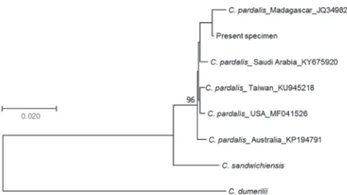
Molecular identification of the specimen was conducted by using the DNA sequences (547 bp) of COI gene. Ge- nomic DNA extraction and PCR were performed accord- ing to Chang et al. (2016). The DNA sequence of the spec- imen was deposited at the National Center for Biological
Information (NCBI) databases (Accession no.: MF959468).
This DNA sequence was compared with those of C. par- dalis (Saudi Arabia, KY675920; Taiwan, KU945218;
USA, MF041526; Madagascar, JQ349820; Australia, KP194791), C. dumerilii (Polyneisia, JQ431522) and C.
sandwichiensis (Polynesia, JQ431523). The phylogenetic tree (Fig. 2) was constructed and genetic distances were estimated by using the program MEGA 7.0 (Kumar et al., 2016) to confirm the relationships between the DNA se-
Fig. 1. Cantherhines pardalis, JNU-0765, 105.4 mm SL, gill net, Busan, Korea, June 20, 2012.
Fig. 2. A photograph showing the first dorsal spine(A), non-mobile pelvic fin rudiment(B), polygonal spots(C), and black lines(D).
quence from the specimen and those depositing in NCBI.
Cantherhines pardalis (Rüppell), 1837 (Korean name: Yuk-gak-mu-nui-jwi-chi)
(Fig. 1; Table 1)
Monacanthus pardalis Rüppell, 1837: 57 (type locality:
El-Tor, coastal waters of Sinai, Egypt).
Cantherhines pardalis: Hutchins, 1977: 55 (Australia);
Matssura in Masuda et al. 1984: 360 (Japan); Hutchins, 1986: 884 (South Africa); Randall, 1995: 396 (Oman);
Myers, 1999: 285 (Micronesian); Allen & Adrim, 2003:
64 (Indonesia).
Material examined. JNU-0765, 105.4 mm in standard length (SL), gill net, Busan, Korea. June 20, 2012.
Description. Measurements of morphological traits for the present specimen are shown in Table 1. Measure- ments are revealed as a percentage against SL: Body depth, 45.5; body width, 13.3; head length, 31.2; snout length, 26.6; eye dimeter, 7.5; interorbital width, 9.8;
length of gill opening, 10.3; snout to origin of dorsal fin, 32.3; snout to origin of anal fin length, 70.0; prepectoral fin length, 30.3; interdorsal space, 32.3; length of longest dorsal fin ray, 9.2; length of pectoral fin ray, 10.0; length of longest anal fin ray, 8.9; caudal peduncle length, 11.9;
length of dorsal fin base, 37.2; length of anal fin base, 30.4; length of caudal fin, 21.6.
Body moderately compressed and deep, head body covered with minute slender spinules. Head rather acute;
upper profile of snout prominently concave; lower pro- file slightly convex; gill slit is located below middle of eye; mouth small, terminal, upper jaw protruding slightly lower jaw, lips not obviously fleshy; consisting of three outer and two inner teeth on each side of upper jaw, ex- tremities of inner teeth projecting between outer ones;
first dorsal spine originating over anterior half of eye, armed with very small posteriorly directed barbs along each posterolateral edge (Fig. 2a), received partly into a deep groove in back when depressed; soft dorsal and anal fin rays about equal in height; pelvic fin rudiment non-mobile (Fig. 2b), and not prominently projecting rearward of ventral flap; no dense patch of bristles on caudal peduncle; caudal fin rounded.
Coloration. When fresh, body and head dark brown with a network of close-set polygonal spots; blue lines running from eye and gill slit to mouth; fins pale yel- low-orange, the caudal rays brownish; a white spot was present at rear base of both second dorsal and anal fins.
After preservation, body and head dark brown with a net- work of close-set polygonal spots (Fig. 2c); black lines
running from eye and gill slit to mouth (Fig. 2d); dorsal and anal fins pale yellow-white, the caudal rays brown- ish; a white spot not existing at rear base of both second dorsal and anal fins.
Distribution. Red Sea to South Africa and northeast- ward to Korea (Busan, present study), southern Japan and to southeastern Oceania (Francis, 1993; Myers, 1999).
Also eastern Atlantic including Gulf of Guinea, Anno- bon Islands, south coast of Africa (Harmenlin-Vivien and Quéro, 1990).
Remarks. The present specimen was identified as C.
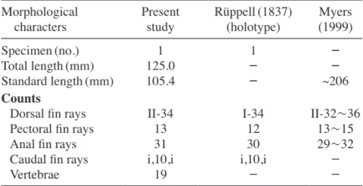
pardalis based on morphological characters in 34 dorsal fin rays, 31 soft anal fin rays, posterior end of pelvic with encasing scales distinctly more protruding than ventral flap, caudal peduncle without large spines, and distinct spine row on the lateral side of first dorsal spine (Hayashi, 2002). Meristic characters of the present specimen were compared with those of C. pardalis reported by Rüp- pell (1837) and Myers (1999) (Table 1). Although slight differ ences can be seen in pectoral fin rays (13 in present specimen vs. 12 in holotype) and anal fin rays (31 vs. 30 in holotype), these traits were well agreed with those of C.
pardalis given by Myers (1999). Therefore, these differ- ences seem to be due to intraspecific variation. In addition, we adopted molecular identification method based on the DNA sequences of COI gene to make sure of the correct- ness of species identification. The result indicated that DNA sequence from the present specimen was very sim- ilar to those of C. pardalis obtained from NCBI (99.3~
99.5% similarity), but was different from C. dumerilii and C. sandwichiensis (Fig. 3).
C. pardalis is known as having one out of three kinds of basic body color patterns including mottled grey and brown, dark brown, or grey with a network of close-set polygonal spots (Fischer and Bianchi, 1984). The pres- ent specimen has a dark brown pattern with polygonal spots. As the caudal peduncle of adult male is armed with
Table 1. Morphological traits compared between present and previ- ous studies on C. pardalis
Morphological
characters Present
study Rüppell(1837)
(holotype) Myers (1999)
Specimen(no.) 1 1 -
Total length(mm) 125.0 - -
Standard length(mm) 105.4 - ~206
Counts
Dorsal fin rays II-34 I-34 II-32~36
Pectoral fin rays 13 12 13~15
Anal fin rays 31 30 29~32
Caudal fin rays i,10,i i,10,i -
Vertebrae 19 - -
a dense patch of fine bristles, naked in adult female and juvenile (Fischer and Bianchi, 1984), we think that the collected individual is female.
The specimen is morphologically quite similar to C.
dumerilii inhabiting the coastal waters of Jejudo Island, Korea. However, the latter can be easily distinguished from the former in having the caudal peduncle without large spines (two pairs of large spines for C. dumerilii) (Hayashi and Hagiwara, 2013). We herein propose the Korean name, “Yuk-gak-mu-nui-jwi-chi” for this spe- cies because it has a network of hexagonal pattern on its body.
ACKNOWLEDGMENTS
This study was funded by a grant from the National In- stitute of Fisheries Sciences (No. R2017031), Korea.
REFERENCES
Allen, G.R. and M. Adrim. 2003. Coral reef fishes of Indonesia. Zo- ological Studies, 42: 1-72.
Chang, C.H., K.T. Shao, H.Y. Lin, Y.C. Chiu, N.Y. Lee, S.H. Liu and P.L. Lin. 2016. DNA barcodes of the native ray-finned fishes in Taiwan, Mol. Eco. Resour., 17: 796-805.
Fischer, W. and G. Bianchi, 1984. FAO species identification sheets for fishery purposes. Western Indian Ocean Fishing Area 51. Food and Agriculture Organization of the United Na-
tions, Volume 3.
Francis, M.P. 1993. Checklist of the coastal fishes of Lord Howe, Norfolk, and Kermadec Island, southwest Pacific Ocean.
Pac. Sci., 47: 136-170.
Froese, R. and D. Pauly. 2017. Fishbase. World Wide Web electron- ic publication. www.fishbase.org. version(06/2017).
Harmelin-Vivien, M.L. and J.-C. Quéro, 1990. Monacanthidae. In:
Quero, J.C., J.C. Hureau, C. Karrer, A. Post and L. Sal- danha(eds.), Check-list of the fishes of the eastern tropical Atlantic(CLOFETA). JNICT, Lisbon; SEI, Paris; and UN- ESCO, Paris. V. 2: p. 1061-1066.
Hayashi, M. and K. Hagiwara. 2013. Monacanthidae. In: Nakabo, T.(ed.), Fishes of Japan with pictorial keys to the species.
Third edition, Tokai Univ. Press, Kanagawa, pp. 1712-1726.
Hutchins, J.B. 1977. Descriptions of three new genera and eight new species of monacanthid fishes from Australia. Records of the Western Australian Museum, v. 5(pt 1): 3-58.
Hutchins, J.B. 1986. Family No. 264: Monacanthidae. In: Smith, L.B., M.M. Smith, P.C. Heemstra(eds.), Smiths’ Sea Fishes, Macmillan South Africa, pp. 882-887.
Kim, I.S., Y. Choi, C.L. Lee, Y.J. Lee, B.J. Kim and J.H. Kim. 2005.
Illustrated book of Korean fishes. KyoHak Publishing Co Ltd Seoul, 615pp.
Kumar, S., G. Stecher and K. Tamura. 2016. MEGA7: Molecular evolutionary genetics analysis version 7.0 for bigger data- sets. Molecular Biology and Evolution, 33: 1870-1874.
Matsuura, K. 1980. A revision of Japanese balistoid fishes. Bull, Natn. Sci. Mus., Ser. A(Zool.), 6: 27-30.
Matssura, T. 1984. Family Monacanthidae. In: Masuda, H., K. Ama- oka, C. Araga, U. Uyeno and T. Yoshino(eds.), The fishes of the Japanese archipelago, Tokai Univ. Press Tokyo, p.
Myers, R.F. 1999. Micronesian reef fishes. A comprehensive guide 360.
to the coral reef fishes of Micronesia. 3rd revised ed. Coral Graphics, Guam, i-vi + 1-330, 192 pls.
Nelson, J.S., T.C. Grande and M.V.H. Wilson. 2016. Fishes of the world. 5th ed. John Wiley & Sons, New Jersey, 707pp.
Randall, J.E. 1995. Coastal fishes of Oman. Crowford House Pub- lishing Pty Ltd, Bathurst, Australia, i-xvi + 1-439.
Rüppell, W.P.E.S. 1837. Neue wirbelthiere zu der fauna von ab- yssinien gehörig. Fische des Rothen Meeres. Siegmund Schmerber, Frankfurt am Main, i-ii+1-148, Pls. 1-33.
Swainson, W. 1839. On the natural history and classification of fishes, amphibians, & reptiles, or monocardian animals.
Spottiswoode and Co., London, 2: i-vi+1-448.
Fig. 3. The phylogenetic tree inferred using the Neighbor-Joining method.
한국산 쥐치과 어류 1미기록, Cantherhines pardalis
김맹진
·
한송헌·
송춘복1국립수산과학원 서해수산연구소, 1제주대학교 해양과학대학
요약 :
쥐치과에 속하는 Cantherhines pardalis 1개체(표준체장 105.4mm)가 2012년 6월 20일에 부산 연안에서 자망에 처음으로 채집되었다. 이 종은 등지느러미 기조수 II, 34개를 갖는 점, 뒷지느러미 기조수 31개를 갖는 점, 가
슴지느러미 기조수 13개를 갖는 점, 꼬리지느러미 미병부에 큰 가시가 없는 점, 첫 번째 등지느러미 극조 측면에
분명한 거치가 있는 점, 그리고 흉부 말단에 딱딱한 비늘이 돌출되어 있는 특징을 갖는다. 그리고 이 종은 형태적
으로 검은쥐치(C. dumerilii)와 유사하지만 꼬리지느러미 비병부에 가시가 없어서, 2쌍의 가시를 가지는 검은쥐치
와 형태적으로 쉽게 구분된다. 또한 유전학적 방법을 사용하여 이 표본이 C. pardalis임을 확인하였다. 우리나라에
서 처음 보고되는 이 종을 한국 어류상 목록에 추가하였고, 국명으로 “육각무늬쥐치”를 제안하였다.