IM-9 세포에 있어서 세라마이드에 의한 세포주기 변화와 아포프토시스
윤 기 호 • 최 관 수 • 김 원 호 * • 최 경 희 * • 김 미 영 *
중 앙 대 학 교 약 학 대 학 , * 중 앙 대 학 교 이 과 대 학
(Received November 12. 1995)
C e ll C y c le A lt e r a t io n a n d A p o p to s is In d u c e d b y C e r a m id e i n IM-9 C e lls
K i H o Y o o n , K w a n S o o C h o i, W o n H o K im * , K y u n g H ee C h o i* a n d M ie Y o u n g K im휴
College of Pharmacy, Chung-Ang University, Seoul 156-756y Korea
*College of Natural Science, Chung-Ang University, Seoul 156-756, Korea
Abstract— Sphingolipids play important roles in cell regulation and signal transduction. Recently, a sphingomyelin cycle has been described in which activation of neutral sphingomyelinase leads to the breakdown of sphingomyelin and the generation of ceramide. Ceramide, in turn, has emerged as a candidate intracellular mediator for the action of certain cell agonists and has multiple biologic actions.
Ceramide is a potent suppressor of cell growth and an inducer of apoptosis. The present studies show that exposure of IM-9 cells to ceramide resulted in intemucleosomal cleavage of DNA, yielding laddered patterns of oligonucleosomal fragments characteristic of apoptosis. DNA fragmentation induced b y ceramide was also confirmed by diphenylamine assay. Tlie effect of ceramide on cell cycle progression was also studied. The addition of ceramide increased Gi phase distribution in cell cycle. Cell cycle-re
lated cyclin Di gene ejq^ression was decreased in a time-dependent manner. These results suggest that apoptosis induced by ceramide is related to cell cycle associated with the alteration of cell cycle in IM-9 cells.
Keywords_n Ceramide. 늙 ptosis, Cell Cycle. Cyclin Di.
Sphingolipid와 그 분 해 산 물 은 세 포 기 능 의 조 절 에 중
요 한 역 할 을 하 며 또 한 signal transductio n에 도 관 여 하 는 것 으 로 알 려 져 있 다 . 이 와 관 련 하 여 최 근 에 는
sphingom yelin cycle이 발 견 되 었 고 ^^ 세 포 자 극 제 에 의 하 여 sphingom yelin의 기 수 분 해 가 촉 진 되 며 . 이 때 생 성 된 ceram ide는 생 물 학 적 활 성 물 질 임 이 밝 혀 졌 다 .""®
C eram ide는 세 포 막 sphingom yelin이 sphingo- m yelinase에 의 하 여 가 수 분 헤 되 어 생 성 되 며 vitam in T>i\ TN F-oc, 74nterferon잇. in te rle u k irT r*녹 용 의 여
* 본 논문에 관한 문의는 이 저자에게로
(전화) 02-820-5607 (팩스) 02-816-7338
러 세 포 기 능 조 절 외 m ediator로 알 려 져 있 다 . C era m - ide에 의 하 여 매 개 되 는 세 포 내 작 용 으 로 서 는 세 포 분 화 촉 진 , 세 포 중 식 억 제 , apoptosis. protein tra ffick in g의 조 절 및 염 증 반 응 동 이 보 고 되 어 있 다 . " 특 허 세 포 증
식 억 제 작 용 은 m yeloid, lym phoid, fib ro b last세 포 에 서 보 고 되 어 있 으 며 최 근 에 이 르 러 ceram ide의 세 포 증 식 억 제 작 용 은 apoptosis유 발 에 의 한 것 으 로 알 려 지 기 시 작 하 였 다 . Ceram ide는 m yeloid U -937세 포 와 H L - 60세 포 에 서 TNF-OC에 의 한 apoptosis의 m ediator로 밝 혀 졌 는 데 U -937세 포 에 서 ceram ide는 3^ 24시 간 에
D N A frag m entation과 형 태 학 적 변 화 를 나 타 내 며 d i- acylg lycer이 은 ceram ide의 apoptosis효 과 를 저 해 하 는
윤기호•최관수•김원호•최경희• 김미영
것으로 보고되고 있다.®"™ 이상과 같은 연구결과룰 볼 때 일부 세포에 있어서 ceram ide외 세포중식억제작용 은 apoptosis유발결과에 의한 것으로 밝혀졌으나 이의 확실한 작용기전에 대해서는 잘 알려져 있지 않다. 따라 서 본 연구에서는 기존에 보고된 m yeloid cell lin e과는 다른 종류의 세포에서도 apoptosis작용을 가지는지 알 아보기 위하여 B-lym phoblastoid cell인 IM -9 세포를 사용하여 ceram ide의 apoptosis유발효과를 확인하였 으며 이는 세포주기의 G l기 분포의 중가와 관련이 있는 것으로 밝혔다.
실험방법
시약- R P M I 1640과 fe ta l bovine se ru m 은 G ib - co사로부터 구입하였고 C2-ceram id e와 propidium iodide는 Sig m a사로부터 구입했다. 또한 P e n ic illin - Strep to m ycin so lu tio n , X h o 1. K len o w enzym e와 probe la b elin g 시약은 B o h rin g er M ann h eim 로부터 구입했다. (cc"¥ ]d C TP (3 0 0 0 C i/m m o /)는 A m e rsh - am 사에서 구입했고 기타 다른 시약은 최상품에 준하는 것을 사용하였다.
세포 배앙 - IM -9 세포는 hum an B -lym ph o - blasto id cells로써 R P M I 1640 (Gibco) 에 10% heat- in activated feta l bovine serum (G ibco )을 가한 배지 롤 세포 배양액으로 사용하였고, 5% C 02와 습도가 일 정하게 유지되는 37°C incubator에서 2 x 1 0 -1 x1 0 ® cells/m i 농도로 유지시키면서 3일마다 한번씩 배양액을 교환해 주었다.
세포 증식과 Cell V iab ility의 측정 - 2x10® c e lls/
mZ로 심은 세포에 5 나M , 10 나M , 20 jiM 의 ceram - id e를 가하고 일정시간 배양 후 hem acytom eter로 세 포수를 세었다. 세포의 v ia b ility 는 tryp a n blue e x
clu sio n 방법을 사용하였다.
D iphenylam ine assay - 약 2 x 1 0 ®개의 세포를 P B S 용액으로 2번 세척한 후 ly s is b u fferd O m M T r is , pH 7 .5 , 1 m M E D T A , 0.2% T rito n X -100)를 가하고 얼옴에서 15분간 배양한 후 in ta c t D N A 와 frag m ented D N A 틀 분리하기 위하여 4°C에서 20분간 원심분리(1 3 0 0 0 xg )한 다 . 수 층 은 12.5% 의 T C A (trich lo ro a ce tic acid )로 18시간 동안 D N A 를 침전시 키고. p e lle t에는 400 Ji/의 차가운 12.5% T C A 를 가하 고 흔합한다. 이를 4°C에서 5분간 원심분리(1 3 0 0 0 xg )
하고 이 p ellet에 30 띠의 5 m M N a O H , 30 나/의 1 M perchlo ric acid 를 가하고 70°C에서 20분간 D N A 를 주줄한 후, 140 Mi외 d ip henylam ine 시약을 가하고 3 T C 에서 18시간동안 반응시킨다. 여기서 120 를 취 해 96 w e ll p lates (fla t bottom ed)로 옮겨서 E L IS A M icro R ead er (B io -R a d 1350) 를 이용하여 600 nm 에서의 흡광도를 측정하여 fragm ent된 앙을 백분율로 나타낸다.
Fragmented D N A의분리와 Agarose gel 전기영동에 서의 확언 - 2 x 1 0 ®개의 세포를 P B S 용액으로 두번 세 척한 후 4 0 0 띠의 extractio n buffer (1 0 mM T ris . pH 8.0, 0.1 M E D T A , pH 8.0, 20 M-g/m/ R N ase, 0.5% sodium dodecyl su lfate )를 가하여 가볍게 흔들 며 37°C에서 1 시간 반응시키고, 100 [ig/ml protei
nase K 를 가한 후 sh akin g incubator에서 50°C로 1 6 -1 8 시간동안 반응시킨다. 이를 4°C에서 20분간 원 심분리 (20000xg )하고 수층을 취해 phenol/Chloro- form 로 추출하고, E tO H 에서 농축한 후 T E buffer로 충분히 녹여서, 1.5% agarose gel에서 전기영동 한다.®*
이 gel을 ethidium bromide (0.5^lg/m/)로 염색시키고 U V 에서 D N A 를 확인한다.
세포 주기 분석 - 2 X 1 0 ®개의 세포에 15 cer- am id e를 가하고 지시된 시간에 세포를 P B S 용액으로 세척을 한 후, 1 m M E D T A 를 함유하는 P B S 용액으로 재분산시킨 다옴, 70% E tO H 로 고정시킨다. 시료를 실온에서 원심분리한 다옴 상등액을 버린다. 시료에 R N ase A (0.1 mg/mZ)를 처리하고 propidium iodide so lutio n (50 나g/m /)로 염색시킨다. F A C S (B e cto n -D ickin so n 사)로 4 8 8n m 에서 형광을 측정한
다 17)
R N A의분리및 N orthern blo tting - 세포를 4°C 에 서 원심분리하여 p e lle t을 모으고 P B S 용액으로 씻어 준 후 p e lle t에 R N A e xtra ctio n b u ffe r(140 m M N a C l. 1.5 m M M gC lz, 10 m M T r is - C l p H 8 .0 , 0.5% N P -4 0 , 2 m M D T T )을 넣고 원심분리한다. 상 층액을 취하여 S T E b u ffe r(0 .2 M T ris - C l p H 8 .0 , 25 m M E D T A p H 8 .0 , 0.3 M N a C l)를 가하고 phen o l/ch lo ro fo rm 으로 추출하고 에탄올에서 침전 시킨 후 원심분리하고 U V spectrophotom eter를 이 용하여 2 6 0 n m 에서 정량한다. 분리한 R N A 를 1%
fo rm ald eh yd e ag arose gel 상에서 전기영동하여 분 리한 후 n itro ce llu lo se filte rs 에 tra n s fe r시킨다.
TIME (day)
TIME (day)
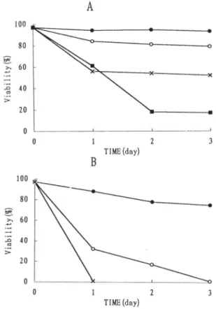
Fig. 1 - Viability of IM-9 cells by ceramide. Cells were grown in 10% FBS-supplemented media (A) or serum-free media supplemented with insulin (5 nig//) and transferrin (5 ing//) (B). Cells were treated with ethanol vehicle or with the in
dicated concentration of C2-ceramide for 3 days.
Percentage viability was determined by trypan blue dye. The results shown are representative of three seperate experiments. ( • —— • : Control, o — o : 5 Ceramide, x — x : 10 Ceram
ide. ■ — ■ : 20 M^M Ceramide).
F ilt e r는 42°C 에 서 2 시 간 동 안 p re h yb rid iza tio n하 고 나 서 random p rim er la b e llin g방 법 을 이 용 하 여 만 든 [ a크 누]-la b elle d 1.3 kb X h o l c y c lin D i probe
을 이 용 하 여 42°C에 서 ■20시 간 h yb rid iza tio n한 다 . F ilt e r를 씻 어 주 고 건 조 시 킨 후 -70°C에 서 :K -ra y film에 노 출 시 킨 다 .
결 과
IM-9세포에 대한 ceramide의 yiability 감소 - 효 과
2X lOVm / 농 도 의 세 포 에 5 10 나M , 20 나 설 의 Ca- cerain id e(N -acetyl-sp h in g o sin e)를 가 하 고 cell v i- a b ility를 3일 동 안 측 정 한 결 과 . ceram ide가 농 도 의 존
0 7 12 20
TIME (hr)
Fig. 2 — Time- and concentration- dependent D N A frag
mentation by Crceramide in IM-9 cells. Frac
tional solubilized DNA was quantitated by D i- phenylamine assay. The results shown are representative of three seperate experiments.
( • — • ■ Control. 0 — 0: 5 (xM Ceramide, x — x : 10 M-M Ceramide, ■ — ■ ■ 20 M-M Ceramide).
적 으 로 세 포 사 멸 을 유 발 하 였 다 . 10 ^iM 이 상 외 ceram ide 에 대 해 1일 후 약 50%의 v ia b ility를 나 타 냈 으 며 5 M-M ceram id e에 서 부 터 v ia b ility의 감 소 효 과 가 나 타 나 기 시 작 했 다 (F ig . 1 A ). 또 한 IM -9세 포 룰 seru m -free m ed ia(5 m g//의 in su lin과 5 m /e//의 tra n s fe rrin을 함 유 하 는 R P M I-1 6 4 0 )에 서 각 각 의 농 도 로 ceram id e룰 처 러 했 을 경 우 C2-ceram id e에 서 도 1일 후 약
30%의 v ia b ility를 나 타 내 었 고 . 2일 후 에 는 거 의 세 포 사 멸 을 일 으 킨 것 을 볼 수 있 었 다 (F ig . I B ) .
D iphenylam ine assay와 agarose gel 전기영동에 의 한 D N A fragm entation Sf인 - C e ra m id e의 apop- to sis 유 발 작 용 을 알 아 보 기 위 하 여 D N A fra g - m e n tatio n정 도 틀 분 석 하 였 다 . 2x10®개 의 세 포 를 5, 10, 20 n M C2-ceram id e를 처 리 하 여 지 시 된 시 간 별 로 세 포 를 모 아 ly s is시 킨 후 in ta c t D N A와 fra g m ented D N A를 분 리 하 여 d ip h e n yla m in e시 약 을 가 하 고 600 n m에 서 흡 광 도 를 측 정 한 결 과 농 도 의 존 적 으 로 D N A frag m en tation 비 율 이 중 가 됨 을 보 여 주 었 다
(F ig . 2 ). 특 히 2(H iM C2-ceram id e률 처 리 한 후 6시 간 뒤 에 대 조 군 에 비 헤 15%의 증 가 를 보 였 으 며 20시 간 후 에 는 frag m ent된 D N A는 50%이 상 중 가 되 었 다 . 또 한 lO u M Cz-ceram ide 존 재 하 에 서 24시 간 배 양 후
1.5% agarose gel 전 기 영 동 으 로 D N A la d d e r band
를 확 인 할 수 있 었 다 (F ig . 3 ).
세포주기의 변화와 cydin D ,유전자 발현 - C e ra m -
0000008642
Sc o
'■ 금
c
을bO
(_C3
(4-1
<
o
윤기호•최관수•김원호•최경희• 김미영
a b
Fig. 3 ― DNA ladder by ceramide in EM-9 cells. Cells were grown in serum-free media for 24 hr in the presence of ethanol vehicle or Ca-ceramide (10 |iM). DNA was analyzed by elec
trophoresis on a 1.5% agarose gel and stained ethidium bromide(0.5 냐g/mZ). (a: Control, b:
Ceramide).
gS
I 1^3
t = l h r
G O / G l S G 2 / M
t = 1 2 h r
건
G O / G l G 2 / M
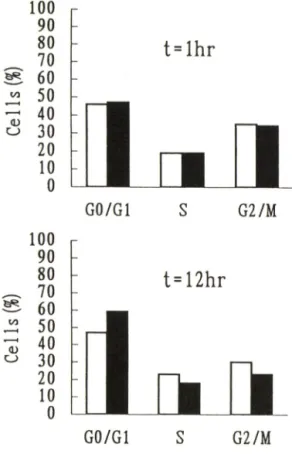
Fig. 4 — Effects of ceramide on cell cycle. IM-9 cells were treated with C2-ceramide(15 ^iM) or ethanol vehicle for the indicated time. Cells were fixed, stained and analyzed described under **MethocT.
.싶 늘 헬
찰
m m
Fig. 5
a b e d
Effects of ceramide on cyclin Di gene ex
pression in IM-9 cells. Cells were treated with C2-cerainide(15 M-M) or ethanol vehicle for the indicated time. Total cellular RNA was iso
lated and analysed by northern blotting, (a: 0 hr. b: 4hr, c- 8hr d: 15 hr).
id e에의한세포중식억제와apoptosis가세포주기의조
절과관련이 있는것인지알아보기위하여 2 X 1 0®개의
세포에C2-ceram ide를처리한후propidium iodide로
세포를염색하여 F A C S를이용하여세포주기를분석하
였다. 이결과대조군은대략46%외G i기. 19%의S기. 35%의Gb/M 기로분포하고있었고10 |xM ceram ide 를처러한후 12시간후에 59%외대기. 18%의S기.
23%의 G / M 기률나타냈다(F ig . 4 ). 또한 G i기에서S
기로의진행에관여하는cyclin 유전자발현을살피
보면 15 (iM C2~ceram ide를가한후4시간후부터 감 소하기시작하였다(F ig . 5 ).
고 찰
C e ram id e는T N F -o t의세포분화작용. 세포중식억 제작용및ap o p to sis유발 작용의 메개물질로서 알려 져 있으며 ceram id e자체가 다양한 생물학적 작용을 가지는것으로보고되어 있다. 지금까지 본연구자등 은4 P공L -6 0 . U -937. S w e i 세포에서세포투과성 ceram id e가세포분화촉진. 세포중식억제작용을가지 며이는c~myc m R N A 수준의 감소와관련이 있다는 결과를얻은바있다. 최근에이르러m yeloid c e ll에서 ceram id e의 세포중식억제작용은 ap o p to sis유발에 의한 것으로 알려졌으나아직 ap o p to sis유발 기전의 연구와 대상세포외 중류가극히제한되어 있다. 본연 구에서 ceram id e가lym p h o id c e ll인I1VI-9세포에서 세포중식의 억제와 ap o p to sis를유발하는 것으로 나 타났다. ceram id e는5 -성(U iM 농도에서 농도 의존 적으로 세포사멸을 촉진하였고 톡히 se ru m -free
m ed ia에서의 억제효과가더욱강하게나타내는 것을
볼수있었다. 최근에 M o lt-4 세포에서 se ru m -d ep -
riv e d 상태에서 ceram id e 의 생성이 증가한다는 보고 가 있는데^^\ seru m -free m ed ia에서의 억제효과가 크게 나타나는 것은 이와 관련이 있을 것으로 사료된 다. A p o p tosis는 ch ro m a tin con d ensatio n , ce ll sh rin k a g e , apo ptic body의 생성등 형 태학적 톡정과 생화학적 특징인 in te rn u cle o so m al D N A fra g - m e n ta tio n 이 일어나 180-■심0 0 base p a irs 크기의 frag m en t가 생성되어 gel e le ctro p h o re sis상에서 D N A la d d e r b an d로 확인된다. 본 연구에서는 ceram id e외 apo pto sis 유발작용을 알아보기 위하여 D N A frag m e n tatio n 을 확인하였다. D N A fra g - m en tatio n 을 확인하기 위해 d ip h e n ylam in e a s - s a y 와 agarose gel 전기영동 실험결과 모두 ce ram - k ie 에 의하여 D N A fra g m e n ta tio n 을 확인할 수 있었 다. D N A frag m en tatio n 은 C a 오'\ 의존성 e n - d o n u clease의 활성 이 증가되어 일어나는 현상으로 알 려져 있다/ " Apopt osi s유발은 G l기 흑은 G2기
a rre s t에 외한 세포주기 조절과 관련이 있는 것으로 밝혀지고 있 다 . * ® 본 연구에서 ceram id e는 G ,기 분 포를 중가시켰으며 c y c lin D i 유전자 발현을 억제한다 는 것으로 나타나 ceram id e에 의한 ap o p to sis생성은 세포주기 조절에 관여하는 유전자가 ceram id e의 직접 또는 간접적 인 ta rg e t일 가능성 이 있다. D -typ e cyc- lin 은 C D K 와 결함하여 R b 단백질을 인산화시켜 G 1기에서 S 기로의 진행을 촉진하는 단백질이다. 많은 암세포에서 c y c lin D i의 과다발현이 관찰되었으며 이 는 u n co n tro lled 세포증식과 관련되 어 있다고 보고되 어 있다/®^ p53은 ap o p to sis유도와 관련된 암 유전자 로서 세포주기를 정지시켜 ap o p to sis유발을 촉진하며 b cl-2 유전자는 이를 억제한다.^ '2"^ p53단백질의 전 사기능조절에 의하여 c y c lin -C D K 의 작용을 억제하는 p21 단백질이 중가되며 ( V I a rre s t을 유발하는 것으 로 알려져 있다. 따라서 ceram id e는 tum o r su p p re sso r lip id 로서의 가능성을 가진다고 볼 수 있으며 앞으로 ceram id e의 ap o p to sis에 유발 작용기 전을 규 명하기 위하여 암 발생억제유전자와의 관계를 알아보 고자 한다.
감사의 말씀
본 연구는 한국과학재단 연구과제(931-0 70 (H )ll-2) 연구비의 지원으로 수행되었으며 이에 감사드럽니다.
문 헌
1) O k a z a k i, T ., B e ll. R . and H a n n u n . Y . A . : Sphingom yelin turnover induced by v ita m in Da in H L-60 cell. I Biol. Chem.264. 19076 (1989).
2) O k a za k i. T .. A . B ie la w sk a , B e ll. R . M . and H an n u n . Y . A . : Role of ceram ide as a lip id m ediator of 1 a , 25-d ih yd ro xyvitam in D s-in - duced H L-60 cell d ifferentiation. I Biol. Chem,
265. 15823 (1990).
3) K im , M . Y ., C . L in a rd ic, L . Obeid, and H an n u n . Y . A .: Id entification of sphingom yelin tu rn o ver as an effector m echanism for the action o f tu m or necrosis factoi^a and *y-inteiferon. J. Biol Chem. 266.484(1991).
4) B allo u , L , R ., Chao, C . P ., M aureen, C .. H o le- n ess, M . A ., B a rk e r, S . C . and R aghow , R .■
In te rle u k in -l-m e d ia te d P G E2 production and sphingom yelin m etabolism . /. Biol Chem. 267, 20044 (1992).
5) D ressier, K . A ., S . M athias, R . N . K o le sn ick . : Tum or necrosis factor^a a ctiva te s th e sp h in gom yelin signal transduction pathw ay in a cell- free system . Science255, 1715 (1992).
6) H an nu n, Y . A . : Th e sphingom yelin cycle and the second m essenger function of ceram ide . J.
Biol. Chem.269, 3125 (1994).
7) H annun, Y . A . and Obeid. L . M . Ceram ide- an in trace llu lar signal for apoptosis. Trends Biochem.
Sci. 20. 73 (1995).
8) Obeid. L . M ., C . M . L in a rd ic , L . A . K a ro la k , and H an nu n. Y . A .: Program m ed cell death by ceram ide. Science259. 1769 (1993).
9) Ja rv is , D . W .. R . N . Ko lesnick, F . A . F o m a ri. R . S . T ra y lo r, D . A .. Gtewritz. and S . G ra n t: In duction of apoptotic D N A degradation and cell death by activatio n of the sphingom yelin p ath w ay. Proc. Natl Acad. Sci. USA.91, 73 (1994).
10) Ja rv is , W . D ., F o m a ri, F . A ., Bro w ning. J . L ., G ew ritz, D . A .. Ko lesnick. R . N and G ra n t, S.- A ttenuatio n o f ceram ide-induced apoptosis by diglyceride in hum an m yeloid leukem ia ce lls. L Biol Chem.269. 31685 (1994).
11) Cohen, J . J ., and D uke, R . C .; Glucocorticoid ac
tivation of a calcium-dependent endonculease in
윤기호•최관수•김원호•최경희• 김미영
theymocyte m uclei leads to cell death. J. Immunol.
132. 38 (1984)
12) C hoi. K . Y .. C ha. J . W . and M . Y . K im .: Effect of ceram ide on c-m yc gene expression during differentiation of U-937 cells. Mol Cells.3, 217
(1993).
13) K im , M . Y .. K im , M . A ., Moon. E . S . and Choi, K . Y .: Effects of sphingom yelin hydrolysis and ceram ide on the inhibitio n of cell growth Kor, Biochem, J.27, 271 (1994).
14) Gerschenson L . E . and Rotello R . J .: Apoptosis
; a different type of cell death. Faseb ].23, 2450 (1993).
15) V a u x, D . L . : Tow ard an understanding of the m olecular m echanism s of physiological death.
Proc. Natl Acad. Sci. USA.90, W illiam s, G . T . and C . A .
786 (1993).
Sm ith.- M olecular
regulation of apoptosis ■ genetic controls on cell death. Cell.74. 777 (1993).
17) Jayad ev, S ., L iu . B ., B ielaw ska, A . E ., Lee, J . Y .. N azaire, F ., pushkareua, M . Y .. Obeid, L . M . and H an nu n, Y . A .* Role for ceramide in cell cycle arrest. ]. Biol. Chem,270. 2047 (1995).
18) H unter. T . and Pines. J .: C yclin s and Cancer II- cyclin D and C D K inhibitors come of age.
Cell. 79, 573 (1994).
19) R ya n . J . J ., D an ish , R ., G ottlieb, C . A . and C la rk e . M . F .: C e ll cycle a n a lysis of p53-in- duced cell death in m urine eryth ro leu kem ia cells. A M Cell. Biol.13. 711 (1993).
20) M iy a sh ita , T . and Reed . J . C . : B cl-2 on
coprotein blocks chem otherapy-induced apop
tosis in a hum an leukem ia cell line. Blood. 81.
151 (1993).