Capsaicin-Induced Apoptosis of FaDu Human Pharyngeal Squamous Carcinoma Cells
Thanh-Do Le,
1* Dong Chun Jin,
1* Se Ra Rho,
1Myung Su Kim,
1Rina Yu,
2and Hoon Yoo
11Department of Pharmacology and Dental Therapeutics, School of Dentistry, Chosun University, Gwangju;
2Department of Food Science and Nutrition, University of Ulsan, Ulsan, Korea.
Received: September 2, 2011 Revised: October 31, 2011 Accepted: November 1, 2011 Corresponding author: Dr. Hoon Yoo, Department of Pharmacology and Dental Therapeutics, School of Dentistry, Chosun University, 375 Seoseok-dong, Dong-gu, Gwangju 501-759, Korea.
Tel: 82-62-230-6894, Fax: 82-62-230-6894 E-mail: [email protected]
*Thanh-Do Le and Dong Chun Jin contributed equally to this work.
∙ The authors have no financial conflicts of interest.
© Copyright:
Yonsei University College of Medicine 2012 This is an Open Access article distributed under the terms of the Creative Commons Attribution Non- Commercial License (http://creativecommons.org/
licenses/by-nc/3.0) which permits unrestricted non- commercial use, distribution, and reproduction in any medium, provided the original work is properly cited.
Purpose: To investigate the anti-tumor effect of capsaicin on human pharyngeal squamous carcinoma cells (FaDu). Materials and Methods: The expression of apoptosis/cell cycle-related proteins (or genes) was examined by reverse transcrip- tase-polymerase chain reaction, western blotting and ELISA methods, while the apoptotic cell population, cell morphology and DNA fragmentation levels were as- sessed using flow cytometry, fluorescence microscopy and agarose gel electropho- resis. Results: Capsaicin was found to inhibit the growth and proliferation of FaDu cells in a dose- and time-dependent manner. Apoptotic cell death was confirmed by observing increases in nuclear condensation, nuclear DNA fragmentation and sub- G1 DNA content. The observed increase in cytosolic cytochrome c, activation of caspase 3 and PARP (p85) levels following capsaicin treatment indicated that the apoptotic response was mitochondrial pathway-dependent. Gene/protein expression analysis of Bcl-2, Bad and Bax further revealed decreased anti-apoptotic Bcl-2 pro- tein and increased pro-apoptotic Bad/Bax expression. Furthermore, capsaicin sup- pressed the cell cycle progression at the G1/S phase in FaDu cells by decreasing the expression of the regulators of cyclin B1 and D1, as well as cyclin-dependent pro- tein kinases cdk-1, cdk-2 and cdk-4. Conclusion: Our current data show that capsa- icin induces apoptosis in FaDu cells and this response is associated with mitochon- drial pathways, possibly by mediating cell cycle arrest at G1/S.
Key Words: Capsaicin, apoptosis, cell cycle arrest, human pharyngeal squamous carcinoma cells
INTRODUCTION
Oral cancers are aggressive tumors arising in the lip, tongue, floor of the mouth, gingivae, palate, buccal mucosa/vestibule or salivary glands, and are associated with a high mortality rate.1,2 Over 300000 cases of oral and pharyngeal cancer are annually reported worldwide with male outnumbering female patients.3 Oral can- cer in Korea, based on the 2008 report of the national cancer center, has an inci- dence of 2.6 cases per 100000. The treatment of oral cancer typically involves che- motherapy in combination with other modalities such as radiation therapy or surgery.4 However, the mechanisms underlying tumor generation in the oral cavity
Polymerase chain reaction (PCR) primers were purchased from Bioneer (Daejeon, Korea). Antibodies were purchased from the following sources: cdk-1, cdk-4, cyclin B1, Bcl-2, Bad, Bax and all secondary antibodies from Santa Cruz Bio- technology (Santa Cruz, CA, USA); cdk-2, cyclin D1 and PARPp85 from Epitomics (Burlingame, CA, USA). All other chemicals were obtained from Sigma.
Cell lines and cell culture
Human pharyngeal squamous carcinoma cells were pur- chased from the Korean cell line bank (KCLB, Seoul, Ko- rea) and were maintained at 37°C in humidified atmosphere at 5% CO2 in MEM supplemented with 10% FBS and anti- biotics/antimycotics.
Growth inhibition
Growth inhibition was assessed via an MTT assay. Briefly, FaDu cells were plated at a density of 1×10⁵ cells/well on 24-well plate. After overnight growth, the cells were treated with various concentrations of capsaicin for 24, 48 and 72 hours, with medium replacement every 24 hours. At the end of treatment, 30 µL of the tetrazolium compound MTT (Sig- ma, St. Louis, MO, USA), and 270 µL of fresh medium were added. After further incubation for 4 hours at 37°C, 200 µL of 0.1 N HCl in 10% SDS was added into each well to dissolve the tetrazolium crystals. Finally, the absorbance at a wavelength of 540 nm was recorded using an ELISA plate reader (Thermo Fisher Scientific, Waltham, MA, USA).
DNA fragmentation
Cells cultured in 100 mm dishes were treated with capsa- icin (100 µM and 300 µM) for 24 hours, trypsinized and collected with ice-cold phosphate-buffered saline (PBS).
After centrifugation at 300 g for 10 minutes at 4°C, the cells were washed with PBS and centrifuged again at 5000 rpm for 5 minutes at 4°C. Cell pellets were resuspended in 0.5 mL of lysis buffer (10 mM EDTA, 50 mM Tris-HCl, pH 8.0, 0.5% SDS, 0.5 mg/mL proteinase K) and incubated over- night at 50°C. The lysate was centrifuged at 14000 rpm for 5 minutes at 4°C to separate the soluble DNA fragment from the intact chromatin pellet. The fragmented DNA was ex- tracted with phenol/chloroform/isoamyl alcohol (25 : 24 : 1) and precipitated with ethanol. The purified DNA was treat- ed with 200 µg/mL of DNase-free RNaseA for 1 hour at 37°C prior to electrophoresis on a 1.8% agarose gel con- taining ethidium bromide.
are not yet fully understood, and the mortality rate for this disease has not improved in recent years.
Capsaicin, N-vanillyl-8-methyl-α-nonenamide, is a spicy component of hot pepper and comprises an aromatic ring and dipolar amide bond in its chemical structure. Recent re- ports have indicated that capsaicin has beneficial effects on certain types of cancers such as tumors of the colon, pros- tate, lung and breast.5-7 Capsaicin has also been shown to inhibit N-methyl-N’-nitro-N-nitrosoguanidine-induced gas- tric carcinogenesis and reduce the incidence of colonic ade- nocarcinomas in an azoxymethane-induced rat colon carci- nogenesis model.8,9 In vitro studies of transformed cells and various types of cancer cells have further shown that capsa- icin induces programmed cell death.10 These include human stomach cancer cells, hepatocarcinoma, glioblastoma and neuroblastoma cells.8,11-13 However, the molecular mecha- nisms underlying capsaicin-induced apoptosis are cell type dependent: capsaicin induces apoptosis in sensory neurons by increasing calcium influx and does so by activating vanil- loid receptors in some transformed cells.14-16 In human colon cancer cells, capsaicin triggers apoptosis through the inhibi- tion of plasma membrane NADH-oxidoreductase activity and/or NADH: coenzyme Q oxidoreductase in the mito- chondrial electron transport system, generating reactive ox- ygen species.5,10,17 Moreover, capsaicin was found to be as- sociated with PPARγ during the regulation of cell growth and apoptotic cell death in breast or colon cancer cells.18 Despite the cumulative evidence for the tumor suppressive effects of capsaicin, however, few studies have been under- taken to date on the effects of capsaicin on cell signaling and the molecular pathways leading to apoptosis in oral cancer cells.19 In the present study, we investigated the effects of capsaicin on FaDu human pharyngeal squamous carcinoma cells and demonstrated that capsaicin induced apoptosis in FaDu cells.
MATERIALS AND METHODS
Materials
Capsaicin was purchased from Sigma Chemical Co. (St.
Louis, MO, USA). Minimum essential medium (MEM), fe- tal bovine serum (FBS), and antibiotics/antimycotics were purchased from Gibco BRL (Grand Island, NY, USA). Cas- pase activity was measured using a caspase cellular activity assay kit (Calbiochem, Darmstadt, Germany). 4, 6-Diamid- ino-2-phenylindole (DAPI) was purchased from Sigma.
tal RNA was then isolated using gene plate from RNAture (Irvine, CA, USA). RT-PCR was performed to estimate the mRNA levels of anti-apoptotic Bcl-2 and pro-apoptotic Bad.
The primer sequences used were 5’-AGGAGCTCTTCAG GGACGG-3’ and 5’-CCAGGTGTGCAGGTGCC-3’ for Bcl-2 and 5’-CCCGAGAGGTCTTTTTCC-3’ and 5’- GCCTTGAGCACCAGTTTG-3’ for Bax. For the detection of glyceraldehydes-3-phosphate dehydrogenase (GAPDH) transcripts, the primer sequences used were 5’-CCCAT CACCATCTTCCAGGAGC-3’ and 5’-CCAGTGAGCT TCCCGTTCAGC-3’. The expected sizes of the PCR prod- ucts were 161 bp (for Bcl-2), 109 bp (for Bax) and 473 bp (for GAPDH). The detected mRNA levels were normalized to the GAPDH using the Bio-Rad Image Master Program (Bio-Rad, Hercules, CA, USA).
Western blotting
Cells grown in 60 mm dishes (5×10⁵ cells/dish) were treat- ed with 200 µM capsaicin for 24 hours, harvested, washed with cold PBS (×2) and then lysed by the addition of 300 µL of lysis buffer [20 mM Tris-HCl (pH 7.5), 150 mM NaCl, 1 mM Na2EDTA, 1 mM EGTA, 1% Triton, 2.5 mM sodium pyrophosphate, 1 mM β-glycerophosphate, 1 mM Na3VO4, 1 µg/mL leupeptin and 1 mM PMSF]. The cell lysates were homogenized by pipetting for 15 minutes on ice and then sonication for 1 minute. The supernatant from each lysate was collected into new tubes after centrifugation at 20000 g for 15 minutes at 4°C. Total protein concentrations were measured using the BCATM protein assay kit (Pierce, Rock- ford, IL, USA). The protein bands were normalized to the beta actin expression level.
RESULTS
Effects of capsaicin on the morphology and viability of human pharyngeal squamous carcinoma cells
FaDu cells were treated in the presence or absence of cap- saicin (200 µM) for 12 or 24 hours and the cell morpholo- gies were analyzed under the microscope at different time periods. The density of the cells on the capsaicin-treated dish surfaces was reduced without change in shape when treated with 200 of capsaicin for 12 and 24 hours (Fig. 1A).
To evaluate the effects of capsaicin on cell growth and pro- liferation, the cells (4×10⁴/well) in 48-well plates were ex- posed to a series of capsaicin concentration for 24, 48, and 72 hours, and the percentage of living cells was determined Nuclear staining
Cells were incubated in 6-well plates with 200 µM capsa- icin for 24 hours. After washing with PBS, the cells were fixed with 4% formaldehyde and dried briefly. The fixed cells were washed again with PBS, air dried and stained with the DNA-specific fluorochrome, DAPI. The cells were washed with PBS, air dried and mounted with 90% glycer- ol. The plates were then observed under a fluorescence mi- croscope (Olympus Optical Co., Tokyo, Japan).
Flow cytometry analysis
Cells treated with various concentrations of capsaicin for 24 hours were harvested by brief trypsinization and centrif- ugation (at 300 g), washed in ice-cold PBS and fixed in 70% ethanol for 2 hours at -20°C. The fixed cells were then stained with PI (20 µg/mL propidium iodide, 200 µg/mL DNase-free RNase-A in PBS) for 15 minutes at 37°C. The DNA content of cells stained with propidium iodide was measured using a FACScan instrument and Cell Quest soft- ware (Becton Dickinson, Franklin Lakes, NJ, USA).
Analysis of cytochrome c release
The levels of cytochrome c in the cytosol were detected us- ing an ELISA kit (Bender MedSystem, Wien, Austria). Cells were cultured in medium in the presence of 50, 100 and 200 µM of capsaicin for 12 hours. After treatment for 12 hours, the cells were lysed and centrifuged at 14000 rpm to obtain the cytosol fraction. The supernatants were transferred into a new tube and diluted 50-fold in an assay buffer in accordance with the manufacturer’s instructions. The OD values were read on Multiskan EX Microplate Photometer (Thermo Fish- er Scientific, Waltham, MA, USA), using 450 nm as a prima- ry wavelength and 620 nm as a reference wavelength.
Cellular caspase-3 enzyme activity assay
Caspase activity in capsaicin-treated FaDu cells was ana- lyzed using a caspase cellular activity assay kit, which in- cludes human recombinant caspase-3, cell lysis buffer, an Ac-DEVD-pNA colorimetric substrate, a calibration stan- dard (p-nitroaniline), a caspase-3 inhibitor and assay buf- fer. Caspase 3 activity in the cell lysates was determined colorimetrically in accordance with the manufacturer’s in- structions.
Reverse transcriptase-polymerase chain reaction (RT-PCR)
Cells were treated with 200 µM capsaicin for 24 hours. To-
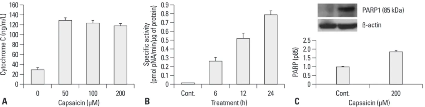
(data not shown). These results indicate that capsaicin-in- duced apoptosis is mediated by cytochrome c triggered cas- pase 3 activation through the mitochondrial pathway. The increase in caspase 3 activity was further evidenced by de- tection of an increased level of inactivated PARP fragment (p85), a main substrate of executor caspases, by western blotting (Fig. 2C).
The effects of capsaicin on apoptosis related gene/
protein expression
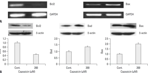
The expression of the Bcl-2 and Bax genes was analyzed by using RT-PCR. As shown in Fig. 3A, the expression of Bcl-2 was found to be decreased after treatment with capsa- icin, whereas the expression of Bax was increased. The pro- tein levels of apoptosis-associated proteins also followed a similar pattern, i.e. a decrease in Bcl-2 and an increase in Bad and Bax (Fig. 3B), indicating that the apoptotic re- sponse in FaDu cells is associated with the functions of mi- tochondria.
via an MTT assay. Cells treated with 50, 100, 200, and 300 µM capsaicin showed an augmented decrease in cell growth as the capsaicin dose increased. In addition, the percentage of viable cells decreased as the incubation time increased.
The observed IC50 value was around 150 µM (Fig. 1B).
The effects of capsaicin on mitochondrial cytochrome c release into cytoplasm and caspase-3 activity
The release of cytochrome c from the mitochondria was confirmed by measuring the absorbance of these molecules in cytosolic extracts after treatment of FaDu cells with 50, 100 and 200 µM capsaicin for 12 hours. A high level of re- leased cytochrome c (about four-fold higher than untreated control) was detectable even at 50 µM capsaicin treatment (Fig. 2A). Caspase-3 activation was confirmed using a cel- lular caspase-3 enzyme activity assay. The activity of cas- pase 3 after capsaicin treatment increased in a time-depen- dent manner (Fig. 2B) and capsaicin-induced caspase 3 activity was blocked by pretreatment with Ac-DEVD-CHO
Fig. 1. Morphologic changes and growth inhibition in FaDu cells following exposure to capsaicin. (A) As the incubation time increases, the cell density is re- duced, reflecting the inhibitory effects of capsaicin on cell growth. Untreated FaDu cells at 12 h (a) and 24 h (b) and FaDu cells treated with 200 µM capsa- icin for 12 h (c) and 24 h (d). (B) The cells were treated with various concentrations of capsaicin for 24, 48 and 72 h. Cell viability was determined by MTT as- say. The IC50 of capsaicin against FaDu cells was measured at around 150 µM. The vertical bars in (B) indicate the means and standard errors (n=3).
Fig. 2. The effects of capsaicin on the mitochondrial cytochrome c release into cytoplasm and caspase activity. (A) Analysis of mitochondrial cytochrome c release: cells were treated with 50, 100 and 200 µM capsaicin for 12 h. The level of cytochrome c in the cytosol was detected by using ELISA kit, BS263 (Bender MedSystem, Wien, Austria). (B) Caspase activity was assayed by using a colorimetric substrate DEVD-pNA after treating cells with 200 µM capsa- icin for indicated time intervals. Caspase activity was expressed as pmol cleaved/min/µg of protein. Each value is the mean of triplicate experiments. (C) Expression of PARP cleaved form (p85) after treating cells with 200 µM capsaicin for 24 h. The bar graphs represent arbitrary units of relative density after normalized with beta actin. Vertical bars indicate means and standard errors (n=3).
0 20 40 60 80 100 120
% L ive cells
0 50 100 150 200 250 300 350
Capsaicin (µM)
24 h 48 h 72 h CAP-
CAP+
A B
a
c
b
d
0 0.51.01.52.02.50
0 0.10.2 0.3 0.4 0.50.6 0.7 0.80.9
20 40 60 80 100 120 140 160
Cytochrome C (ng/m/L) PARP (p85)
Specific activity (pmol pNA/min/µg of protein)
0 50 100 200 Cont. 6 12 24 Cont. 200
Capsaicin (µM) Treatment (h) Capsaicin (µM)
A B C
PARP1 (85 kDa) ß-actin
Flow cytometric analysis
Apoptotic cell death in FaDu cells was further confirmed by analyzing the sub-G1 DNA content using flow cytome- try. Cells were treated with various concentrations of capsa- icin for 24 hours, resuspended in propidium iodide dye, and subjected to flow cytometric analysis. As shown in Fig. 5, plots of the cell numbers versus propidium iodide intensity showed that the cell population undergoing apoptosis (in the sub-G1 peak) increased to 31.3% at 100 µM concentra- tion of capsaicin and to 55.1% at 200 µM capsaicin, com- pared with 6.22% in untreated cells (Fig. 5).
Effects of capsaicin on cell cycle regulator proteins To further evaluate the effects of capsaicin on cell cycle regulators such as cyclins and cyclin-dependent protein ki- nases (CDKs), FaDu cells were treated with/without 200 µM capsaicin for 24 hours. Capsaicin strongly suppressed the expression of G1/S regulators such as cdk-2, cdk-4 and cyclin D, indicating that capsaicin induces a cell cycle ar- rest at G1/S. The expression of cyclin B1 and cdk-1 showed a similar pattern, although the degree of inhibition was rela- tively weaker in this instance (Fig. 6).
DISCUSSION
Capsaicin, a spicy component of hot pepper, is widely con- sumed as a spice or food additive in Korea and other coun- tries. Although there have been many previous reports of the Capsaicin-induced DNA condensation and apoptosis in
FaDu cells
To confirm that capsaicin-induced apoptosis is initiated via damage to cellular DNA, the effects of capsaicin on nuclear chromatin were investigated by staining the cells with DAPI.
FaDu cells treated with 200 µM capsaicin for 24 hours showed condensed nuclei, seen by fluorescence microsco- py (Fig. 4A). In addition, nuclear DNA fragmentation was additionally assessed by extracting nuclear DNA after treat- ment of the cells with 100 or 300 µM capsaicin for 24 hours.
Capsaicin was found to induce internucleosomal degrada- tion of DNA, resulting in ladder-shaped nucleosomal DNA fragments (Fig. 4B).
Fig. 3. The effects of capsaicin on apoptosis related gene/protein expression. (A) mRNA expression of Bcl-2 and Bax was assessed by RT-PCR. (B) Protein expression of Bcl-2, Bad and Bax was evaluated by western blotting. FaDu cells were treated with 200 µM capsaicin for 24 h and processed for RT-PCR and western blotting. The bar graphs indicate arbitrary units of relative density that have been normal- ized using beta actin. The vertical bars indicate the means and standard errors (n=3). RT-PCR, reverse transcriptase-polymerase chain re- action; GAPDH, glyceraldehydes-3-phosphate dehydrogenase.
Fig. 4. Capsaicin-induced DNA condensation and damage in FaDu cells. (A) DAPI staining indicating apoptotic nuclei. FaDu cells treated with capsaicin for 24 h were observed by fluorescence microscopy using a 488 nm filter.
(B) Nuclear DNA fragmentation assessed on a 1.8% agarose gel. FaDu cells (1.2×10⁶/well) were incubated with capsaicin for 24 h or untreated. Lane 1, untreated control cells; lane 2, FaDu cells treated with 100 µM capsaicin;
lane 3, FaDu cells treated with 300 µM capsaicin. DAPI, 4, 6-Diamidino-2- phenylindole.
0 0 0
0.2 0.5
0.4 0.5
0.6 1.0 1.0
0.8 1.5
1.0 1.5 2.0
1.2 2.0 2.5
Bcl2 Bad Bax
Cont. 200 Cont. 200 Cont. 200
Capsaicin (µM) Capsaicin (µM) Capsaicin (µM)
ß-actin
GAPDH GAPDH
ß-actin ß-actin
Bcl2
Bcl2 Bax
Bad Bax
Control
Capsaicin (200 µM)
A B
A
B
of caspase-3 was found to increase proportionally with the exposure time to capsaicin, suggesting that the cytochrome c release into the cytosol induced the apoptotic response by activating caspase-3, probably through an autocatalytic mechanism mediated by caspase-9.
The leakage of cytochrome c from the internal mitochon- drial membrane into the cytosol initiates a “death wheel”
involving active caspase-9.20-24 In our data, the cytochrome c levels in the cytosol of FaDu cells rapidly increased after exposure to a relatively low concentration of capsaicin (50 µM) and persisted even at high capsaicin doses, possibly due to cell death at these higher concentrations. An increase tumor suppressive effects of capsaicin in human cancer cells,
few studies have been undertaken to elucidate capsaicin-in- duced apoptosis in human oral cancer cells. In our current study, we demonstrated that capsaicin induces apoptosis in FaDu human pharyngeal squamous carcinoma cells via mi- tochondrial pathways. Capsaicin inhibits the growth and proliferation of FaDu cells in a dose- and time-dependent manners, and the apoptotic response was evidenced by char- acteristics such as cytochrome c release into cytosol, active caspase-3 generation, the expression of pro-apoptotic (or anti-apoptotic) markers, chromatin condensation in the cel- lular nucleus, and nuclear DNA fragmentation. The activity
Fig. 5. Flow cytometric assay. Cells were untreated or treated with 100 or 200 µM capsaicin for 24 h and then harvested for PI staining.
The cellular DNA contents were monitored by flow cytometry. M1 indicates apoptotic bodies in the sub-G1 population. (A) Control cells.
(B) 100 µM capsaicin treated cells. (C) 200 µM capsaicin treated cells.
Fig. 6. The effects of capsaicin on cell cycle proteins. FaDu cells were treated with 200 µM capsaicin for 24 h. Capsaicin was found to af- fect the protein expression of cdk-1, cdk-2, cdk-4, cyclin B1 and cyclin D1. The bar graphs represent arbitrary units of relative expression normalized to beta actin. The vertical bars indicate the means and standard errors of three to five independent experiments.
Marker Events % Total
All 10000 100.00
M1 622 6.22
Marker Events % Total
All 10000 100.00
M1 3131 31.31
Marker Events % Total
All 10000 100.00
M1 5514 55.14
100
0 0 0
40 40 40
80 80 80
120 120 120
160 160 160
200 200 200
100 100
101
M1 M1 M1
101 101
102 103 104 102 103 104 102 103 104
Counts Counts Counts
0
0
0
0
0 0.2
0.2
0.2 0.4
0.4
0.4
0.4 0.2
0.2
0.4 0.6
0.6
0.8
0.8 0.6
0.6 0.8
0.8
0.8 0.6 1.0
1.0
1.0
1.0
1.0 1.2
1.2
1.2
1.2
1.2
Cdk-1Cyclin B1 Cdk-2Cyclin D1 Cdk-4
Cont.
Cont.
Cont.
Cont.
Cont.
200
200
200
200
200 Capsaicin (µM)
Capsaicin (µM)
Capsaicin (µM)
Capsaicin (µM)
Capsaicin (µM) ß-actin
ß-actin
ß-actin
ß-actin
ß-actin Cdk-1
Cyclin B1
Cdk-2
Cyclin D1
Cdk-4
A B C
capsaicin dose for the oral cancer chemoprevention is un- clear at this point and remains to be answered in future in- vestigation.
In conclusion, capsaicin induces apoptosis in FaDu cells and this response is associated with mitochondrial path- ways. The apoptosis may be mediated via a cell cycle arrest at the G1/S phase.
ACKNOWLEDGEMENTS
This research was supported by the National Research Foun- dation of Korea funded by the Ministry of Education, Sci- ence and Technology (Grant No. R13-2008-010-00000-0).
REFERENCES
1. Chen AY, Myers JN. Cancer of the oral cavity. Curr Probl Surg 2000;37:633-731.
2. Figuero Ruiz E, Carretero Peláez MA, Cerero Lapiedra R, Espar- za Gómez G, Moreno López LA. Effects of the consumption of alcohol in the oral cavity: relationship with oral cancer. Med Oral 2004;9:14-23.
3. Pintos J, Black MJ, Sadeghi N, Ghadirian P, Zeitouni AG, Viscidi RP, et al. Human papillomavirus infection and oral cancer: a case- control study in Montreal, Canada. Oral Oncol 2008;44:242-50.
4. Day TA, Davis BK, Gillespie MB, Joe JK, Kibbey M, Martin- Harris B, et al. Oral cancer treatment. Curr Treat Options Oncol 2003;4:27-41.
5. Yang KM, Pyo JO, Kim GY, Yu R, Han IS, Ju SA, et al. Capsaicin induces apoptosis by generating reactive oxygen species and dis- rupting mitochondrial transmembrane potential in human colon cancer cell lines. Cell Mol Biol Lett 2009;14:497-510.
6. Oyagbemi AA, Saba AB, Azeez OI. Capsaicin: a novel chemopre- ventive molecule and its underlying molecular mechanisms of ac- tion. Indian J Cancer 2010;47:53-8.
7. Huang SP, Chen JC, Wu CC, Chen CT, Tang NY, Ho YT, et al.
Capsaicin-induced apoptosis in human hepatoma HepG2 cells.
Anticancer Res 2009;29:165-74.
8. Kim JD, Kim JM, Pyo JO, Kim SY, Kim BS, Yu R, et al. Capsa- icin can alter the expression of tumor forming-related genes which might be followed by induction of apoptosis of a Korean stomach cancer cell line, SNU-1. Cancer Lett 1997;120:235-41.
9. Yoshitani SI, Tanaka T, Kohno H, Takashima S. Chemoprevention of azoxymethane-induced rat colon carcinogenesis by dietary cap- saicin and rotenone. Int J Oncol 2001;19:929-39.
10. Morré DJ, Chueh PJ, Morré DM. Capsaicin inhibits preferentially the NADH oxidase and growth of transformed cells in culture.
Proc Natl Acad Sci U S A 1995;92:1831-5.
11. Jung MY, Kang HJ, Moon A. Capsaicin-induced apoptosis in SK- Hep-1 hepatocarcinoma cells involves Bcl-2 downregulation and caspase-3 activation. Cancer Lett 2001;165:139-45.
12. Lee YS, Nam DH, Kim JA. Induction of apoptosis by capsaicin in A172 human glioblastoma cells. Cancer Lett 2000;161:121-30.
in caspase 3 activity was further supported by the detection of inactivated PARP fragment (p85), a principal substrate of executor caspases, on a western blot. The appearance of p85 fragment indicates that the DNA repair system in the cell has been damaged. Expression analysis further re- vealed a decrease in the anti-apoptotic Bcl-2 gene and an increase in the pro-apoptotic Bax gene. In addition, western blotting revealed a reduction of Bcl-2 with the enhanced expression of the pro-apoptotic Bax and Bad genes. Capsa- icin-induced apoptotic cell death was further shown by the presence of nuclear condensation, DNA fragmentation and increased sub-G1 DNA content. Hence, our present findings confirm that capsaicin-induced apoptosis is mediated via mitochondrial-dependent pathways. Capsaicin-induced apoptosis was found to be mediated via the inhibition of cell cycle progression, since FaDu cells treated with capsa- icin showed reduced expression of cyclin and cyclin-depen- dent protein kinases. Of particular note, the expression of all G1/S regulators such as cdk-2, cdk-4 and cyclin D, was found to be strongly affected by capsaicin, indicating that capsaicin induces a cell cycle arrest at G1/S, thereby sup- pressing cell cycle progression.
Previous studies of capsaicin-induced apoptosis in human cancer cells have revealed the involvement of various molec- ular mechanisms, depending on the tumor cell-types.16,17,19,25
However, effects of capsaicin on cell signaling mechanisms or on the molecular pathways of apoptosis in oral cancer cells are hardly known. To the best of our knowledge, the only report thus far is a recent study of apoptosis in SCC-4 human tongue cancer cells by Ip, et al.,19 exploring the mo- lecular basis of the effects of capsaicin on apoptosis through mitochondria-dependent and -independent pathways. In this study, the apoptotic response in SCC-4 cells was associ- ated with an increase in reactive oxygen species, Ca+2 gener- ation and a disruption of the mitochondrial transmembrane potential. Our current results on the capsaicin-induced apop- tosis in FaDu carcinoma cells are consistent with those of Ip, et al.19 in terms of the role of the mitochondrial pathway, al- though further investigations regarding the possible associ- ation of mitochondria-independent pathways remain to be conducted. Both studies clearly indicate that capsaicin has chemopreventive activities in cancer cells of different region of oral cavity, and thus has a great potential as a future ther- apeutic agent for oral cancer. The level of capsaicin (200 µM) in the present study is similar to the dose range of cap- saicin used for the detection of non-neuronal cancer cells in other reports including SCC-4 cells.18 The physiological
in HT-29 human colon cancer cells. J Med Food 2004;7:267-73.
19. Ip SW, Lan SH, Huang AC, Yang JS, Chen YY, Huang HY, et al.
Capsaicin induces apoptosis in SCC-4 human tongue cancer cells through mitochondria-dependent and -independent pathways. En- viron Toxicol 2010. [Epub ahead of print]
20. Green DR, Reed JC. Mitochondria and apoptosis. Science 1998;
281:1309-12.
21. Desagher S, Martinou JC. Mitochondria as the central control point of apoptosis. Trends Cell Biol 2000;10:369-77.
22. Liu X, Kim CN, Yang J, Jemmerson R, Wang X. Induction of apoptotic program in cell-free extracts: requirement for dATP and cytochrome c. Cell 1996;86:147-57.
23. Borner C. The Bcl-2 protein family: sensors and checkpoints for life-or-death decisions. Mol Immunol 2003;39:615-47.
24. Susin SA, Lorenzo HK, Zamzami N, Marzo I, Snow BE, Brothers GM, et al. Molecular characterization of mitochondrial apoptosis- inducing factor. Nature 1999;397:441-6.
25. Gil YG, Kang MK. Capsaicin induces apoptosis and terminal dif- ferentiation in human glioma A172 cells. Life Sci 2008;82:997- 1003.
13. Lee JM, Moehlenkamp JD, Hanson JM, Johnson JA. Nrf2-depen- dent activation of the antioxidant responsive element by tert-butyl- hydroquinone is independent of oxidative stress in IMR-32 human neuroblastoma cells. Biochem Biophys Res Commun 2001;280:
286-92.
14. Szallasi A, Blumberg PM. Vanilloid (Capsaicin) receptors and mechanisms. Pharmacol Rev 1999;51:159-212.
15. Cho YS, Park SY, Lee CK, Lee EY, Shin JH, Yoo B, et al. En- hanced cough response to hyperpnea with cold air challenge in chronic cough patients showing increased cough sensitivity to in- haled capsaicin. Allergy 2003;58:486-91.
16. Amantini C, Mosca M, Nabissi M, Lucciarini R, Caprodossi S, Arcella A, et al. Capsaicin-induced apoptosis of glioma cells is mediated by TRPV1 vanilloid receptor and requires p38 MAPK activation. J Neurochem 2007;102:977-90.
17. Kim MY, Trudel LJ, Wogan GN. Apoptosis induced by capsaicin and resveratrol in colon carcinoma cells requires nitric oxide pro- duction and caspase activation. Anticancer Res 2009;29:3733-40.
18. Kim CS, Park WH, Park JY, Kang JH, Kim MO, Kawada T, et al.
Capsaicin, a spicy component of hot pepper, induces apoptosis by activation of the peroxisome proliferator-activated receptor gamma