Copyright ⓒ 2011, The Korean Academy of Oral Biology
129
Journal of Oral Biology
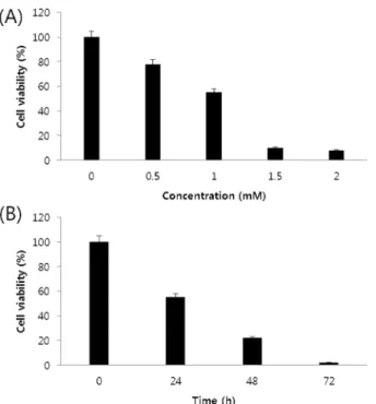
S Phase Cell Cycle Arrest and Apoptosis is Induced by Eugenol in G361 Human Melanoma Cells
Byul-Bo Ra Choi, Sang-Hun Shin
1, Uk-Kyu Kim
1, Jin-Woo Hong
2*, and Gyoo-Cheon Kim*
Department of Oral Anatomy, School of Dentistry, Pusan National University, Yangsan 626-870, Korea
1
Department of Oral & Maxillofacial Surgery, School of Dentistry, Pusan National University, Yangsan 626-870, Korea
2