INTRODUCTION
All somatic cells proliferate using common mechanism of mitosis following progression through the cell cycle. In addi- tion, apoptosis or programmed cell death appears to be a ubiq- uitous process in multicellular organisms to remove unwant- ed or damaged cells (1, 2). As it is essential to identify and eli- minate cells proliferating inappropriately, apoptosis and pro- liferation are tightly coupled, and cell cycle regulators can influ- ence both cell division and cell death (1). It is well understood that cell cycle checkpoints monitor the timing and order of cell cycle events, and these occur at the G1/S phase boundary, in S phase, and during the G2 and M phases (2). These check- points ensure that important events in each phase of the cell cycle are completed before a new phase is initiated, thereby preventing the formation of genetically abnormal cells. Dur- ing G1, the cell decides whether to continue through the cell cycle and divide, or leave the cell cycle and enter a state of qui- escence (G0). Pardee further introduced the term restriction (R) to define the point in G1, after which cells can prolifer-
ate independent of mitogenic stimuli, and also suggested the importance of the R point in malignant transformation (3).
Proteins, which are involved in the G1 checkpoint include p53, pRb, and the E2F family of transcription factors (4). The p53 protein is thought to be an essential mediator that reg- ulates the R point (5). The p53 protein is induced by DNA damaging chemicals as well as by ionizing radiation. p53 is required for G1 arrest and/or apoptosis following DNA dam- age (6). p53 activates the transcription of several genes and the best studied target molecule is p21WAF1/Cip1, which has a role in p53-mediated G1 arrest (2). In addition, the expression of the proapoptotic gene, bax, is upregulated by p53 induced after DNA damage, and this upregulation causes apoptosis (7).
p53 induces apoptosis as well as G1 arrest, but G1 arrest is not a prerequisite for p53 induced apoptosis. Cells from p21WAF1/Cip1 (-/-) mice, for example, exhibit a defect in p53 induced G1 arrest, but show normal p53-mediated apoptosis (8). There- fore, p53 induction may cause G1 arrest or apoptosis, depend- ing on the cell type.
Cells are able to undergo apoptosis in response to various Weon Seo Park, Kyeong Cheon Jung�, Doo Hyun Chung*, Woo-Dong Nam�, Won Jin Choi�, Youngmee Bae
Department of Pathology, �Orthopaedic Surgery, and �General Surgery, Kangwon National University College of Medicine, and Clinical Research Institute of Kangwon National University Hospital, Chuncheon; �Department of Pathology, Hallym University College of Medicine, Chuncheon;
*Department of Pathology, Seoul National University College of Medicine, Seoul, Korea
Address for correspondence Youngmee Bae, D.Phil.
Department of Pathology, Kangwon National University College of Medicine, 192-1 Hyoja-dong, Chuncheon 200-701, Korea
Tel : +82.33-250-8844, Fax : +82.33-242-7571 E-mail : [email protected]
*This work was supported by grant (R11-2002-098- 00000-0) form Korea Science & Engineering Foundation through the RRC (Rheumatism Research Center) at Catholic University, and a Korea Research Foundation Grant (2000-042-F00017).
467
Distinct Patterns of Cleavage and Translocation of Cell Cycle Control Proteins in CD95-induced and p53-induced apoptosis
Apoptotic cell death induced by p53 occurs at a late G1 cell cycle checkpoint termed the restriction (R) point, and it has been proposed that p53-induced apoptosis caus- es upregulation of CD95. However, as cells with defective in CD95 signaling path- way are still sensitive to p53-induced apoptosis, CD95 cannot be the sole factor result- ing in apoptosis. In addition, unlike p53-induced apoptosis, the relationship between CD95-mediated apoptosis and the cell cycle is not clearly understood. It would there- fore be worth investigating whether CD95-mediated cell death is pertinent with p53- induced apoptosis in view of cell cycle related molecules. In this report, biochemical analysis showed that etoposide-induced apoptosis caused the induction and the nuclear translocation of effector molecules involved in G1 cell cycle checkpoint. How- ever, there was no such translocation in the case of CD95-mediated death. Thus, although both types of apoptosis involved caspase activation, the cell cycle related proteins responded differently. This argues against the idea that p53-induced apop- tosis occurs through the induction of CD95/CD95L expression.
Key Words : Etoposide; Cell Cycle; Nuclear Translocation; Caspase; DNA Damage; Apoptosis
Received : 20 February 2003 Accepted : 18 March 2003
stimuli (2, 4), including death receptor ligation, cytotoxic drugs, ionizing radiation, and DNA damaging chemicals.
Among receptors that induce apoptosis, CD95 (Apo-1 or Fas) is a member of the TNF receptor family, and is expressed on various types of cells including thymocytes (9) and activated T cells (10, 11). When CD95 interacts with its ligand (CD95L), CD95 is trimerized and the trimer recruits an adaptor protein Fas associated death domain (FADD), which in turn activates caspase 8. This upstream or initiator caspase has two death effector domains (DED) at its N-terminus (12). This leads to the activation of other caspases, resulting in the cleavage of multiple vital substrates and apoptosis (13). Many studies have been focused on CD95-mediated death, but its relation- ship with the cell cycle is still controversial. For instance, Dao et al. reported that CD95-mediated death happens in G1 phase suggesting that CD95-mediated death may mimic death at the R point, while Boehme and Lenardo suggested that acti- vation induced cell death (AICD) of T cells occurs in S phase (14, 15).
It has been shown that p53 activation induces CD95 and/
or its ligand through a translation-independent pathway (16) and transiently sensitizes cells to CD95-induced apoptosis (16). However, this cannot be a general mechanism as etopo- side-induced apoptosis can be CD95-independent (17, 18). For example, cells without FADD or that express a dominant-neg- ative FADD, and which are therefore defective in CD95 sig- naling, are still sensitive to ionizing radiation and DNA-dam- aging chemicals (19, 20). Furthermore, Chen and Tan report- ed that induction of CD95 expression alone was not sufficient to induce apoptosis in irradiated cells (18). Thus, the relation- ship between CD95-induced and R checkpoint-based apop- totic mechanisms needs to be understood. These findings pro- mpted us to compare the biochemical basis of p53-induced and CD95-induced apoptosis in T cells, with respect to cell cycle control proteins.
Etoposide was used in this study to induce apoptosis, as it is known to inhibit topoisomerase II and cause DNA double strand breaks, which result in apoptosis mediated through p53 induction (21). We chose Jurkat cells as target cells to induce apoptosis as they are known to be sensitive to both CD95 and etoposide mediated apoptosis (22). Thus, we have studied the possible convergence of caspase-based and cell cycle checkpoint-based apoptotic mechanisms in response to two stimuli, CD95 ligation and etoposide treatment. We reasoned that, if a CD95-based mechanism were the principal down- stream effector of p53 induction, both etoposide-induced and CD95-induced apoptosis would result in similar patterns of substrate changes. However, the patterns we observed were quite different. We found that DNA damage induced p53 and showed strong mobilization of cell cycle R checkpoint- associated proteins to the cell nucleus, while no such move- ments were seen in CD95-mediated apoptosis. This argues against the idea that p53-induced apoptosis occurs through the induction of CD95/CD95L expression.
MATERIALS AND METHODS Cells and Reagents
Jurkat cells were maintained in RPMI1640 containing 10%
fetal calf serum (FCS, Hyclone Logan, UT, U.S.A.), 2 mM L-glutamine, 50 M -mercaptoethanol, 10 mM HEPES, and 100 U/mL each of penicillin and streptomycin. Polyclonal or monoclonal antibodies against a number of cell cycle control proteins were purchased from Santa Cruz Biotechnology (Santa Cruz, CA, U.S.A.). They were anti-p53 (sc-126), anti-E2F-1 (sc-193), anti-E2F-4 (sc-866), anti-p21WAF1/Cip1(sc-397), anti- p27Kip-1(sc-776), anti-CDK4 (sc-601), anti-p107 (sc-250), and anti-pRb (sc-50) antibodies. Anti-mouse or anti-rabbit Ig anti- serums coupled to horseradish peroxidase (HRP) were also from Santa Cruz Biotechnology. Etoposide was purchased from Sig- ma (St. Louis, MO, U.S.A.). The caspase inhibitors, z-VAD- fmk and DEVD-CHO, were from Calbiochem (La Jolla, CA, U.S.A.).
Cell Culture and Flow Cytometric Analysis
Jurkat cells were suspended at 0.5×10⁶cells/mL in a final volume of 10 mL of RPMI culture medium, and layered over control or CD95L-expressing fibroblasts (14) in 10 cm dish- es (Falcon 35-3003, Becton-Dickinson, Franklin Lakes, NJ, U.S.A.). At the indicated time points, non-adherent Jurkat cells were harvested and then tested for apoptosis (using PI staining to detect sub-diploid cells) or lysed for western blot analysis. To detect sub-diploid cells, a suspension of cells were incubated with propidium iodide (PI) at 50 g/mL (Sigma Chemical Co.) for 30 min in 0.1% sodium citrate, 0.3% Non- idet P-40, and 50 g/mL RNAse. PI stained cells were analyzed using a FACScalibur (Becton-Dickinson, Mountain View, CA, U.S.A.) and data were analyzed using Cellquest software.
Immunoblotting Analysis
Jurkat cells were centrifuged and then the cell pellet was washed and lysed in the Laemmli buffer containing a protease inhibitor cocktail (Boehringer Mannheim, Manheim, Ger- many). In order to separate cytoplasmic and nuclear proteins, Jurkat T cells were incubated with CD95L expressing fibro- blasts, or treated with 50 M of etoposide, and subcellular frac- tionation was performed as described previously (23). Briefly, cells were lysed in a hypotonic buffer (10 mM HEPES-KOH, pH 7.9; 10 mM KCl; 1.5 mM MgCl2; 0.5 mM dithiothreitol) containing 0.1% Triton X-100, and fractionated by low speed centrifugation into a low-salt-soluble supernatant for cytoplas- mic proteins and an insoluble nuclear pellet. The insoluble nuclear pellet fraction was extracted further with ELB (50 mM HEPES, pH 7.2; 250 mM NaCl; 2 mM EDTA; 0.1% NP-40).
The protein concentration was checked by using BioRad solu- tion (Bio-Rad DC Protein Assay Kit I, BioRad) and the same
amount of proteins were loaded onto 6, 10, 12, or 15% SDS- PAGE gels, electrophoresed, and transferred to PVDF Immo- bilon membranes (Millipore, Bedford, MA, U.S.A.) using an electrotransfer apparatus from Bio-Rad (Hercules, CA, U.S.A.).
Transferred membranes were incubated with primary anti- bodies recognizing each of the cell cycle related proteins for 1-3 hr at room temperature, followed by extensive washing.
Immune complexes were tagged using goat anti-rabbit or rabbit anti-mouse HRP, then visualized using the enhanced chemiluminescent (ECL) kit from Amersham Life Sciences (Buckinghamshire, U.K.).
RESULTS
In order to determine the role of caspases in CD95 or etopo- side-mediated apoptosis in Jurkat cells, the pan-caspase inhi- bitor, z-VAD-fmk was added to cells subjected to either of two death signals: CD95-engagement or etoposide treatment.
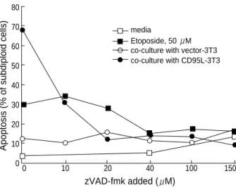
CD95 is known to activate caspase 8 through interaction of its death domain with FADD while etoposide, a DNA dam- aging agent, causes p53-induced cell death. In both cases, Jur- kat cells underwent apoptosis which was verified by PI stain- ing (Fig. 1). Upon CD95 ligation, Jurkat cells responded rapid- ly as ~52% cells were subdiploid after the 5 hr incubation with CD95L expressing fibroblasts. Etoposide induced cell death was as extensive as that of CD95 ligation but it took 18 hr to cause the same percentage of apoptotic cells (Fig. 2). In order
to examine whether caspases were the mediators of apoptosis in both cases, the caspase-inhibitor, z-VAD-fmk, was added at various concentrations. As shown in Fig. 1, z-VAD-fmk was able to block apoptosis in both cases. However, CD95-induced apoptosis was more sensitive to z-VAD-fmk inhibition, since even 10 M z-VAD-fmk caused significant blockade of CD95- induced Jurkat cell death, while significant blockade of etopo- side-induced death required 40 M z-VAD-fmk.
In the case of CD95 mediated death, z-VAD-fmk blocks the caspase 8 pathway and the subsequent cascade of caspase activation. In etoposide treatment, it is known that p53 induc-
Apoptosis (% of subdiploid cells)
zVAD-fmk added ( M) media
Etoposide, 50 M co-culture with vector-3T3 co-culture with CD95L-3T3 80
70 60 50 40 30 20 10
00 10 20 40 100 150
Fig. 1.Apoptosis induced by CD95 ligation or etoposide treatment in Jurkat cells. 50 M etoposide or co-culture of CD95L express- ing fibroblasts was applied to induce apoptosis as described in Material and Methods. 10-150 M of z-VAD-fmk or same volume of DMSO (control) was added to block the caspase activation. After 8 hr incubation, the degree of apoptosis was determined by count- ing subdiploid cells by using a PI staining and analyzed by FAC- Scalibur. open circle, Jurkat cells co-cultured with control fibrob- lasts (vector 3T3 cells); closed circle, co-cultured with CD95L trans- fected fibroblasts; open square, cultured in media containing DMSO;
and closed square, culture with 50 M of etoposide.
Fig. 2.The expression of cell cycle related proteins upon treatment with apoptotic stimuli. For etoposide-induced apoptosis, Jurkat cells were incubated in media containing 50 M etoposide for 18 hr.
Media alone was used as a control. To induce CD95 mediated de- ath, Jurkat cells were co-cultured with CD95L-transfected fibrob- lasts or control fibroblasts (vector-3T3). Apoptotic cells were cal- culated by counting subdiploid cells using PI staining as described in Material and Methods. In order to block caspase action, caspase inhibitors, 50 M of either z-VAD-fmk or DEVD-CHO was added before the induction of apoptosis. 30 g of protein extracts from each sample was subjected to SDS-PAGE. The arrows indicate each cell cycle protein. Overexposure of the p53 blot was used to verify the induction of p53 in CD95-mediated apoptosis. p107 has a caspase recognition site producing a 15 kDa C-terminal fragment which is recognized by anti-p107 ab (sc-250) and is indeed visu- alized by immunoblotting (p107 fragments).
p53 cell death (%, subdiploid)
p53
Etoposide, 50 M
6 67 52 47 7 52 75 12 72 CD95 Iigation
media etoposide + zVAD 50 M + DEVD 50 M vector-3T3 5 hr overnight + zVAD 50 M + DEVD 50 M
(longer exposure)
p21WAF1/Cip1
p27Kip-1
E2F-4
p107 (intact) p107 (fragment)
tion leads to bax expression that activates apoptosis (24).
In addition, it was reported that the expression of CD95/
CD95L was upregulated by p53 induced by DNA damaging agents (16). However, it is controversial whether p53-medi- ated death occurs through CD95-upregulation followed by its engagement by simultaneously up-regulated CD95L. Further- more, it is not known at which phase of the cycle cells become targets for CD95 mediated apoptosis, nor how cell cycle-relat- ed molecules would respond during apoptosis due to CD95 ligation in T cells. In the case of p53-mediated death, it is well documented that apoptosis occurs at the R point which is G1/
S boundary (2). We reasoned that, if a CD95-based mechanism were the principal downstream effector of p53 induction, both etoposide induced and CD95-induced apoptosis would result in similar pattern of substrate cleavage. In view of the contro- versy surrounding the relationship between apoptosis and cell cycle control, we focused on the cell cycle proteins that regu- late G1 phase. These include p53, E2F-4, the cyclin-depen- dent kinase inhibitors, p21WAF1/Cip1and p27Kip-1, and the pock- et protein, p107.
Fig. 2 shows the expression of these molecules in response
to CD95 ligation or treatment with etoposide. As expected, strong induction of p53 was found in etoposide-treated cells, while very little induction was seen in CD95 mediated death even when a longer exposure time was used. This finding con- firmed that etoposide induces classic p53-mediated apoptosis.
Furthermore, p21WAF1/Cip1was induced in both CD95 and etopo- side mediated death, although the amount induced was much higher in the case of etoposide treatment. This is also thought to be p53 dependent. For p27Kip-1, both stimuli led to the de- gradation of this protein. This could be caused by ubiquiti- nation, or through caspase activity. In fact, it was shown that p27Kip-1is rapidly ubiquitinated during the G1-S transition (25). However, it was recently reported that p27Kip-1is cleaved by caspase action, and that this resulted in increased cell death in human epithelial cells (26). In Jurkat cells, caspase-medi- ated breakdown of p27Kip-1also appeared to be important, since the loss of p27Kip-1was inhibited by z-VAD-fmk.
The E2F family consists of 6 members, which act as tran- scription factors. They are believed to be critically positive regulators of cell cycle progression. Among them, E2F-1 either induces or protects cells from apoptosis, depending on the cell type and the apoptotic stimuli (27, 28). Although E2F-4 is expressed in the early phases of the cell cycle and the expres- sion continues throughout the cycle (29), the role of E2F-4 in apoptosis is poorly understood. We were only able to detect only trace amounts of E2F-1 in Jurkat cells, but E2F-4 was abundant (Fig. 3). We therefore focused on the expression of E2F-4, which might regulate G1 progression. In the exper- iment shown in Fig. 2, there was not much difference in the expression of E2F-4, although the density of the hyperphos- phorylated form of the protein was slightly reduced. This was possibly a result of cleavage by caspases, as there is a conserved caspase-3 cleavage site at the C-terminus in all E2F family members.
DISCUSSION
The pocket proteins are the partners of E2Fs. The retinoblas- toma protein pRb interacts with E2F-1 to inhibit the func- tion of E2F-1 from acting as a transcription factor, while the pRb-related proteins, p107 and p130, bind to E2F-4. There- fore, E2F-4 is thought to play a role in the transition from the G0 to the G1 phase. As both CD95 and etoposide induce apoptosis in G1 phase cells (2, 6, 13, 14, 30), we hypothesized that E2F-4 could be involved in both apoptotic stimuli. Con- sequently, p130 and/or p107 could be involved in both cases.
However, p130 is known to act on the G0-G1 transition, while our main focus of interest is the G1/S transition. We there- fore evaluated the expression and modulation of p107. All pocket proteins have caspase-3 target sites, i.e., the DxxD motif, and it is already known that pRb is cleaved during TNFR1 and CD95-mediated apoptosis (31). As shown in Fig. 2, a 15 kDa C-terminal cleavage product of p107 was detected
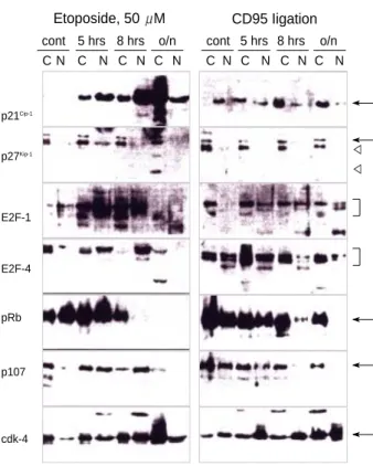
Fig. 3.Nuclear translocation of G1 cell cycle proteins. Etoposide and CD95 ligation were applied as described in Fig. 2. At the indi- cated time point, Jurkat cells were pelleted and cytoplasm and nu- clear proteins were fractionated as described in Material and Meth- ods. 30 g of each sample (either cytoplasm or nuclei) were sep- arated and immunoblotting was performed with the antibodies indi- cated. C: cytoplasmic extracts, N: nuclear extracts. Arrows indicate each cell cycle protein and arrowheads in the p27Kip-1blot are be- lieved to indicate caspase-cleaved fragments.
p21Cip-1
p27Kip-1
Etoposide, 50 M
C N C N C N C N C N C N C N C N cont 5 hrs 8 hrs o/n cont 5 hrs 8 hrs o/n
CD95 Iigation
E2F-1
E2F-4
pRb
p107
cdk-4
△△
during apoptosis due to either CD95 ligation or treatment with etoposide. This seemed to be produced by caspase activ- ity, as in CD95-ligated cells the cleavage was completely inhib- ited by z-VAD-fmk. This is the first demonstration that, in addition to pRb, p107 is one of the substrates of caspase(s) activated upon CD95 ligation.
There were subtle differences between CD95-induced and etoposide mediated apoptosis in Jurkat T cells (Fig. 2). One noteworthy detail was that the breakdown of p107 was inhib- ited completely by 50 M z-VAD-fmk in the case of CD95- induced apoptosis, but only inhibited partially by the same concentration of z-VAD-fmk in etoposide mediated apopto- sis. This is a hint that a different spectrum of caspases might be involved in the two different types of apoptosis.
The biological activity of cell cycle control proteins at the G1/S checkpoint is associated with their translocation into the nucleus (1, 5). To test whether this mechanism was operating in Jurkat cells undergoing apoptosis, we examined nuclear vs.
cytoplasmic lysates of Jurkat cells during CD95-induced and etoposide-induced apoptosis. Fig. 3 showed the differential translocation of several proteins upon etoposide treatment or CD95 ligation. Interestingly, there was no translocation of p27Kip-1into the nucleus although it has a nuclear localization signal. This suggests that other proteins such as cyclin and/or CDK might regulate the intracellular location of p27Kip-1. The E2F-1 protein did not move into the nucleus upon apoptotic signals, and therefore, it was probably not able to act as a tran- scription factor under these conditions. In accordance with this, the natural interaction partner of E2F-1, pRb, did not move into the nucleus in either case. In addition, pRb disap- peared over 8 hr during etoposide mediated cell death. There- fore, the E2F-1/pRb complex was unlikely to be a mediator in either of these apoptotic signals. This is surprising, as E2F- 1-induced cell death is thought to be important in T cells (27).
E2F-1 deficient mice exhibit a defect in T lymphocyte devel- opment leading to an excess of mature T cells due to defective apoptosis in the thymus (27). Furthermore, it was reported that E2F-1 overexpression causes increased S phase and apop- tosis (32), while pRb inhibits both processes (4, 33). However, it is possible that the apoptotic activity of E2F-1 is restricted to certain forms of apoptosis that occur in S phase. As CD95 and p53-mediated apoptosis are supposed to occur in G1 phase or during the G1-S transition, cell cycle proteins regulating G1 phase would be more likely to play a role in apoptosis due to these causes. We therefore think that at least in Jurkat cells, E2F-1-mediated cell death is not the same as G1/S checkpoint death.
There is no concrete evidence that specifically implicates E2F-4 in apoptosis. As mentioned above, p107 is expressed and functional in the G1 phase of the cycle (4). Therefore, it would be informative to determine whether there is differen- tial regulation of this molecule in response to the two distinct apoptotic signals. Among the cell cycle control proteins test- ed, three molecules were differentially translocated in the two
different types of apoptosis: p21WAF1/Cip-1, E2F-4 and p107.
Those molecules were translocated to the nucleus in etopo- side-induced apoptosis, but not in CD95-induced apoptosis.
In the case of etoposide treatment, the translocation of those molecules could be the result of a G1/S checkpoint, induced through p53. In the mouse system, TCR mediated death is thought to happen in late G1 phase (30), although this is con- troversial (15) and there is evidence that CD95-mediated death also happens in G1 (13, 14). However, at least in Jurkat cells, our data show that there is no movement of the G1 cell cycle proteins during CD95-induced apoptosis. This implies that CD95-mediated death does not involve the same mechanism as apoptosis associated with the G1-checkpoint. While sever- al previous reports document that etoposide treatment induces CD95/CD95L expression, this may not be the general mech- anism of etoposide-induced death in most cell types. In sup- port of this argument, the cells from FADD deficient mice are apoptotic in response to DNA damaging agents (19, 20).
Also, thymocytes from p53 deficient mice are sensitive to CD95-mediated death (34).
In summary, we compared the response of cell cycle control proteins to two different apoptotic stimuli, CD95 ligation and treatment with etoposide. Although both pathways were mediated by caspases, the translocation of cell cycle control proteins, especially the G1/S checkpoint-associated G1 mo- lecules, was different. Etoposide induced p53 and showed the p53 dependent, G1/S checkpoint-associated pattern of nucle- ar translocation of E2F-4, p21WAF1/Cip-1, and p107. In contrast, in CD95 mediated death, there was no translocation of G1/S cell cycle proteins. This suggests that CD95 ligation does not involve the G1/S checkpoint cell cycle control proteins to induce cell death. In addition, while p53 induction may result in CD95/CD95L expression in some systems, it is not the main pathway through which p53 induces apoptosis.
REFERENCES
1. Meikrantz W, Schlegel R. Apoptosis and the cell cycle. J Cell Biochem 1995; 58: 160-74.
2. King KL, Cidlowski JA. Cell cycle regulation and apoptosis. Annu Rev Physiol 1998; 60: 601-17.
3. Pardee AB. A restriction point for control of normal animal prolifer- ation. Proc Natl Acad Sci USA 1974; 71: 1286-90.
4. Almasan A, Yin Y, Kelly RE, Lee EY, Bradley A, Li W, Bertino JR, Wahl GM. Deficiency of retinoblastoma protein leads to inappropri- ate S-phase entry, activation of E2F-responsive genes, and apoptosis.
Proc Natl Acad Sci USA 1995; 92: 5436-40.
5. Zetterberg A, Larsson O, Wiman KG. What is the restriction point?
Curr Opin in Cell Biol1997; 7: 835-42.
6. Cox LS, Lane DP. Tumour suppressors, kinases and clamps: how p53 regulates the cell cycle in response to DNA damage. Bioessays 1995;
17: 501-8.
7. Miyashita T, Reed JC. Tumor suppressor p53 is a direct transcription-
al activator of human bax gene. Cell 1995; 80: 293-9.
8. Deng C, Zheng P, Harper JW, Elledge SJ, Leder P. Mice lacking
p21cip1/WAF1undergo normal development, but are defective in G1 check-
point control. Cell 1995; 82: 675-84.
9. Nagata S. Apoptosis by death factor. Cell 1997; 88: 355-65.
10. Crispe IN. Fatal interactions: Fas-induced apoptosis of mature T cells.
Immunity 1994; 1: 347-9.
11. Chung DH, Jung KC, Park WS, Lee I-S, Choi WJ, Kim C-J, Park SH, Bae Y. Costimulatory effect of Fas in mouse T lymphocytes. Mol Cells 2000; 10: 642-6.
12. Chinnaiyan AM, O’Rourke K, Tewari M, Dixit VM. FADD, a novel death domain-containing protein, interacts with the death domain of Fas and initiates apoptosis. Cell 1995; 81: 505-12.
13. Bae Y, Crispe IN. Differential regulation of cell cycle related proteins by CD95 engagement in thymocytes and T cell leukemic cell line, Jurkat.
J Cell Biochem 2001; 80: 328-38.
14. Dao T, Huleatt JW, Hingorani R, Crispe IN. Specific resistance of T cells to CD95-induced apoptosis during S phase of the cell cycle. J Immunol 1997; 159: 4261-7.
15. Boehme SA, Lenardo MJ. Propricidal apoptosis of mature T lympho- cytes occurs at S phase of the cell cycle. Eur J Immunol 1993; 23: 1552- 60.
16. Bennett M, Macdonald K, Chan SW, Luzio JP, Simari R, Weissberg P. Cell surface trafficking of Fas: a rapid mechanism of p53-mediated apoptosis. Science 1998; 282: 290-3.
17. Fuchs EJ, McKenna KA, Bedi A. p53-dependent DNA damage-induced apoptosis requires Fas/APO-1-independent activation of CPP32beta.
Cancer Res 1997; 57: 2550-4.
18. Chen YR, Tan TH. Lack of correlation in JNK activation and p53- dependent Fas expression induced by apoptotic stimuli. Biochem Bio- phy Res Comm 1999; 256: 595-9.
19. Rehemtulla A, Hamilton CA, Chinnaiyan AM, Dixit VM. Ultravio- let radiation-induced apoptosis is mediated by activation of CD95 (Fas/APO-1). J Biol Chem 1997; 272: 25783-6.
20. Yeh W-C, Pompa JL, McCurrach ME, Shu H-B, Elia AJ, Shahinian A, Ng M, Wakeham A, Khoo W, Mitchell K, El-Deiry WS, Lowe SW, Goeddel DV, Mak TW. FADD: essential for embryo development and signaling from some, but not all, inducers of apoptosis. Science 1998; 279: 1954-8.
21. Davis PL, Shaiu WL, Scott GL, Iglehart JD, Hsieh TS, Marks JR. Com- plex response of breast epithelial cell lines to topoisomerase inhibitors.
Anticancer Res 1998; 18: 2919-32.
22. Zhou BB, Li H, Yuan J, Kirschner MW. Caspase-dependent activation
of cyclin-dependent kinases during Fas-induced apoptosis in Jurkat cells. Proc Natl Acad Sci USA 1998; 95: 6785-90.
23. Hinds P, Tiemann F. Induction of DNA synthesis and apoptosis by regulated inactivation of a temperature-sensitive retinoblastoma pro- tein. EMBO J 1998; 17: 1040-52.
24. Brady HJM, Salomons GS, Bobeldijk RC, Berns AJM. T cells from bax alpha transgenic mice show accelerated apoptosis in response to stimuli but do not show restored DNA damage-induced cell death in the absence of p53. EMBO J 1996; 15: 1221-30.
25. Shirane M, Harumiya Y, Ishida N, Hirai A, Miyamoto C, Hatakeyama S, Nakayama, K, Kitagawa M. Down-regulation of p27Kip1by two me- chanisms, ubiquitin-mediated degradation and proteolytic processing.
J Biol Chem 1999; 274: 13886-93.
26. Levkau B, Koyama H, Raines EW, Clurman BE, Herren B, Orth K, Roberts JM, Ross R. Cleavage of p21Cip1/Waf1and p27Kip1mediates apop- tosis in endothelial cells through activation of cdk2: role of a caspase cascade. Mol Cells 1998; 1: 553-63.
27. Field SJ, Tsai F-Y, Kuo F, Zubiaga AM, Kaelin WG Jr, Livingston DM, Orkin SH, Greenberg ME. E2F-1 functions in mice to promote apoptosis and suppress proliferation. Cell 1996; 85: 549-61.
28. Phillips AC, Bates S, Ryan KM, Helin K, Vousden KH. Induction of DNA synthesis and apoptosis are separable functions of E2F-1. Genes Dev 1997; 11: 1853-63.
29. Sardet C, Vidal M, Cobrink D, Geng Y, Onufryk C, Chen A, Wein- berg RA. E2F-4 and E2F-5, two members of the E2F family, are ex- pressed in the early phases of the cell cycle. Proc Natl Acad Sci USA 1995; 92: 2403-7.
30. Lissy N A, Dyk LFV, Becker-Hapak M, Vocero-Akbani A, Mendler JH, Dowdy SF. TCR antigen-induced cell death occurs from a late G1 phase cell cycle check point. Immunity 1998; 8: 57-65.
31. Tan X, Wang JYJ. The caspase-Rb connection in cell death. Trends in Cell Biol 1998; 8: 116-20.
32. Kowalik TF, DeGregori J, Leone G, Jakoi L, Nevins JR. E2F-1-spe- cific induction of apoptosis and p53 accumulation, which is blocked by Mdm2. Cell Growth Differ 1998; 9: 113-8.
33. Hsieh JK, Fredersdorf S, Kouzarides T, Martin K, Lu X. E2F1-induced apoptosis requires DNA binding but not transactivation and is inhib- ited by the retinoblastoma protein through direct interaction. Genes Dev 1997; 11: 1840-52.
34. Li T, Ramirez K, Palacios R. Distinct patterns of Fas cell surface ex- pression during development of T- or B-lymphocyte lineages in nor- mal, scid, and mutant mice lacking or overexpressing p53, bcl-2, or rag-2 genes. Cell Growth Differ 1996; 7: 107-14.