Silibinin Inhibits Cell Growth and Induces Apoptosis through Cell-cycle Arrest in PC-3 Prostate Cancer Cells
Sang-Hun Kim
1, Kwang-Youn Kim
1, Sun-Nyoung Yu
1, Hyun-Joo Jeon
1, Young Rang Jin
3, Chang-Min Lee
2and Soon-Cheol Ahn
1,3*
1
Department of Microbiology and Immunology, Pusan National University School of Medicine, Yangsan 626-870, Korea
2
Department of Internal Medicine, Yale University School of Medicine, New Haven 06520-8057, USA
3
Medical Research Institute, Pusan National University, Yangsan 626-870, Korea
Received September 2, 2011 /Revised November 10, 2011 /Accepted November 11, 2011
Milk thistle (silybum marianum) is a famous dietary supplement widely used in the United States and Europe. Silbinin is a major biologically active compound of milk thistle and has strong antioxidant and radical scavenger activities. Anticancer activities, as well as chemopreventive effects on various cancer cell lines, including prostate, lung, colon, skin, and bladder, have also been reported in silbinin.
In the present study, we investigated the anticancer effects of silibinin and apoptosis through cell cy- cle arrest on prostate cancer cell PC-3. We performed cell viability by MTT assay and western blotting to confirm cell cycle check point proteins such as cyclin A/D1/E and cyclin-dependent kinase (CDK) 2/4/6. To quantify silibinin-induced apoptotic cell death of PC-3, Annexin V and PI double staining was performed by flow cytometry, by which its cell distribution was determined. As a result, silibinin inhibited the cell growth of PC-3 cells in a time- and dose-dependent manner, and its treatment re- sulted in cell cycle arrest at the G
1phase. Also the level of cell cycle check point proteins (cyclin, CDK) was decreased by silibinin in a dose-dependent manner. Taken together, we suggest that apop- tosis of prostate cancer cell line PC-3 induced by silibinin is associated with cell cycle arrest through decrease of cell cycle check point proteins, caspase-3 activation and poly (ADP-ribose) polymerase (PARP) cleavage.
Key words : Silibinin, apoptosis, cell cycle arrest, prostate cancer cells
*Corresponding author
*Tel:+82-51-510-8092, Fax:+82-55-382-8090
*E-mail : [email protected]
서 론
Silymarin은 Milk thistle (Silybum marianum)과 식물의 열 매나 씨앗에서 분리된 플라보노이드계 생리활성 성분 중 하나 이다. Silymarin은 silibinin, siliychristin, silydianin 등의 입체 이성질체를 포함하고 있으며, 현재까지 전립선암, 폐암, 대장 암, 피부암, 방광암, 유방암 등 다양한 암세포에 대하여 항암 효과가 있다고 보고되어 있다[1-2,6,8-9,19,21,26,29,30-31,33- 34]. 특히 silibinin은 urokinase plasminogen activator (u-PA), matrix metalloproteinase-2 (MMP-2), mitogen-activated pro- tein kinase (MAPK)의 활성을 조절하여 암세포의 증식, 침윤 및 전이에 영향을 미치고[4-5], 세포주기조절 및 세포사멸 유 도에 관한 활성이 보고되어 있다[7,11,17-18,25,28].
세포사멸은 세포가 병리학적인 요인들에 의해 노출되어 세포 내∙외부의 다양한 신호전달에 의해 세포가 죽음에 이르 는 생리학적 과정으로 세포질 및 염색질 응축, 세포막 수포 화, DNA 단편화 등으로 나타날 수 있으며, 세포 내 단백질 인 Bcl-2 family 및 caspase 활성화, poly (ADP-ribose) poly-
merase (PARP) 분절들에 의해 조절된다[10,13,15]. 최근에는 암세포의 세포주기조절을 통해 세포사멸을 유도한다는 연구 가 많이 진행되고 있다[16,24]. 세포의 성장과 분열에는 G
1, S 및 G
2/M기로 나눠지는 세포주기가 존재하는데 각 단계에서 의 조절을 통해 세포가 증식할 수 있고, 각각의 단계는 광학 현미경을 통해 관찰 할 수 있다. 보통 암세포는 정상세포에 비해 더 활발한 세포증식을 하는 것이 보고되어 있고, 이것 을 바탕으로 세포주기 또한 매우 활발히 진행됨을 알 수 있 다[14]. 세포의 증식과 사멸은 세포주기 조절인자인 cyclins, cyclin dependent kinases (CDKs), cyclin dependent kinase inhibitor 등에 의해 결정된다[3]. 세포주기 G
1기에서 S기로의 진행에는 cyclin A/cyclin D/cyclin E와 이들과 결합하는 CDK2/CDK4/CDK6에 의해서 조절된다[27,32]. Cyclin/
CDK complex의 활성에 의해 세포주기가 조절되고 불활성 에 의해 세포증식 억제 및 세포사멸을 유도하는 것으로 알려 져 있다[12,22-23].
본 연구에서는 silibinin이 인간 전립선암 세포주인 PC-3세
포에 대하여 세포주기조절과 이에 따른 세포사멸의 기전에
대하여 조사하여 얻어진 결과를 보고하고자 한다.
drochloride (DAPI)는 Sigma chemical Co. (St. Louis, MO, USA)에서 구입하였다. 각각의 antibody는 Santa Cruz Biotechnology, lnc. (Santa Cruz, Ca, USA)와 Cell Signaling Technology (Beverly, MS, USA)에서 구입하였다. 인간 전립 선암 세포주인 PC-3 세포는 American Type Culture Collection (ATCC, Manassas, VA, USA)로부터 분양 받아 10% fetal bovine serum (FBS) (WelGene Inc., Korea), 100 unit/ml penicillin, 100 μg/ml streptomycin (WelGene Inc., Korea)이 첨가된 DMEM 배지 (WelGene Inc., Korea)에서 37℃, 5% CO
2조건에서 배양하였다.
MTT assay를 이용한 세포 증식률 측정 및 세포형태 변화 의 관찰
PC-3 세포를 세포배양용 48-well plate를 이용하여 1×10
4cells/well로 분주하고 24시간 동안 안정화시킨 후, 50-250 μM 의 농도로 12, 24, 36 및 48 시간 동안 처리하였다. 이후 MTT를 0.5 mg/ml 농도가 되게 처리하여 37℃에서 4 시간 반응하였 다. 생성된 보라색의 formazan을 dimethyl sulfoxide (DMSO) 로 녹인 후 ELISA reader (VERSA
MAXmicroplate reader, Molecular Devices, Toronto, Canada)로 540 nm에서 흡광도 를 측정하였다. 또한 세포형태 변화 관찰을 위해서 세포배양 용 dish에 24 시간 동안 안정화 시킨 다음 50-250 μM 농도로 48 시간 동안 배양한 후, 광학현미경에서 각 농도에 따른 형태 변화를 관찰하였다.
세포주기 분석
PC-3 세포를 세포배양용 6-well plate를 이용하여 5×10
4cells/well로 분주하고 24 시간 동안 안정화시킨 후, 다양한 농도로 24 시간 동안 배양하였다. 그 후, silibinin이 처리된 PC-3 세포를 분리하여 phosphate-buffered saline (PBS)에 세 척하여 70% ethanol로 4℃에서 12 시간 고정한 후, RNase A를 37℃ 에 1 시간 동안 처리하고 DNA staining dye인 PI로 염색 하였다. 염색된 DNA는 flow cytometry (Becton Dickinson Co., Franklin Lakes, NJ, USA)를 이용하여 분석하였다.
세포사멸 측정
세포에서 세포사멸에 대한 신호전달이 일어나면 세포막 내
Western blot을 이용한 단백질 발현 분석
Silibinin을 처리한 PC-3 세포를 모아 PBS로 세척한 후 lysis buffer [150 mM NaCl, 10 mM Tris (pH 7.4), 5 mM EDTA (pH 8.0), 1% Triton X-100, 1 mM PMSF, 20 mg/ml aprotinin, 50 μg/ml leupetin, 1 mM venzaidine, 1 mg/ml pepstain]를 첨가하여 4℃ 에 30 분간 반응시킨 후, 13,000 rpm으로 30 분간 원심분리하여 상층액을 취하였다. Bradford 법을 이용하여 단 백질을 정량한 뒤, 50 μg의 단백질을 sodium dodecyl sul- phate (SDS)-polyacryamide gel을 이용하여 분리하였다. 그 후, poly-vinylidene difluoride membrane에 transfer하여 5%
skim milk가 함유 된 TBS-T buffer [20 mM Tris (pH 7.4), 150 mM NaCl, 0.1% Tween-20]를 이용하여 실온에서 1 시간 blocking 하였다. 각각의 1차 항체를 4℃에서 12 시간 반응하 였으며, 2차 항체를 상온에서 2 시간 반응하여 enhanced chemilluminescence (ECL) kit (Amersham pharmacia Biotech)를 이용하여 측정하였다.
통계처리
모든 실험의 표시된 결과는 3번 수행하였으며, 통계분석은 mean±S.D로 표시하였고, ANOVA에 의해 분석하였다. 통계 적 유의성은 p≤0.05로 판정하였다.
결과 및 고찰
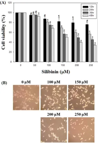
Silibinin의 세포 독성 및 세포형태 변화 측정
Silibinin은 다양한 암세포에 세포증식과 세포독성에 영향
을 미치는 것으로 보고 되었다[1,6,33-34]. 본 연구에서는 PC-3
전립선 암세포에서 silibinin에 의한 세포 독성을 측정하기 위
해 MTT assay를 수행하였다. 그 결과, 농도 및 시간 의존적으
로 silibinin에 의해 유의적으로 생존률이 감소되는 것을 관찰
하였다(Fig. 1A). 특히 48시간 처리군 중 150 μM 농도에서 비
처리군에 비하여 50% 이상 세포 독성을 나타내었고, 이는 최
근 선행연구 결과와 유사한 경향을 나타냄을 확인할 수 있었
다[20]. 또한 세포형태 변화에 미치는 영향을 확인하기 위하여
silibinin을 다양한 농도로 처리하여 48시간 배양하여 광학현
미경으로 관찰한 결과, 농도 의존적으로 세포밀도 감소와 형
태변화를 관찰 할 수 있었다(Fig. 1B). 이들 결과들을 종합해
Fig. 1. Growth inhibitory effect of silibinin on PC-3 cells. (A) PC-3 cells were treated with silibinin for 12 hr, 24 hr, 36 hr, and 48 hr. Cell viability was determined by MTT assay. (B) PC-3 cells were incubated with silibinin (100-250 μM) for 48 hr. Cell morphology was visualized by inverted microcopy. Data are presented as mean±SD (n=3 in each group).#
p
<0.05,¶p
<0.01, *p
<0.001 vs. the control group.볼 때, silibinin은 인간 전립선암 세포주인 PC-3 세포의 성장 을 억제하고 특히 세포 형태의 변화를 통해 세포 사멸이 나타 냄을 알 수 있었다.
Silibinin의 세포주기 조절 분석
세포주기는 cyclin/CDK complex의 활성과 불활성의 의해 조절되는데, 먼저 silibinin이 PC-3 전립선 암세포의 세포분열 주기상에 미치는 영향을 flow cytometry로 조사하였다. Fig.
2A에서와 같이 정상세포의 경우 G
1, S, G
2/M기에 해당하는 세포는 각각 43.77%, 28.07%, 28.15% 정도의 분포를 보였다.
반면에 silibinin이 150 μM의 농도로 처리된 세포의 분포는 G
1기가 56.66%로 증가하였고, G
2기는 18.54% 로 감소하는 것 을 관찰하였다. 이상의 결과로 보아 G
1기의 세포분포는 증가 하고 G
2기의 세포가 상대적으로 줄어드는 것을 확인함으로써
Fig. 2. Effect of silibinin on cell cycle distribution in PC-3 cells.
(A) Intracellular DNA contents with PI staining and cell cycle arrest analysis were observed by flow cytometry.
PC-3 cells were treated with 150 μM of silibinin for 24 hr. The cells were harvested, washed, and then fixed with ice-cold 70% (v/v) ethanol. After that, PI (1 mg/ml) was stained at 4℃ for 5 min. (B) The percentage of the cell cycle distribution data in histograms.
silibinin은 세포주기 중 G
1기를 억제함으로써 세포의 증식에 영향을 미치는 것을 알 수 있었다.
또한, G
1기와 관련한 세포주기에 관여하는 cyclin과 CDK의 발현 양을 western blot을 통해 확인하였다. Cyclin A, D1, E와 CDK 2, 4, 6의 발현양은 시간 및 농도 의존적으로 감소함을 알 수 있었다(Fig. 3A). 이와 같은 결과를 통해 cyclin과 CDK의 발현 양의 감소를 통해 세포주기 중 G
1기가 억제됨을 알 수 있었다.
Silibinin의 세포 사멸 분석
세포사멸을 유도하는 것을 Annexin V-FITC apoptosis de-
tection kit를 이용하여 정량화 할 수 있다. 본 연구에서는 sili-
binin을 100-200 μM의 농도로 24, 48시간 처리한 뒤, 세포막
외부로 노출되는 phosphatidylserine (PS)과 세포막의 파괴로
인해 노출되는 핵을 각각 Annexin-V와 PI를 염색하여 flow
cytometry로 측정하였다. Silibinin 처리시 세포사멸의 양이 농
도 및 시간 의존적으로 증가하는 것을 확인하였다. 특히, 48
시간 처리시, 1.11%에서 44.19%로 농도 의존적으로 증가하는
것을 확인함으로써 silibinin이 PC-3 세포에서 세포사멸을 유
도하는 것을 알 수 있었다(Fig. 4A). Fig. 4B는 세포사멸이 유도
된 세포를 백분율로 나타내었다.
Fig. 4. Effect of silibinin on apoptosis in PC-3 cells. (A) Annexin-V/ propi- dium iodide (PI) double staining assay detected by flow cytometry.
PC-3 cells were treated with silibi- nin (100-200 μM ) for 24 hr, 48 hr.
(B) The percentage of apoptotic cell data in histograms. Data are pre- sented as mean±SD (n=3 in each group). 
p
<0.01, *p
<0.001 vs. the control group.Fig. 3. Effect of silibinin on expression of cell cycle-related proteins. Cells were treated with silibinin (100-200 μM) for 24-48 hr. (A) Expression of cyclin D1, cyclin E and cyclin A. (B) Expression of CDK 2, CDK 4 and CDK 6. β-Actin was used as a loading control.
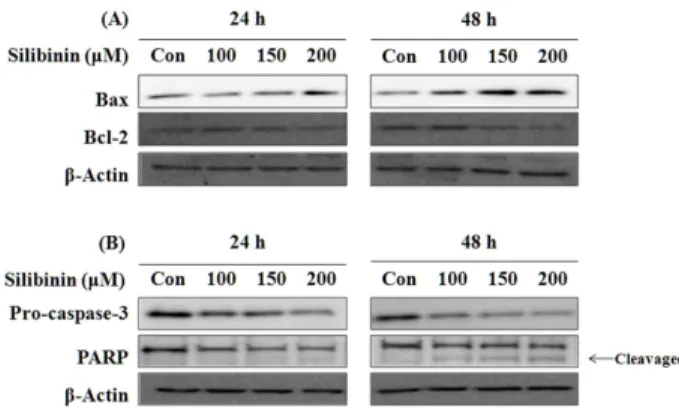
Silibinin의 세포사멸 관련 단백질인 Bcl-2 family의 발현과 caspase-3 및 poly(ADP-ribose) polymerase (PARP)의 활성화
세포 내에서 세포사멸의 조절은 관련 단백질의 조절에 의해 일어 날 수 있는데 대표적으로 Bcl-2 family와 caspase-3, PARP 등이 가장 잘 알려져 있다. 그 중 Bcl-2 family는 세포
pro-apoptotic protein인 Bax의 발현양은 증가하는 것을 확인 하였다. 또한 pro-caspase-3의 발현이 시간, 농도 의존적으로 감소하였고, caspase-3의 기질로 알려진 PARP의 분절형태가 증가하는 것을 확인 하였다. 이와 같은 결과를 통해 silibinin이 PC-3 세포에서 Bcl-2 family의 변화를 유도하여 caspase-3 경 로를 통해 세포사멸을 유도하는 것을 알 수 있었다.
이상의 결과들을 통해 silibinin이 인간 전립선 암세포 PC-3
세포의 증식을 억제하여 세포사멸을 유도하는 것을 알 수 있
었다. 특히 flow cytometry analysis와 세포주기 G
1기 관련 cy-
clin과 cyclin dependent kinase 등의 단백질 발현이 감소하는
것을 확인함으로써 G
1arrest를 하여 증식을 억제하는 것을
알 수 있었다. 또한 Bcl-2 family, caspase-3, PARP 등의 단백질
의 발현을 확인함으로써 세포사멸을 유도하는 것을 확인하였
다. 본 연구는 세포주기와 관련하여 silibinin의 전립선 암에
대한 세포사멸기전을 확인하였으나 앞으로의 전립선암에 대
한 치료제로서의 사용 가능성을 나타내기 위해서는 이에 대한
Fig. 5. Effects of silibinin on expression of apoptosis-related pro- teins in PC-3 cells. Cells were treated with silibinin (100-200 μM) for 24-48 hr. (A) Expression of Bcl-2 and Bax proteins. (B) Expression of procaspase-3 and PARP.
β-Actin was used as a loading control.
더 자세한 항암기전에 관한 연구가 더 진행 되어야 할 것이다.
감사의 글
본 연구는 한국 연구 재단 일반연구자 지원사업으로 수행되 었습니다(MOEHRD) (KRF-2009-0071732).
References
1. Agarwal, C., A. Tyagi, M. Kaur, and R. Agarwal. 2007.
Silibinin inhibits constitutive activation of Stat3, and causes caspase activation and apoptotic death of human prostate carcinoma DU145 cells.
Carcinogenesis
7, 1463-1470.2. Agarwal, C., R. P. Singh, S. Dhanalakshmi, A. K. Tyagi, M.
Tecklenburg, R. A. Sclafani, and R. Agarwal. 2003. Silibinin upregulates the expression of cyclin-dependent kinase in- hibitors and causes cell cycle arrest and apoptosis in human colon carcinoma HT-29 cells.
Oncogene
22, 8271-8282.3. Aktas, H., H. Cai, and G. M. Cooper. 1997. Ras links growth factor signaling to the cell cycle machinery via regulation of cyclin D1 and the Cdk inhibitor p27KIP1.
Mol. Cell Biol
. 17, 3850-3857.4. Chen, P. N., Y. S. Hsieh, C. L. Chiang, H. L. Chiou, S. F.
Yang, and S. C. Chu. 2006. Silibinin inhibits invasion of oral cancer cells by suppressing the MAPK pathway.
J. Dent. Res.
85, 220-225.
5. Chen, P. N., Y. S. Hsieh, H. L. Chiou, and S. C. Chu. 2005.
Silibinin inhibits cell invasion through inactivation of both PI3K-Akt and MAPK signaling pathways.
Chem. Biol.
Interact.
156, 141-150.6. Chu, S. C., H. L. Chiou, P. N. Chen, S. F. Yang, and Y.
S. Hsieh. 2004. Silibinin inhibits the invasion of human lung cancer cells via decreased productions of urokinase-plasmi- nogen activator and matrix metalloproteinase-2.
Mol.
Carcinog
. 40, 143-149.7. Deep, G., N. H. Oberlies, D. J. Kroll, and R. Agarwal. 2008.
Identifying the differential effects of silymarin constituents on cell growth and cell cycle regulatory molecules in human prostate cancer cells.
Int. J. Cancer
123, 41-50.8. Dhanalakshmi, S., G. U. Mallikarjuna, R. P. Singh, and R.
Agarwal. 2004. Dual efficacy of silibinin in protecting or en- hancing ultraviolet B radiation-caused apoptosis in HaCaT human immortalized keratinocytes.
Carcinogenesis
25, 99-106.9. Dhanalakshmi, S., R. P. Singh, C. Agarwal, and R. Agarwal.
2002. Silibinin inhibits constitutive and TNFalpha-induced activation of NF-kappaB and sensitizes human prostate car- cinoma DU145 cells to TNFalpha-induced apoptosis.
Oncogene
21, 1759-1767.10. Eom, K. S., H. J. Kim, H. S. So, R. Park, and T. Y. Kim.
2010. Berberine-induced apoptosis in human glioblastoma T98G cells is mediated by endoplasmic reticulum stress ac- companying reactive oxygen species and mitochondrial dysfunction.
Biol. Pharm. Bull.
33, 1644-1649.11. Flaig, T. W., L. J. Su, G. Harrison, R. Agarwal, and L. M.
Glodé. 2007. Silibinin synergizes with mitoxantrone to in- hibit cell growth and induce apoptosis in human prostate cancer cells.
Int. J. Cancer
120, 2028-2033.12. Jackson, S. J. and K. W. Singletary. 2004. Sulforaphane in- hibits human MCF-7 mammary cancer cell mitotic pro- gression and tubulin polymerization.
J. Nutr.
134, 2229-2236.13. Jin, C. Y., G. Y. Kim, and Y. H. Choi. 2008. Induction of apoptosis by aqueous extract of
Cordyceps militaris
through activation of caspases and inactivation of akt in human breast cancer MDA-MB-231 cells. J. Microbiol. Biotechnol
. 18, 1997-2003.14. Kim, D. W., S. L. Kwang, M. K. Kim, Y. H. Cho, and C.
H. Lee. 2001. Inhibitory mechanisms of cell cycle regulation induced by indole-3-carbinol in hepatocellular carcinoma HepG2 cells.
Korean J. Appl. Microbiol. Biotechnol
. 29, 181-185.15. Kong, E. H., Y. J. Kim, Y. J. Kim, H. J. Cho, S. N. Yu, K.
Y. Kim, J. H. Chang, and S. C. Ahn. 2008. Piplartine induces caspase-mediated apoptosis in PC-3 human prostate cancer cells.
Oncol. Rep
. 20, 785-792.16. Liu, M. J., Z. Wang, Y. Ju, R. N. Wong, and Q. Y. Wu. 2004.
Diosgenin induces cell cycle arrest and apoptosis in human leukemia K562 cells with the disruption of Ca2+homeostasis.
Cancer Chemother. Pharmacol
. 55, 79-90.17. Li, L., Y. Gao, L. Zhang, J. Zeng, D. He, and Y. Sun. 2008.
Silibinin inhibits cell growth and induces apoptosis by cas- pase activation, down-regulating survivin and blocking EGFR–ERK activation in renal cell carcinoma.
Cancer Letters
272, 61-69.18. Mateen, S., A. Tyagi, C. Agarwal, R. P. Singh, and R.
Agarwal. 2010. Silibinin inhibits human nonsmall cell lung cancer cell growth through cell-cells arrest by modulating expression and function of key cell-cycle regulators.
Mol.
Carcinog.
49, 247-58.19. Mohan, S., S. Dhanalakshmi, G. U. Mallikarjuna, R. P. Singh, and R. Agarwal. 2004. Silibinin modulates UVB-induced apoptosis via mitochondrial proteins, caspases activation,
초록:인간 전립선 암세포 PC-3 세포에서 Silibinin의 세포주기조절을 통한 세포사멸 유도 효과 김상헌
1․김광연
1․유선녕
1․진영랑
3․전현주
1․이창민
2․안순철
1,3*
(
1부산대학교 의학전문대학원 미생물학 및 면역학 교실,
2예일대학교 호흡기내과,
3부산대학교 의학연구소) Silibinin은 milk thistle에서 분리된 주된 생리활성 성분으로 강력한 항산화제, 항암활성에 대해서 보고되어 있 다. 하지만 항암활성에 대한 정확한 기전에 대해서는 밝혀져 있지 않다. 본 연구에서는 인간 전립선 암세포주인 PC-3 세포를 이용하여 세포사멸의 기전을 조사하였다. MTT assay를 통해 세포독성을 확인하였고, PI 염색을 통 해 세포주기를 확인하고, Annexin-V/PI 염색을 통한 세포사멸을 확인하였다. 뿐만 아니라 western blot을 이용하 여 세포주기 및 세포사멸에 관련된 단백질 발현 정도를 확인하였다. 본 연구의 결과에서 silibinin은 인간 전립선 암세포주인 PC-3 세포에서 세포주기관련 단백질의 발현을 조절하여 세포주기 진행을 억제함으로써 세포사멸을 유도 함을 알 수 있었다.
22. Parnaud, G., P. Li, G. Cassar, P. Rouimi, J. Tulliez, L.
Combaret, and L. Gamet-Payrastre. 2004. Mechanism of sul- foraphane-induced cell cycle arrest and apoptosis in human colon cancer cells.
Nutr. Cancer
48, 198-206.23. Pham, N. A., J. W. Jacobberger, A. D. Schimmer, P. Cao, M. Gronda, and D. W. Hedley. 2004. The dietary iso- thiocyanate sulforaphane targets pathways of apoptosis, cell cycle arrest, and oxidative stress in human pancreatic cancer cells and inhibits tumor growth in severe combined im- munodeficient mice.
Mol. Cancer Ther
. 3, 1239-1248.24. Prasad, S., J. Kaur, P. Roy, N. Kalra, and Y. Shukla. 2007.
Theaflavins induce G2/M arrest by modulating expression of p21waf1/cip1, cdc25C and cyclin B in human prostate carcinoma PC-3 cells.
Life Sci.
81, 1323-1331.25. Raina, K., S. Rajamanickam, R. P. Singh, G. Deep, M.
Chittezhath, and R. Agarwal. 2008. Stage specific inhibitory effects and associated mechanisms of silibinin on tumor pro- gression and metastasis in TRAMP Model.
Cancer Res
. 68, 6822-6830.26. Sharma, G., R. P. Singh, D. C. Chan, and R. Agarwal. 2003.
Silibinin induces growth inhibition and apoptotic cell death in human lung carcinoma cells.
Anticancer Res
. 23, 2649-T47D human breast carcinoma cells involves caspase-8 acti- vation and mitochondrial pathway.
Cancer Invest.
1, 12-20.30. Tyagi, A. K., C. Agarwal, R. P. Singh, K. R. Shroyer, L. M.
Glode, and R. Agarwal. 2003. Silibinin down-regulates sur- vivin protein and mRNA expression and causes caspases activation and apoptosis in human bladder transitional-cell papilloma RT4 cells.
Biochem. Biophys. Res. Commun.
312, 1178-1184.31. Tyagi, A., C. Agarwal, G. Harrison, L. M. Glode, and R.
Agarwal. 2004. Silibinin causes cell cycle arrest and apopto- sis in human bladder transitional cell carcinoma cells by reg- ulating CDKI-CDK-cyclin cascade, and caspase 3 and PARP cleavages.
Carcinogenesis
25, 1711-1720.32. Weinberg, R. A. 1995. The retinoblastoma protein and cell cycle control.
Cell
81, 323-330.33. Yang, S. H., J. K. Lin, W. S. Chen, and J. H. Chiu. 2003.
Anti-angiogenic effect of silymarin on colon cancer LoVo cell line.
J. Surg. Res
. 113, 133-138.34. Zhu, W., J. S. Zhang, and C. Y. Young. 2001. Silymarin in- hibits function of the androgen receptor by reducing nuclear localization of the receptor in the human prostate cancer cell line LNCaP.