Anti-proliferation, Cell Cycle Arrest, and Apoptosis Induced by Natural Liquiritigenin from Licorice Root in Oral Squamous Cell Carcinoma Cells
Ah-Won Kwak
1, Goo Yoon
1, Jung-Il Chae
2* and Jung-Hyun Shim
1,3*
1
Department of Pharmacy, College of Pharmacy, Mokpo National University, Jeonnam 58554, Korea
2
Department of Dental Pharmacology, School of Dentistry, BK21 Plus, Chonbuk National University, Jeonju 54896, Korea
3
The China-US (Henan) Hormel Cancer Institute, Zhengzhou, Henan, 450008, P.R. China Received November 8, 2018 /Revised December 28, 2018 /Accepted February 12, 2019
Liquiritigenin (LG) is a chiral flavonoid isolated from the roots of licorice. It exhibits multiple bio- logical activities including anti-oxidant, anti-cancer, and anti-inflammatory effects. In particular though, the anti-cancer activity of LG in oral squamous cell carcinoma has yet to be elucidated, and LG-in- duced apoptosis in oral squamous cell carcinoma remains poorly understood. In the present study, we tested the role of LG in inducing apoptosis in oral squamous cell carcinoma cells. LG treatment of HN22 cells resulted in a dose-dependent inhibition of cell viability as detected by a 3-(4,5-dime- thylthiazol-2-yl)-2,5 diphenyltetrazolium bromide assay. The induction of apoptosis in terms of Annexin V/7-Aminoactinomycin D staining, sub-G1 population, and multi-caspase activity were assessed with a Muse
TMCell Analyzer. Flow cytometric analysis revealed that LG treatment resulted in G2/M arrest in cell cycle progression and downregulation of cyclin B1 and CDC2 expression in a concentration- dependent manner. It also resulted in significant upregulation of p27. In addition, LG was seen to trigger the generation of reactive oxygen species and induce CCAAT/enhancer-binding protein ho- mologous protein and 78-kDa glucose-regulated protein in concentration-dependent upregulation. The LG treatment of HN22 cells led to a loss of mitochondrial membrane potential (△Ψm); it also reduced the levels of anti-apoptotic protein and increased the expression of apoptotic protease activating fac- tor-1, cleaved poly (ADP-ribose)polymerase and Bax. Overall, our results indicate that the pro-apop- totic effects of LG in HN22 cells depend on the activation of both intrinsic and extrinsic signaling pathways. Thus, our results suggest that LG constitutes a natural compound with a potential role as an anti-tumor agent in oral squamous cell carcinoma.
Key words : Apoptosis, Liquiritigenin; oral squamous cell carcinoma, reactive oxygen species
*Corresponding authors
*Tel : +82-61-450-2684, Fax : +82-61-450-2689
*E-mail : [email protected] (Jung-Hyun Shim)
*Tel : +82-63-270-4024, Fax : +82-63-270-4037
*E-mail : [email protected] (Jung-Il Chae)
This is an Open-Access article distributed under the terms of the Creative Commons Attribution Non-Commercial License (http://creativecommons.org/licenses/by-nc/3.0) which permits unrestricted non-commercial use, distribution, and reproduction in any medium, provided the original work is properly cited.
Journal of Life Science 2019 Vol. 29. No. 3. 295~302 DOI : https://doi.org/10.5352/JLS.2019.29.3.295
서 론
전세계적으로 암은 인간의 사망원인 중 많은 부분을 차지하 며, 그 중 구강암은 인체에 발생하는 전체 암종의 3~5% 정도를 차지한다[36]. 구강암은 다른 부위의 암종과 비교하여 발현빈 도는 낮으나 악성도가 높고 5년 생존율이 비교적 낮다[17]. 구 강암중에서 90% 이상을 차지하는 구강편평세포암종은 주변 조직으로 쉽게 침습하여 잦은 전이를 보인다[22]. 최근 구강암 의 치료를 위해 크게 수술, 방사선, 항암화학요법 등의 3가지 치료방법이 사용되고 있다[4, 16]. 주요 치료법 중에서 1980년
대부터 cisplatin등의 약제가 구강암을 포함한 두경부 암종에 효과적으로 작용한다는 보고가 알려지면서 항암 화학요법이 증가되었지만, 골수기능저하나 소화기계 합병증 및 면역력저 하 등과 같은 부작용이 나타나고 있다[1]. 따라서, 최근에는 합성화학물에 비해 상대적으로 부작용이 적은 천연물의 활성 연구가 진행되고 있다[27].
천연물인 감초에서 추출한 Liquiritigenin (LG)은 플라보노 이드 구성원이다[21]. LG는 항암, 항산화, 항염증, 항고지혈증 및 항알레르기작용을 포함하여 다양한 생화학적 활성을 가지 고 있다[1, 37]. LG는 포유류 세포주에서 세포사멸을 통한 항 종양 활성을 나타낸다는 연구 결과가 있다[14, 21, 37]. 그렇지 만 인간 구강편평세포암종 세포 내에서 LG이 어떠한 분자적 기작을 통하여 세포사멸에 의한 항 종양 활성을 나타내는지에 대한 연구가 필요하다.
세포사멸은 발달과 노화과정에서 정상적으로 일어나며, 면
역반응이나 질병 및 유해 물질에 의해 세포가 손상 될 때와
같은 방어 기작으로 발생한다[8]. Apoptosis는 크게 외인적 경
로(death receptor-mediated)와 내인적 경로(mitochon-
drial-mediated)로 구분된다[9]. 외인적 경로는 세포막에 존재
하는 death receptor에 death ligand가 결합하여 death com- plex를 형성하고, death domain-containing protein [Fas-asso- ciated protein with death domain (FADD)]와 결합하여 cas- pase-8을 포함한 initiator caspase를 활성화 시켜 이루어진다 [6, 9, 30]. 내인성 경로는 미토콘드리아의 막투과성과 관련있 으며, Bcl-2 family protein의 비활성화를 통하여 cytochrome C (cyto C)의 방출을 유도한다[11]. 방출된 cyto C는 apoptotic protease activation factor-1 (Apaf-1)과 initiator caspase인 caspase-9과 결합하여 apoptosome 복합체를 형성한다[31].
Apoptosome은 caspase-9을 활성화 시켜 effector caspase (caspase-3/-7)의 활성을 통해 apoptosis를 유발한다[9]. 본 논 문에서는 LG가 구강편평상피암 세포(HN22)의 성장 억제 및 세포사멸 기전을 확인하였다.
재료 및 방법
재료 및 항체
본 실험에 사용된 LG은 목포대학교 약학대학의 윤구 교수 에게 제공받아 실험을 수행하였다[24]. 세포의 배양에 사용된 DMEM 배지은 Welgene (Daegu, Korea)에서 구입하였다.
Fetal bovine serum (FBS), penicillin/streptomycin, Phos- phate buffered saline (PBS) 및 trypsin은 Hyclone (Logan, UT, USA)에서 구입하였다. 세포증식 측정을 위해 사용된 Dimethyl sulfoxide 및 3-(4,5-dimethylthiazol-2-yl)-2,5-diphe- nyltetrazolium bromide (MTT)은 Sigma-Aldrich (St.Louis, MO, USA)에서 구입하였다. 단백질 분석을 위하여 사용된 모 든 항체는 Santa Cruz Biotechnology (Santa Cruz, CA, USA) 에서 구입하였다.
세포배양
인간 구강편평상피암 HN22 (RRID:CVCL_5522) 세포는 단 국대학교에서 제공받았다[29]. HN22 세포는 10% FBS 및 1%
penicillin/streptomycin을 포함하는 DMEM 배지에 5% CO
2가 공급되는 37°C incubator에서 배양하였다.
Cell viability assay
HN22 세포를 96-well plates에 각 well 당 1.5×10
3으로 100 μl의 배지와 함께 분주하고 5% CO
2가 공급되는 incubator 에 서 배양 후, 50, 100 및 150 μM 농도의 LG를 24시간 및 48시간 동안 처리하였다. 배양된 배지를 제거하고 MTT 30 μl/well 넣고 incubator에서 1시간 30분간 반응 뒤에 상층액을 제거하 고 DMSO를 100 μl/well 넣고 5분간 반응시켰다. 흡광도는 Spectrophotometer (Thermo Fisher Scientific, Vantaa, Fin- land)을 사용하여 570 nm에서 측정하였다.
세포주기변화 분석
HN22 세포에서 LG의 세포주기에 미치는 영향을 알아보기 위하여, 6-well plate에 HN22 세포를 5.4×10
4cell/3 ml 씩 분 주하여 배양한 후, 다양한 농도(0, 50, 100 및 150 μM)의 LG를 처리하였다. 48시간 동안 처리 후 세포를 회수하여 PBS로 세 척한 다음 70% 에탄올을 첨가하여 -20°C에서 하루 동안 고정 하였다. 고정된 세포를 다시 PBS로 세척하고 13,000 rpm으로 20분간 원심 분리하였다. PBS를 제거한 후 Muse Cell cycle reagent (Merck Millipore, Darmstadt, Germany)를 200 μl 첨 가하여 상온에서 30분간 반응하였다. HN22 세포의 세포주기 는 Muse
™Cell Analyzer (EMD Millipore, Billerica, MA, USA)를 이용하여 분석하였다.
Annexin V 염색에 의한 apoptosis 관찰
LG 처리에 따른 HN22 세포의 apoptosis 유도를 관찰하기 위하여 Muse
TMAnnecin V & Dead Cell Kit (Merck Millipore, Darmstadt, Germany)를 사용하였다. HN22 세포를 6-well plate 배양한 후, LG를 다양한 농도로 48시간 동안 처리하였 다. 약물 처리 후 세포를 회수하여 Muse
TMAnnecin V & Dead reagent 100 μl를 첨가하여 실온에서 30분간 반응하였다. 염색 한 세포는 Muse
™Cell Analyzer로 분석하였다.
Mitochondrial membrane potential (MMP)의 분석 HN22 세포의 MMP 변화 정도를 측정하기 위하여 HN22 세포를 6-well plate에 배양하고, LG를 0, 50, 100 및 150 μM의 농도로 48시간 처리하였다. LG를 처리한 세포를 회수하여 1X Assay buffer (Merck Millipore, Darmstadt, Germany)로 세척 한 후, MiroPotential working solution (Merck Millipore, Darmstadt, Germany) 95 μl를 첨가하여 37°C에서 20분간 반 응시켰다. 다음 세포에 7-aminoacinomycin D (Merck Milli- pore, Darmstadt, Germany)를 5 μl 처리하여 상온에서 10분간 반응시킨 후 Muse
™Cell Analyzer로 측정하였다.
활성산소종 분석
HN22 세포 내 활성산소종 생성 변화를 확인하기 위하여, HN22 세포를 6-well plate에 24시간 동안 배양한 후, LG를 다양한 농도로 처리하였다. LG 처리 후, 세포를 회수하여 PBS 로 세척한 후 Muse Oxidative Stress Reagent working sol- ution (Merck Millipore, Darmstadt, Germany)을 처리하여 37°C에서 30분간 반응시켰다. 세포는 Muse
™Cell Analyzer를 이용하여 분석하였다.
단백질 발현 분석
LG의 항암 활성 기전을 밝히기 위해 apoptosis 조절에 관여 하는 단백질 발현을 확인하고자 Western blot을 수행하였다.
LG를 농도 별로 처리한 HN22 세포를 PBS로 세척한 후, RIPA
24 hr 48 hr
A
B
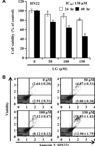
Fig. 1. Effects of LG on the cell viability and apoptosis in human oral squamous carcinoma HN22 cells. HN22 cells were treated with the indicated concentration of LG for 24 and 48 hr. (A) Cell viability was assessed by MTT assay. (B) The apoptotic status was evaluated by Annexin V/7-ami- noacinomycin D assay. The lower right part was per- ceived as early stage of apoptotic cells and top right part was considered as late stage of apoptotic cells. The sig- nificance was presented as mean ± SD, p<0.05 compared to control group.
lysis buffer (iNtRON, Korea)를 첨가하여 sonication 하였다.
용해된 단백질을 13,000 rpm으로 30분간 원심 분리하여 상층 액을 회수하였다. 추출한 단백질 농도를 Bio-Rad DC Protein Assay (Bio-RAD, Hercules, CA, USA)로 결정한 후 SDS- PAGE로 전기 영동 하였다. 분리된 단백질을 polyvinylidene fluoride로 전이시킨 후 0.1% tween-20을 함유한 PBS (PBST) 에 녹인 탈지유를 이용하여 blocking하였다. PBST로 2차례 세 척한 후 1차 항체를 처리하여 4°C에서 14시간 동안 반응하였 다. Polyvinylidene fluoride를 PBST로 30분간 세척한 후에 Horse radish peroxidase가 표지된 2차 항체를 처리하여 상온 에서 2시간 동안 반응하였다. 단백질은 Western blotting lu- minol reagent (Santa Curz Biotechnology, Santa Curz, CA, USA)를 이용해 ImageQuant LAS 500 (GE Healthcare, Uppsala, Sweden)로 분석하였다.
통계분석
모든 실험 결과는 평균값(mean values) ± 표준편차(SD)로 나타내었다. 분석된 실험 데이터의 통계적 유의성은 분산분석 (Analysis of Vatiance, ANOVA)을 사용하여 검증하였고, p value<0.05인 경우 유의성이 있는 것으로 판단하였다.
결과 및 고찰
LG 처리에 의한 HN22 세포의 증식 억제 및 apoptosis 유발
플라보노이드 화합물은 항산화제, 향균제 및 항암제와 같은 많은 유익한 활성을 나타낸다[20, 40]. 이전 연구에서 Hepato- cellular carcinoma 세포인 HepG2 및 PLC/PRF/5 세포에 24 시간 동안 LG를 처리한 하였을 때, IC
50값은 각각 495.5 μM 및 372.5 μM으로 나타났다[37]. 또한, Glycyrrhizae radix에서 추출한 플라보노이드 중 하나인 LG을 pituitary adenoma 세 포인 MMQ 세포와 GH3 세포에 48시간 처리하였을 때 IC
50값이 각각 289.2 μM 및 272.9 μM로 나타났다[38]. 감초에서 추출된 LG가 구강편평상피암세포 HN22에서의 세포 생존율 에 미치는 영향을 확인하기 위하여 HN22 세포에 LG (50, 100 및 150 μM)를 24, 48시간 동안 처리한 후 MTT assay를 실시하 였다. Fig. 1A에 나타낸 결과에서 알 수 있듯이, LG를 처리하 였을 때 시간 및 농도 의존적으로 HN22 세포의 증식이 유의적 으로 감소되었다. HN22에 LG를 24시간 동안 처리하였을 때 IC
50값은 204 μM이며, 48시간 동안 처리하였을 때 IC
50값은 HN22에서 138 μM이었다. 기존의 논문에서 보고한 바와 같이, 다양한 암종에서 LG의 항암 효능을 확인할 수 있는 IC
50농도 보다는 구강편평상피암세포에서 더 낮은 IC
50농도로 세포증 식을 억제함을 확인하였다. Apoptosis가 유발되면 염색질 응 축, 핵 분열, 세포자멸체 형성, 세포 수축, 막 소포 및 세포 내막에 존재하던 phosphatidylserine의 노출과 같은 뚜렷한
형태학적 특징이 나타난다[18, 39]. 세포증식 억제가 apoptosis 유발과 관련성이 있는지의 여부를 조사하기 위하여, Annexin V/7-aminoacinomycin D 이중 염색을 통해 Muse
™Cell Analyzer로 측정하였다. 실험 결과, 대조군에서 자연적으로 apoptosis가 유발된 세포의 빈도가 5.55±0.23%로 나타났으며, 고농도인 150 μM에서는 31.84±0.44%로 농도의존적으로 apo- ptosis가 유발된 세포의 빈도가 증가하였다(Fig. 1B). 이는 LG 처리에 의한 HN22 세포의 증식 억제가 apoptosis 유발과 연관 되어 있음을 확인하였다.
HN22 세포의 세포주기 조절 및 G2/M기 조절 단백질의 발현에 미치는 LG의 영향
세포주기의 진행은 세포가 DNA를 복제, 분열 및 증식시키
는 일련의 신호전달에 의해 조절된다[34]. Cyclin D1과 CDK4
A
B
C
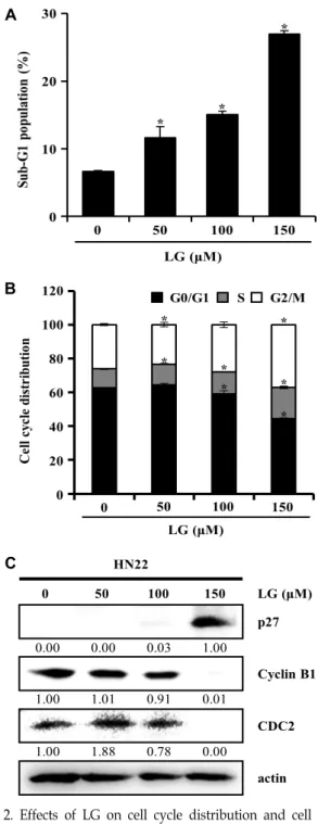
Fig. 2. Effects of LG on cell cycle distribution and cell cycle regulation protein expression in HN22 cells. Cells was treated with 0, 50, 100 and 150 μM of LG for 48 hr.
Following incubation, cells were harvested, fixed and stained with Muse
TMCell cycle reagent and analysed by Muse
TMcell analyzer. (A) Bar diagram showing the percentage of apoptotic cells in Sub-G1 phase of cell cycle. (B) The proportions (%) in each phase (G0/G1, S and G2/M) of the HN22 cells. Each point represents the mean of three independent experiments, expressed by mean ± SD. *p<0.05 significantly different from control. (C) The cells were lysed and performed to west- ern blotting. The expression level of actin was used as internal controls.
및 CDK6의 결합은 G1 단계를 거치면서 진행되며, cyclin E 및 cyclin A가 CDK2와 결합하여 중심체 G1/S와 S 단계 동안 중심체의 복제를 조절하며, cyclin A/cyclin B/ CDK1복합체 는 G2/M checkpoint를 조절한다[5, 34]. p27, p21 및 p57은 각각의 표적과 CDK의 상호 작용을 차단하여 세포주기 정지를 일으킨다[25, 34]. LG에 의한 세포 증식 억제 효과가 세포주기 조절에 영향이 있는지 확인하기 위하여, HN22 세포에 LG를 48시간 동안 농도별로 처리한 후 Muse
™Cell Analyzer를 통해 세포주기를 측정하였다. 그 결과 Apoptosis를 나타내는 sub-G1 기의 세포 비율은 대조군 경우 6.63± .12%로 나타났으 며, 고농도인 30 μM에서 27.00±0.44%로 세포 비율이 증가되는 것을 확인할 수 있었다(Fig. 2A). 또한 G2/M 기는 대조군일 때 26.7±0.60%였던 세포 빈도가 고농도인 30 μM에선 37.10±
0.26%로 농도 의존적으로 증가된 것을 확인했다(Fig. 2B). cy- clin B1은 CDC 2를 활성화 시켜 복합체를 형성해 G2/M기 전이를 조절한다[35]. 반면, cip/kip family은 cyclin/CDKs 활 성 억제제이며, 세포 주기의 진행을 막는다[42]. HN22 세포에 서 LG 처리에 의한 G2/M arrest 유발에 관련된 기전 확인을 위하여, G2/M 기 주요 세포주기 조절인자들의 단백질 발현 변화를 Western blot으로 확인하였다. Fig. 2C에 나타낸 바와 같이 LG처리에 의해 세포주기 양성 조절 인자인 cyclin B1의 발현이 감소 되었으며, cyclin 단백질들과 결합하여 세포주기 조절에 중요한 역할을 하는 CDKs인 CDC2의 발현 또한 감소 하였다. cip/kip family 구성원인 p27의 발현이 증가하였다.
따라서, LG가 HN22 세포에서 sub-G1기의 증가 및 G2/M ar- rest를 통하여 세포사멸 유도함을 시사한다.
LG에 의한 apoptosis 유발에서 활성산소종 및 MMP의 역할
활성산소종은 생체 내에서 일어나는 대사과정 중 발생된다
[7]. 높은 수준의 ROS는 세포에 DNA 손상, 미토콘드리아 기
능 장애 및 apoptosis를 일으켜 암에서 치료 효과를 가져온다
[15]. 많은 항 종양 약물은 암세포의 활성산소종 생산과 미토콘
드리아 경로에 영향을 주어 세포사멸을 유도한다고 보고되어
있다[23]. LG의 처리에 따른 활성산소종 생성에 미치는 영향
을 확인하기 위하여, 48시간 동안 LG를 처리한 HN22 세포로
Muse
™Cell Analyzer를 통해 분석하였다. Fig. 3A에 나타낸
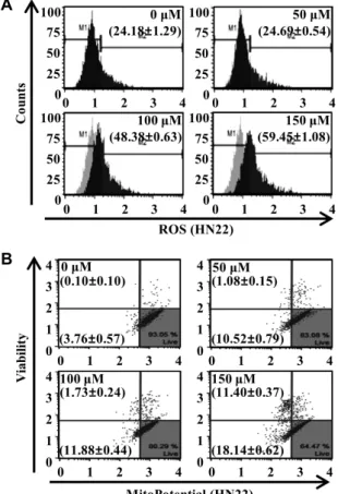
결과, 대조군에서의 M2 값은 24.18±1.29%로 나타났다. LG
(50, 100 및 150 μM) 를 처리한 HN22 세포의 M2 값은 각각
24.69±0.54%, 48.38±0.63%, 59.45±1.08%로 농도의존적으로 증
가하는 것을 볼 수 있었다. MMP는 mitochondrial apoptosis
경로의 중요한 초기 결정 인자이다[23]. 활성산소종의 생성이
증가하면 미토콘드리아 막전위의 손실이 일어나 apoptosis를
유발한다[23]. LG 처리에 따른 HN22 세포에서의 apoptosis
유발에 있어서 미토콘드리아 기능 소실에 관여하는지를 확인
하기 위하여 MMP의 변화 정도를 조사한 결과는 Fig. 3B에
A
B
Fig. 3. Generation of ROS and defect of MMP by LG treatment in HN22 cells. After treatment with various concen- trations of LG for 48 hr, the generation of ROS (A) and change of MMP (B) were analyzed by Muse
TMcell analyzer. The results are presented as the mean ± SD of three independent experiments.
A
B
Fig. 4. Effects of LG on protein expression and multi-caspases activation related to apoptosis. The HN22 cells were in- cubated for 48 hr in the presence of LG (0, 50, 100 and 150 μM) concentrations. (A) The cell lysed and performed to western blotting using specific antibodies. Expression of CHOP, Bax, Bcl2, Apaf-1 and C-PARP are shown.
Actin was used as an internal control. (B) Multi-caspase (caspase-1, -3, -4, -5, -6, -7, -8 and -9) activity was meas- ured by Muse
TMcell analyzer. The values are expressed as the mean ± SD of three independent experiments.
나타난 바와 같다. 정상적인 미토콘드리아 막전위를 가지는 대조군에서는 살아있는 세포가 93.24±0.33%로 나타났다. LG (50, 100 및 150 μM)를 처리한 HN22는 11.61±0.93%, 13.61±
0.26%, 29.54±0.31%로 처리한 LG의 농도가 증가할수록 미토 콘드리아 막전위의 소실 정도가 증가하였다. 내인성 apopto- sis 경로는 MMP 소실 및 ROS 생산과 같은 내부 자극에 의한 세포 사멸을 일으키는 것으로 입증되었다[33, 39]. 이 결과는 LG 처리에 의한 HN22 세포의 apoptosis 유발에는 ROS의 생 성과 이를 통한 미토콘드리아 기능 소실이 관여함을 보여준 다.
HN22 세포에서 apoptosis 관련 단백질의 발현 및 Multi- caspase 활성화에 대한 LG의 효과
LG의 처리에 의한 HN22 세포의 세포사멸 효과 기전을 알 아보기 위하여, apoptosis 관련 단백질들의 발현양상을 확인 하고자 Western blotting을 실시하였다(Fig. 4A). 과도한 ROS 가 생성됨에 따라 ER stress에 의하여 apoptosis가 유도된다 [10]. 78-kDa glucose regulated protein (GRP78) 및 CCAAT/
enhancer-binding protein homologous protein (CHOP)은 ER
stress의 maker로서 사용되며, GRP78은 anti-apoptotic 인자
로서 작용하고 CHOP은 ER stress에 의해 유발되는 세포사의
주요 전사 인자이다[2, 41]. Western blotting을 실시한 결과,
LG처리에 의해 ER stress marker인 GRP78과 CHOP의 발현
수준이 농도의존적으로 증가하였다. Bcl-2 family에는 apopto-
sis를 유도하는 pro-apoptotic protein (Bad, Bax, BID 및 BIM
등)과 apoptosis를 억제하는 anti-apoptotic protein (Bcl-2,
Bcl-XL, Bcl-W 및 Mcl-1 등)으로 구성되어 있다[13, 19]. Pro- apoptotic Bcl-2 family 로서 Bax는 전압 의존성 음이온 채널의 개방을 자극하고 Bad는 Bcl-2 및 Bcl-xL과 상호 작용하는 것을 돕는다[28, 32]. 전압 의존성 음이온 채널이 열리면 caspase와 proteolytic protein의 활성화가 촉진되어 세포사멸을 유발한 다[32]. 따라서, LG이 HN22 세포에서 apoptosis를 유발하기 위하여 Bcl-2 family 단백질의 발현 양상을 Fig. 4A에 나타냈 다. 제시된 결과에서 알 수 있듯이 LG 처리에 의하여 Bcl-2 family 단백질 중 대표적인 pro-apoptotic protein에 해당하는 Bax의 발현이 농도의존적으로 증가하였다. Bax를 억제하는 anti-apoptotic protein인 Bcl-2 및 Bcl-xL의 경우에는 감소할 것으로 추측할 수 있으며 이에 대한 추가적인 연구가 필요할 것으로 보인다. 이는 Bax의 발현 증가가 mitochondria-medi- ated intrinsic pathway 활성에 기여하였을 것으로 추정할 수 있다. 아울러 미토콘드리아 막전위의 손실로 인해 세포질 내 로의 cyto C가 방출됨에 따라 Apaf-1 및 caspase 9 과 결합하여 apoptosome을 형성하고 caspase들을 활성화시켜 apoptosis 을 유도한다[12]. Aoptosome이 pro-apoptotic marker 인 cleaved Poly (ADP-Ribose) Polymerase (C-PARP)을 유도하 여 DNA의 복구를 막아 세포사멸을 일으킨다[26]. 따라서, HN22 세포에 LG 처리한 결과 Apaf-1과 C-PARP의 발현이 농도 의존적으로 증가였다. Caspase는 세포가 세포사멸 유발 신호를 받을 때 내부 aspartate 잔기에서 단백질 분해 과정에 의해 활성화 되어 세포사멸을 촉진시킨다[3]. LG가 caspase 미치는 영향을 알아보기 위하여, multi-caspase (caspase-1, -3, -4, -5, -6, -7, -8, -9)의 활성도를 Muse
TMCell Analyzer로 측정 하였다(Fig. 4B). 대조군에서는 5.60%로 나타났으며, 고농도로 갈수록 caspase의 활성화가 10.82%, 16.48%, 30.07%로 나타났 다. HN22세포에서 LG의 처리에 의해 caspase들의 활성화가 농도의존적으로 증가함을 확인하였다. 이는 LG가 HN22 세포 에서 외인적 및 내인적 경로를 통하여 세포사멸을 유도한다는 것을 시사한다. 이상의 결과를 종합해 보면 LG에 의한 인간 구강상피세포암종인 HN22 세포의 증식 억제가 세포사멸 유 도에 의한 것임을 알 수 있었다. 또한, LG은 sub-G1기의 세포 수 증가, G2/M기 cell cycle arrest 현상, 활성산소종에 의한 소포체 스트레스, 미토콘드리아 막전위 소실 및 세포사멸 단 백질들을 조절함으로써 내·외인적 경로를 통해 세포사멸을 유 도하였다. 따라서, LG의 임상적 실현 가능성을 확인하기 위해
in vivo 실험을 수행할 수 있지만, 다른 약물에 비하여 분리와정제 과정이 어렵다. 그러므로 향후 충분한 양의 LG를 분리한 후 생물학적 영향에 대한 효과를 확인하기 위하여 본 in vitro 연구결과를 토대로 in vivo 실험을 진행하여 암세포 사멸기전 에 대한 상세한 기전 규명, 항암 효과에 대한 연구 및 향후 항암제로써 활용하기 위한 동물실험 등의 추가적인 연구가 필요할 것으로 사료된다.
감사의 글
본 성과물은(논문) 농촌진흥청 연구사업(Project No. PJ 013842)의 지원에 의해 이루어진 것임.
References