약학회지 제42권 제 3 호 324^329(1998) Yakhak Hoeji Vol. 42, No. 3
글리시르허전이 생쥐에 이식된 L1210 세포의 아포프토시스에 미처는 영향
은 재 순* • 권 진 • *오 찬 호
우석대학교약학대학. •자연과학대학 (Received April 30, 1998)
Effect of Glycyrrhizin on Apoptosis of Transplanted-L 1210 cells in mice
Jae-Soon Eun*, Jin Kwon and Chan-Ho Oh*
College o f Pharmacy, ^College o f Natural Science, Woosuk University, Samrye, 565-701 Korea
Abstract— These experiments were conducted to investigate effects of glycyrrhizin (GL) on apoptosis of transplanted-L1210 cells in mice. GL induced apoptosis of transplanted-L1210 cells. GL increased nitric oxide production from peritoneal macrophages of L1210 cells-transplanted mice. N0C12, nitric oxide donor, induced apoptosis of L1210 cells in vitro. The apoptosis of L1210 cells were enhanced by co-cul
ture of the peritoneal macrophages of GL-administeredmice and L1210 cells in vitro, and was inhibited by L-NMMA. These results suggest that the apoptosis of transplanted-L1210 cells is partly induced by nitric oxide produced from peritoneal macrophages in GL-administered mice.
Keywords □ Glycyrrhizin, i^ p to s is . Macrophage, Nitric oxide.
Glycyrrhizin(GL)은 감 초(Glycyrrhizae Radix)의 주 성 분 으 로 항 염 작 용,*> 항 궤 양 작 용,® 항 알 러 지 작 용 ."■ 신 항 암 작 용.®® 항 바 이 러 스 작 용^® 및 면 역 조 절 작 용 » 등 다 양 한 작 용 이 있 옴 이 보 고 되 었 다. 특 히 G L에 는'pro- tein kinase C의 활 성 억 제 작 용 간 암 세 포 주 의 증 식 억 제 작 용,*® lymphoma의 전 이 억 제 작 용*® 등 이 있 옴 이 밝 혀 졌 다.
최 근 GL은 T-lymphocyte로 부 터 f l F N외 생 성"^*®
및 peritoneal macrophage로 부 터 nitric oxide(NO)
생 성 을 촉 진 하 는 것 으 로 알 려 져 있 다"®. 한 편 t I F N는 복 강 macrophage로 부 터 endotoxin에 의 해 유 도 된
N O의 생 성 을 촉 진 하 며,*^ macrophage에 서 생 성 된
NO는 apoptosis를 유 도 하 는 것 으 로 보 고 되 었 다.*® 이 들 보 고 는 G L의 암 세 포 에 대 한 작 용 이 T-lymphocyte
및 macrophage와 도 관 련 되 어 있 음 을 시 사 하 는 것 이 다.
" 본 논문에 관한 문의는 이 저자에게로
( 전화) 0652-290-1569 ( 팩스) 0652-290-1567
따 라 서 본 연 구 에 서 는 leukem ia 세 포 주 인 L1210 세 포 를 생 쥐 에 이 식 하 고 GL을 투 여 하 였 을 때, 이 식 된 L 1210 세 포 의 apoptosis가 유 도 됨 을 확 인 하 고, 이 의 작 용 기 전 에 macrophage에 서 생 성 되 는 nitric oxide가 관 여 하 고 있 는 지 의 여 부 롤 확 인 하 고 자 실 험 하 였 다.
실 험 방 법
실 험 동 물 - 실 험 동 물 은 생 후 8 주 령 된 BALB/c 계 통 의 응 성 생 쥐 를 대 한 실 험 동 물 에 서 구 입 하 여 사 용 하 였 으 며, 실 험 에 사 용 할 때 까 지 온 도 22±3°C, 습 도 50±
5% . 명 암 주 기 가 12 시 간 인 사 육 실 에 서 고 형 pellet 사 료 와 물 을 자 유 로 이 섭 취 케 하 면 서 사 육 하 였 다.
시 약 및 기 기 - 시약은glycyrrhizin, MEM, RPMI- 1640, propidium iodide, carbamoyl cyanide m- chlorophenylhydrazone(mClCCP), MTT, 3,3"-dihe- xyloxacarbocyanine iodidelDiOCe), N°-monome- thyl L-arginine(L-NMMA), Y-interferon(Y-IFN), li-
324
글리시르히진이 생쥐에이식된 L1210 세포외 아포프토시스에 미처는영향 325
popolysaccharide(LPS 0 5 5:B5) 는 Sigma Co.. FBS, thioglycoUate는 Difco Co., NOC12는 Wako Co. 둥 을 사 용 하 였 으 며 기 타 시 약 은 세 포 배 양 용 및 1급 시 약 을 사 용 하 였 다. 기 기 는 flow cytometer(Coulter, EPICS- XL), CO2 incubator(Vision Co.), inverted micro- scope(Nikon Co.) 등 을사 용 하 였 다.
DNA fragmentatioii의 측정 - 생 쥐 에 L1210 세 포
(2x1 0® cells/mouse) 룰 복 강 내 투 여 하 고, 7일 간 GL(0.
1 mg/mouse) 을 경 구 투 여 한 다 옴 생 쥐 를 경 추 탈 구 시 켰 다. PBS용 액 (10m//mouse) 을 복 강 에 주 입 하 여 이 식
한 L1 2 1 0 세 포 률 복 강 으 로 부 터 회 수 하 여 세 척 하 고
C0 2 배 양 기 내 에 서 1시 간 동 안 배 양 하 여 부 착 세 포 를 제 거 한 다 옴 세 포 현 탁 액 을 조 제 하 여 세 포 수 를 1x1 0"
cells/m/로 조 정 하 였 다. 세 포 분 획 에 PI(l(ULg/m/)20
를 주 입 하 여 염 색 (4°C, 30 분 ) 하 고 flow cytometer (excitation: 488 nm, emission- 620 nm) 를 이 용 하 여
DNA fragmentation(sub-Gi peak) 을 측 정 하 였 다 /이
Mitochondrial transmembrane potential의촉정 -
위 와 동 일 한 방 법 으 로 L1210 세 포 를 회 수 한 후, 1 X 10®
cells/well이 되 도 록 세 포 수 를 조 정 하 여 3,3'-dihexy- loxacarbocyanine iodide롤 최종 농 도 가 40 nM이 되 도 록 P B S에 희 석 해 서 염 색 하 고 37°C에 서 15분 간 반 응 시 킨 다 옴 flow cytom eter(excitation: 488 nm; em ission; 525nm) 로 즉 정 하 였 으 며. 이 때 negative contr이 로 는 uncoupling agent로 서 carbonyl cyan
ide m-chlorophenylhydrazone 50 mM을 가 하 여 측 정 하 였 다.
복강 macrophage로부터 nitric oxide의측정 - 생 쥐 에 L1 2 1 0 세 포(2x1 0" cells/mouse) 를 복 강 내 투 여 하 고. 7일 간 GL(0.1 mg/mouse) 을 경 구 투 여 한 다 옴.
최 중 투 여 3일 전 에 멸 균 한 3% thioglycoUate 2 m/를 복 강 에 투 여 하 고 3 일 후 에 생 쥐 를 경 추 탈 골 하 여 도 살 시 킨 후. 복 강 에 cold P B S 10 m/를 주 업 하 여 복 강 세 포 를 수 집 하 였 다. 4 T;에 서 1,300 rpm으 로 10분 간 원 심 분 러 하 여 RPMI 배 지 로 2회 세 척 한 후, 직 경 120 mm pe
tri dish에 분 주 하 여 CO2 incubator에 서 배 양 한 다 옴.
2시 간 후 에 부 착 되 지 않 온 세 포 를 제 거 하 고, 부 착 한 세 포 만 을 cell scraper로 모 아 macrophage로 사 용 하 였 다. 분 리 한 macrophage를 24 well plate에 well당
1 X 10' cells을 분 주 한 후 각 w e lH LPS 1나g/m;와
y-IFN 25 units/m/를 첨 가 하 고 37T) CX Vincu- bator에 서 24 시 간 배 양 한 후 생 성 된 N O양 을 Griess
시약 2 을 이 용 하 여 측 정 하 였 다 즉 배 지 1 0 0IX/와
Griess reagent(1% sulfanilam ide+0.2% N -N a - phthylethylenediam ine 2HC1 + 2 .5 % H3P O4) 100
|x/롤 흔 합 하 여 96 well plate에 넣 고 570 nm에 서 m i- croplate reader로 흡 광 도 를 측 정 하 여 미 리 작 성 한
NaNOs의 검 량 선 에 외 해 NO 양 을 측 정 하 였 다. NO donor에의한 L1210 세포의 apoptosis 측정 - 배 양 중 인 L1210 세 포 를 5 x l0 ^ e lls / m/로 조 정 하 여 24 well plate에 0.5 m/씩 분 주 한 후. N O donor인
N 0 C 12를 1. 10 및 100 |iM 첨 가 하 고 12시 간 C0 2 in - cubator에 서 배 양 하 였 다.^^ 배 양 후 L1 2 1 0 세 포 를
P B S로 세 척 하 여 eppendorff tube에 넣 고 위 와 동 일 한 방 범 으 로 D N A fragmentation을 측 정 하 였 다.
복강 Alacrophage와 L1210 세포의 Co-culture시어 식된 L1210 세포의 apoptosis 측정 - 위 와 동 일 한 방 범 으 로 macrophage를 분 리 하 여. 분 리 한 m acrop- hage를 24 well plate에 well당 I x 1 0® cells을 분 주 하 고. 1시 간 후 에 L1210 세 포 를 w ell당 1x 1 0^ cells씩
transw ell에 넣 어 L PS 1 |ig/m/와 7-IF N 25 units/
m/를 첨 가 하 고 37°C CCVincubator에 서 24 시 간 co- culture 하 였 다. Co-culture 후 L1210 세 호 를 수 거 하 여 위 와 동 일 한 방 법 으 로 apoptosis(Sub-G l p eak) 를 측 정 하 였 으 며, N O S inhibitor 처 리 시 에 는 L -N M - M A 0.5 mM/well을 사 용 하 였 다.
결 과 및 고 찰
G L이이식된 L1210 세포의 DNA fragmentation에
미 처 는 효 과 - 생 쥐 복 강 에 L1 2 1 0 세 포 를 이 식 하 고
7일 간 G L을 투 여 한 후 이 식 된 L1210 세 포 의 D N A fragm entation를 측 정 한 결 과, 대 조 군 의 D N A frag- m entation는 16.9+ 2.1% 이 었 으 며 G L투 여 군 에 서 는
29.0±3.1% 로 D N A fragm entation이 족 진 되 었 다
(Fig. 1). 이 는 G L이 이 식 된 L1210 세 포 의 apopto- sis를 촉 진 하 고 있 음 을 의 미 하 는 것 이다.
G L어 이식된 L1210 세포의 mitochondrial tran
smembrane potential에 D|처 는 효 과 - 이 식 된 L1 2 1 0 세 포 의 mitochondrial transm em brane potential을 죽 정한 결 과, 대 조 군 외 mitochondrial transm em brane potential은 89.2 ± 1.2%이 었 으 며, G L 투 여 군 에 서 는 mitochon(irial transm em brane potential이
67.3± 1.6% 로 감 소 하 였 다(Fig. 2). 생 체 에 서 세 포 사 외
Fig, 2 — Effect of GL on mitochondrial transmembrane potential of transpIanted-L1210 cells in mice.
The cells were stained with DlOCe, and the po
tential was determined with flow cytometer.
Each bar represents the m e a n iS .E . from 5 ex
periments. *Significantly different from control group (p<0.01).
IFN을 첨 가 하 지 않 았 을 때 의 NO 양 은 15.5±1.8 fiM로 대 조 군 에 비 해 증 가 하 였 으 며. L PS와 r~IFN을 첨 가 하 였 을 때 는 25.7±2.6^lM로 현 저 히 중 가 하 였 다. L1210 세 포 를 이 식 한 생 쥐 에 G L을 투 여 하 고 분 리 한
macrophage에 L P S와 y lF N을 첨 가 하 지 않 았 을 때 의 NO 양 은 58.2±3.2 |xM로 L1210 세 포 만 을 이 식 한 군 에 비 해 증 가 하 였 으 며 , L PS와 r I F N을 첨 가 하 였 을 때 는 72.8±3.9|aM로 현 저 히 중 가 하 였 다. 이 때 NOS inhibitor인 L-NMM A를 처 리 하 면 각 각 1.8±0.1, 5.5
±0.4, 4 5 + 0 .4 및 9.9± 1.5 나M로 NO 생 성 이 차 단 되 었 다(Fig. 3). 이 결 과 는 G L이 복 강 macrophage로 부 터 NO 생 성 을 촉 진 한 다 는 Kondo 등"®의 결 과 와 도 동 일 한 결 과 이 며, GL을 투 여 한 생 쥐 의 macrophage에
LPS와 t I F N를 처 리 하 지 않 아 도 NO 생 성 이 촉 진 되 었 다 는 것 은 G L이 투 여 되 었 을 때 T-lymphocyte에 서
Y4FN 등 의 생 성 이 촉 진 되 어 macrophage로 부 터
NO 생 성 이 증 가 된 결 과 라 추 정 된 다. L1210 세 포 만 를 이 식 하 였 을 때 도 NO 생 성 이 촉 진 되 었 다 는 것 은 암 세 포 가 생 체 에 들 어 오 면 macrophage가 자 국 을 받 아
NO 생 성 을 촉 진 하 여 암세포■에 대 해 저 항 력 을 나 타 내 는 것 이 아 닌 가 추 정 된 다. 또 한 L1210 세 포 를 이 식 하 고
GL을 투 여 하 면, L1210 세 포 이 식 또 는 G L만 을 단 득 투 여 하 였 을 때 에 비 해. NO 생 성 이 현 저 히 증 가 하 였 는
J. Pharm. Soc. Korea GL
III*
CONTROL
.1 im
GL
iiH
CONTROL
GL
R CONTENT GL
Fig. 1 Effect of glycyrrhizin on DNA fragmentation of transplanted-L1210 cells in mice. L1210 (2 x 10®
cells/mouse) were transplanted to BALB/c mice, and glycyrrhizin (GL, 0.1 mg/mouse) was administered p.o. for 7 days. The cells were stained with propidium iodide, and DNA content was determined with flow cytometer.
Each bar represents the m e a n iS .E from 5 ex^
periments. *Significantly different from control group (p<0.01).
과 정 에 는 일 련 의 단 계 적 인 순 서 가 있 는 데, 특 히 mi
tochondria 외 swelling이 일 어 나 기 전 단 계 에 서 는 mi
tochondrial transmembrane potential외 감 소 가 선 행 되 는 것 으 로 알 려 져 있 다.2® 생 쥐 에 GL을 투 여 하 여 이 식 된 L1210 세 포 의 apoptosis가 유 도 되 었 는 데, 이 때
mitochondrial transmembrane potential도 감 소 하 였 다 는 것 은 G L이 생 체 내 에 서 이 식 된 L1210 세 포 의
mitochondrial transmembrane potential을 감 소 시 켜 apoptosis롤 촉 진 하 고 었 옴 을 시 사 하 는 것 이 며, 또 한 이 식 된 L1210 세 포 가 G L에 의 해 apoptosis가 유 도 되 고 었 옴 을 확 인 하 여 주 는 결 과 라 할 수 있 다.
G L이 L1210 세 포 를 이 식 한 생 쥐 의 복 강 macrop
hage 로 부 터 NO 생 성 에 미 처 는 효 과 - 대 조 군 의 NO
생 성 양 은 L P S와 r I F N을 첨 가 하 지 않 았 을 때 는 1.5±
0.1 이 었 으 나 ,L P S와 r I F N을 첨 가 하 였 을 때 는
11.2+0.6 nM로 NO 생 성 이 증 가 하 였 다. G L을 투 여 한 생 쥐 에 서 분 러 한 macrophage에 L PS와 y l F N을 첨 가 하 지 않 았 을 때 외 NO 양 은 28.2±2.8 |xM로 대 조 군 에 비 해 증 가 하 였 으 며 , L PS와 t I F N을 첨 가 하 였 을 때 는 43.6±2.4 ^01로 현 저 히 중 가 하 였 다. L1210 세 포 를 이 식 한 생 쥐 에 서 분 리 한 macrophage에 L P S와 y-
는 ■■고,
은재순• 권 진 ■오찬호
0
0
0 0
9
8 1
0
5
0
5
0 3
2
2
1
1
0x>>conenldul6sd
<z o
D LP»*rlFH(-)
E3 LP»*rlPN{*)
CONTROL
G L L- NMMA
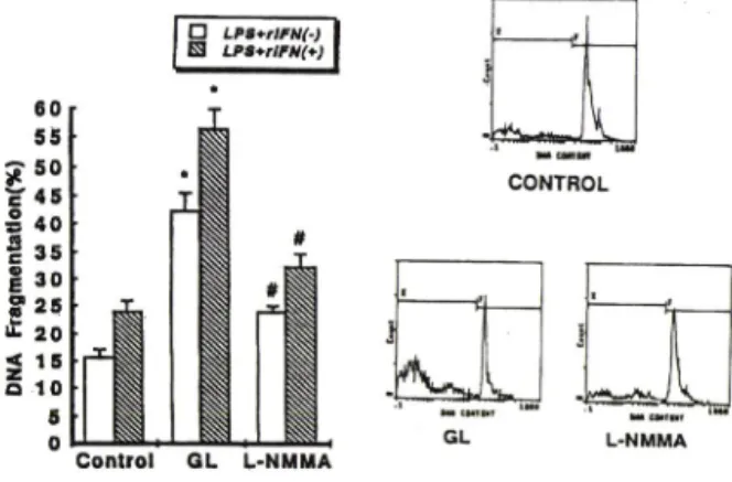
Fig. 5 — Effect of GL on DNA fragmentation of L1210 cells in coculture of peritoneal macrophages ob
tained from GL-administered mice and L1210 cells in vitro. GL (0.1 mg/mouse) was ad
ministered p.o for 7 days, and then m a
crophages were collected. Macrophages were cultured with LPS and y-IFN in 24 well plate, and L1210 cells were cultured in trans well.
Each bar represents the m ean+ S.E. from 5 ex- priments. ^Significantly different from control group (p<0.01), ^Significantly different from GL- treated group (p<0.01). L-NMMA: 0.5m M / well.
C o n t r o l 1 1 0 1 0 0
N 0 C 1 2 C o n c e n t r a t i o n ( ( i M )
Fig. 4 — Effect of nitric oxide donor on DNA frag
mentation of L1210 cells in vitro. L1210 cells were cultured w ith NOC12 for 12 hrs. in CO2
incubator. Each bar represents the m e a n iS .E . from 4 experiments. *Significantly different from control group (*p<0.05. **p<0.01). NOC12; C6H16N4O2 (Wako 347-0691. NO donor. Half-life : 13 m in).
± 1.2 % 및32.2±2.5%로apoptosis 발생율이감소되 었다(Fig. 5).
GL을투여하고분리한macrophage와L1210 세포 를co-culture 하였을때L P S와 T~IFN을처리하지 않 아도L1210 세포외apoptosis 발생율이 증가하였다는
C o n t r o l G L G L ^ L U I O
Fig. 3 —— Effect of GL on nitric oxide production from per
itoneal macrophages of mice. GL (0.1 m g/
mouse) was administered p.o. for 7 days, and L1210 cells were transplanted i.p. at the first day. Each bar represents the mean±S.E. from 5 experiments. *Significantly different from con
trol group (p<0.01). ^Significantly different from L1210 treated group (p<0.01). LPS and 7^ IFN non-treated group. ( + ): LPS and 7니IFN treated group, L-NMMA: 0.5m]VI/well.
데. 이걸과는 L1210 세포에 의해 자극을 받은m a
crophage 에G L이상승적으로작용하는것이 아닌가 추정되나자세한기전은추후연구되어야할것이다.
NO donor가 L1210 세 포 의 DNA fragmentation 에 미 치 는 효 과 - NO donor인 NOC12를 1. 10 및 100
로각각처리하였을때, 대조군의DNA fragmen
tation 은18.0±1.0% 이었으며, N 0 C 12 처리시25.2+
1.7, 27.9 ±0.3 및33.8±1.5%로대조군에비해농도의
존적으로 L1210 세포의DNA fragmentation이증가되
었다(Fig. 4). 이결과로는N O에외해 L1210 세포의 apoptosis가유도되고있음을확인할수있었다.
GL 투 여 생 쥐 의 복 강 macrophage와 L1210 세 포 의
Co-culture 시 이 식 된 L1210 세 포 의 apoptosis에 미 치 는 효 과 - GL 투여 생쥐의 복강 macrophage와 L 1210 세포를 co-culture 하였을 때 L1210 세포의 apoptosis 발생율은L P S와 "H FN을첨가하지않았을 때의 대조군에서 15.7± 1.3%이었으며. LPS와 T IFN을 철가하였을 때는 23.9±2.2%로 증가하였다. G L을투여하고분리한macrophage와L1210 세포를 co-culture 하였을때L1210 세포의apoptosis 발생율
은L PS와 r I F N을첨가하지않았을때는 42.3+3.2%
이었으며, L PS와 y-IFN을 첨가하였을 때는 56.5±
3.5%로증가하였다. 이때L-N M M A를처리하면23.8 LPS*rlFN(>) LPS*rlFN(*l LPS*rlfN*L^NUMA
글러시르히진이 생쥐에 이식된 L1210 세포의 아포프토시스에 머처는영향 327
0
5
0505050505065544332211
? w
c o l e
« E O
«
£
<
QZ
<%}uolsuaEBeJd
<
QZ
o
I
o o o o o o o
8 7
6 5
4 3
2 1
?x}dplxo
3 Z1
□
□
은재순•권 진 • 오찬호
것 온 G L이 m acrophage를 자 국 하 여 N O 생 성 을 촉 진 하 고. 생 성 된 N O가 직 접 적 으 로 L1210 세 포 의 apop- tosis를 유 도 하 기 때 문 이 라 추 정 된 다. 또 한 이 걸 과 는 복 강 m acrophage로 부 터 생 성 되 는 N O가 중 양 세 포 의 apo ptosis를 유 도 한 다 는 C . S h iju n 등 ^ 액 결 과 와 도 유 사 한 결 과 이 다. C o-culture시 L1210 세 포 외 apop- tosis 발 생 올 아 L - N M M A에 의 해 70% 정 도 감 소 되 었 다 는 것 은. 이 식 된 L1210 세 포 외 apoptosis 유 도 기 전 에 복 강 m acro phag e로 부 터 생 성 된 N O가 주 된 작 용 을 하 고 있 으 며. 이 외 에 또 다 른 factor가 일 부 관 여 하 고 있 옴 을 시 사 하 는 것 이 라 할수 있 다.
결 론
암 세 포 인 L1210 세 포 를 이 식 한 생 쥐 에 G L을 투 여 하 면 복 강 m acrophage로 부 터 N O 생 성 을 촉 진 하 여. 이 식 된 L1210 세 포 의 apoptosis률 일 부 유 도 하 고 있 다 고 사 료 된 다.
감사의 말씀
본 논 문 은 1998년 도 우 석 대 학 교 학 술 연 구 조 성 비 에 의 하 여 연 구 되 었 으 며 이 에 감 사 드 럽 니 다.
문 헌
1) H . Inoue, K. Inoue. T. T akeuchi, N . N agata a n d S. S h ib a ta : In h ib itio n o f ra t acute in flam m atory paw edem a by dihem iphthalate of glycyrrhizinic acid derivatives. /• Pharm. Phar
macol. 45,1067 (1993).
2) K. T akagi and Y . Is h ii: Peptic ulcer in hib itin g properties of a new fraction from Licorice root (FmIOO). Arzneimittel Forschung, 18. 53 (1968).
3) T. Iso. N. N akajim a, H . Suda. H . Y am auchi and K. U da ; Passive anaphylaxis in rat conjunctiva an d topical effects o f anti-allergic agents h y persensitivity in conjunctiva and drug efficacy.
Ophthalmic Res., 12, 9 (1980).
4) H . Inoue, T. M ori, S. S hibata and H . Saito ; P h arm aco lo g ical ac tiv itie s o f G ly cy rrh e tin ic acid derivatives ; Analgesic and anti-type IV a l
lergic effects. Chem. Pharm. B ull., 35. 3888 (1987).
5) H . Abe. N. O hya, K. Fujikaw a, T. Shibuga, S.
A ric h i a n d S. O d a s h im a: E ffe cts o f G ly cy rrhizin and G lycy rrhetinic acid on grow th and m elanogenesis in cultured B16 m elanom a cells. Eur. J. Cancer Clin. Oncol., 23, 1549 (1987).
6) H . O kam o to. D . Y o sh id a. Y . S aito an d S.
M izusaki ; Inhibition of l^trtetradecanoylphorbol- 13~acetate induced induction in epstein-barr virus early antigen in R aji cells. Cancer Letters, 19. 27 (1983).
7) M . Ito. H . N akashim a, M . B aba. R. Pauwells, E . DeClercq, S. Shigeta and N. Y am am oto : In hibitory effect o f G lycyrrhizin on vitro infec- tiv ity and cytopathic activity of the h u m an im m unodeficiency virus. Antiviral Research, 7, 127 (1987).
8) H . N a k a s h im a . T. M a ts u i, O . Y o sh id a. Y . Isowa, Y. Kido. Y . M otoki, S. Shigeta. T. Mori an d N . Y am am o to : A new an ti- hu m an im m unodeficiency virus substance, G lycyrrhizin sulfate : E ndow m ent of G lycyrrhizin w ith re
verse transcriptase inhibitory activity by chem ical m odification. JPN. J. Cancer Res., 78, 767 (1987).
9) J . H . H an, C. H . O h and J . S. E u n : Effect of Glycyrrhizae R adix on the im m une responses (II). Im m uncrregulatoiy action of Glycyrrhizin and G lycyrrhetinic acid. Yakhak Hoeji, 35(3). 174 (1991).
10) H . N ishino, S. S hibata. K. H irabayashi : A n ti
tum or prom oting activity of glycyrrhetic acid-re
lated com pounds. I Kyoto Pref. Univ. Med., 95, 1563 (1986).
11) C-족'^ -
키 행 . Mino. Med. Rev.. 17. 89 (1986).
1 2) t i b /hs: m m , m 롱. m s :
b ^ w z a - y ^ h r n r
^ U fv i/ 'j Biotherapy,
3(6). 1515 (1989).
13) H . Abe, N . O hya, K. F. Y am am oto, T. Shibata.
S. A rich i a n d S. O d a s h im a : Effects of gly
cyrrhizin and glycyrrhetinic acid on growth and melanogenesis in cultured B16 m elanom a cells.
Eur. J. Cancer Clin. Oncol., 23, 1549 (1987).
Pharm. Soc. Korea
글러시르히진이생쥐에이식된 L1210 세포의아포프토시스에미치는영향 329
14) M . S hinada, M . A zum a. H . K aw ai, K. Sazakd, I.
Yoshida, T. Yoshida, T. S u zu tan i and T. Saku- m a ; E nhe ncem ent o f interferon-gam m a pro
duction in glycyrrhizin- treated h u m a n p e ri
p h e r a l ly m p h o c y te s in re s p o n s e to c o n canavalin A and to surface antigen of hepatitis B v iru s. Proc. Soc. exp. Biol. M ed., 181, 205 (1986).
15) N . Abe, T. E b in a and N . Ishid a : Interferon in duction by glycyrrhizin and glycyrrhetinic acid in mice. M icrobiol Im m unol., 26, 535 (1982).
16) Y . Kondo an d F. T akano '■ N itric oxide pro
d u c tio n in m ouse p e rito n e a l m acro phag es enhanced w ith G lycyrrhizin. Biol. Pharm. Bull., 17(5). 759 (1994).
17) M . J . H erriott, H . Jia n g , C. A. Stew art. D. J . F a st an d R . W . L eu : M echanistic DifFemces between M igration Inh ibitory F actor(M IF ) and IFN-y for M acrophage Activation. }. Im m unol., 150. 4524 (1993).
18) J . E. A lbina, S. C ui, R . B. M ateo and J . S. Re- ichner : N itric oxide-mediated Apoptosis in m u rine peritoneal m acrophages. J. Im m unol., 150.
5080 (1993).
19) I. N icoletti, G. M igliorati, M . C. Pagliacci, F.
G rig n a n i an d C. A. R iccardi '■ R a p id an d sim ple m ethod for m easuring thym ocyte apoptosis by propidium iodide stain in g an d flow cytom etiy. J.
Im m unol Methods. 139, 271 (1991).
20) N. Zam zam i, P. M archetti, M . Castedo, C. Zanin, J . L. Vayssiere, P. X . P e tit an d G . Kroemer : Reduction in m itochondrial potential constitutes an early irreversible step of program m ed lym~
phocyte death in vivo. J. Exp. M ed., 181, 1661 (1995).
21) K. A. Rockett. M . M . A w b u m , W . B. Cowden.
a n d I. A. C lark. : K illin g o f P lasm o d iu m fa l
ciparum in vitro by nitric oxide derivatives. In fect Im m unity, 59(9), 3280 (1991).
22) S. M otom u. I. T etsuya, O . A k ito sh i, T. No- bu y u ki, M . T akashi, H . T akashi an d Y . Ik u to : N O C , A n itric oxide-releasing com pound, in duces dose dependent apoptosis in m acrophage.
Bioche. and Biophy. Res. Com. 209(2), 519 (1995).
23) C. S h iju n , S. R. Jo n a th a n , B . M . Rom eo an d E.
A . Jroge : A ctivated m u rin e m acrophages in duce apoptosis in tum or cells throug h n itric ox
ide-dependent m echanism s. Cancer Res. 54' 2462 (1994).