Anterior cervical arachnoid cyst
Asian Spine Journal
Asian Spine Journal
119Copyright Ⓒ 2013 by Korean Society of Spine Surgery
This is an Open Access article distributed under the terms of the Creative Commons Attribution Non-Commercial License (http://creativecommons.org/licenses/by-nc/3.0/) which permits unrestricted non-commercial use, distribution, and reproduction in any medium, provided the original work is properly cited.
Asian Spine Journal • pISSN 1976-1902 eISSN 1976-7846 • www.asianspinejournal.org
Received Jan 19, 2012; Revised May 15, 2012; Accepted Jun 2, 2012 Corresponding author: Abolfazl Rahimizadeh
Department of Neurosurgery, Pars Hospital, No. 83-Keshawarz Blvd, 14154 Tehran, Islamic Republic of Iran Tel: +98-21-88-66-12-44, Fax: +98-21-88-79-85-03, E-mail: [email protected]
Anterior Cervical Arachnoid Cyst
Abolfazl Rahimizadeh, Give Sharifi
Department of Neurosurgery, Pars Hospital, Tehran, Islamic Republic of Iran
This report is composed of two patients with anteriorly located cervical intradural arachnoid cyst and review of 24 cases in English- language literature. Both of our patients were in the first two decades of life with neck pain and motor weakness. With suspicious diagnosis of anterior arachnoid cyst surgery was carried out in both cases, though laminectomy in one and laminoplasty in the other.
The cyst wall was widely fenestrated with subsequent subtotal excision of the cyst. Both cases had good long-term outcome. The review disclosed male predominance. 73% of the patients were diagnosed within the first two decades of life. Neck pain and motor weakness were the dominant signs and symptoms of this pathology. Magnetic resonance imaging showing a cerebrospinal fluid (CSF) containing cyst was the best mode of diagnosis. Wide cyst fenestration with waying CSF into subarachnoid cyst was the most appro- priate and applied surgery with optimal outcome.
Keywords: Cervical; Arachnoid cyst; Intradural; Review of the literature; Spinal
Case Report Asian Spine J 2013;2:119-125 • http://dx.doi.org/10.4184/asj.2013.7.2.119
ASJ A SJ
Introduction
Intradural arachnoid cysts are uncommon and arise in thoracic, lumbar and cervical region in decreasing frequency. Arachnoid cyst are mostly located on the posterior aspect of the cord. This means that anteriorly located arachnoid cyst are exceptional, particularly those occurring in the cervical spinal region [1]. Regarding the rarity of this pathology in this certain location, it should be noted that eight out of nine cases of intradural arach- noid cyst reported by Kendall et al. [1] lied posteriorly in the thoracic region. Moreover, of 17 cases of intradural arachnoid cyst including eight cases of cervical region reported by Alvisi et al. [2], all were located posteriorly. A literature search by Kazan et al. [3] from 1974 to 1999 re- vealed only 10 cases of anterior cervical intradural arach- noid cyst including two cases of their own. However, ap- parently six cases escaped their attention [1,4]. In current
article, besides very careful resurvey of the literature up to 1999, we updated the review yielding 24 cases reported in English literature [1,3-21]. Herein, two additional cases who have undergone surgery with good postoperative neural recovery are presented.
Case Report
1. Case 1
This 2-year-old girl was admitted to our hospital with a 2-week history of torticollis and weakness of the left up- per extremity for a week. She had no history of trauma or infection of upper respiratory tract infection. Her neuro- logical exam showed moderate paresis of the left upper extremity, all reflexes were hyperactive and plater reflex was extensor on the left side. Cervical spine X-Ray and open mouth view were regarded normal. Magnetic reso-
nance imaging (MRI) revealed an intradural hypointense cystic mass anterior to the cord in T1 and hyperintense in T2 images resembling cerebrospinal fluid (CSF) intensity at C5-C6 level which was markedly compressing the cord (Fig. 1). A provisional diagnosis of intradural arachnoid cyst was made.
With the patient in prone position laminectomy of C5 and C6 was done. The dura was tense and on opening the cord was flattened and budged posteriorly into the dural incision. The dentate ligaments at C5-C6 level were cut on the left side revealing a translucent cystic collec- tion anterior to the cord. The cyst was aspirated revealing CSF. Following aspiration, the cyst shrunk and the cord pulsation appeared. With gentle retraction of the cord, the cyst’s capsule could be excised totally. Postoperatively, the child showed motor recovery and her neurological examination three months after surgery was normal.
2. Case 2
This 17-year-old male was referred with dull moder- ate neck pain followed with progressive weakness of the right upper extremity of one month duration followed by gait disturbances in recent days. On clinical survey, he was found to have quadriparesis most prominent in the right upper extremity. Cervical MRI disclosed a ventrally located intradural cystic lesion with apparent cord com- pression at C2-C3 level (Fig. 2).
With the patient in prone position, a cervical midline incision was made at the appropriate levels. En-block laminotomy of C2 and C3, incising the corresponding laminas bilaterally with the aid of high speed drill was done. The dura seemed under considerable pressure.
The techal sac was open in the midline. This revealed a splayed cord under considerable pressure from the ante- rior. After sectioning dentate ligament on the right side
A B C
Fig. 1. (A) T2 magnetic resonance images of case one; sagittal view showing hyperintense mass anterolateral to the cord at C5- C6 level. (B) Axial view, showing the left anterolateral location of the cyst. (C) T1-weighted MR image demonstrating hypointense cyst at C5-C6 level of the same patient.
A B C
Fig. 2. (A, B) T2 magnetic resonance (MR) images of the second case showing an anteriorly located hyperintense mass compatible with a cyst at C2-C3 level. (C) T1-weighted MR image of the second case showing a hypointense mass.
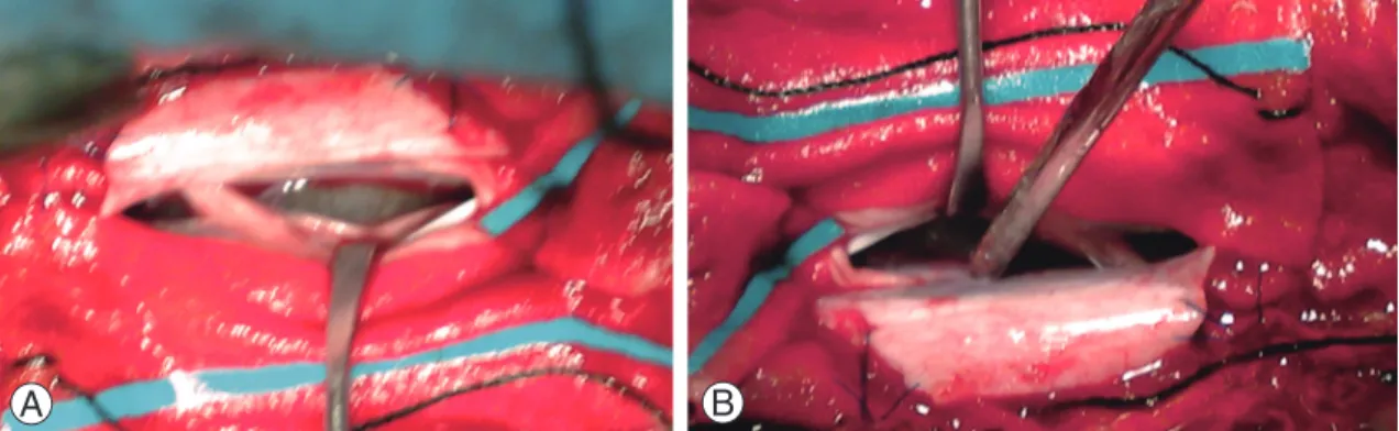
and mild retraction of the cord, a cystic mass with trans- parent wall became visible. The wall of the cyst was mini- mally incised disclosing clear content compatible with CSF. Subsequently, the cyst was widely fenestrated into the subarachnoid space with partial removal of the cap- sule (Fig. 3). The cord was decompressed and returned to its normal position with good pulsation. Later, after dural closure, two level laminoplasty with application of mini plates was done bilaterally (Figs. 4, 5). The patient made uneventful recovery and post operative cervical
MRI showed resolution of the arachnoid cyst (Fig. 6). He returned to his previous activities within six months.
Discussion
Intradural arachnoid cysts are uncommon and most of them are located in the thoracic region followed with the lumbar and the cervical region [1,2]. Majority of these cysts lie posteriorly on the spinal cord and are rarely located anteriorly. Review of the literature revealed that
A B
Fig. 3. (A) Intraoperative view showing a grayish translucent cyst in front of the cord. (B) The cyst is more clearly seen in this view.
Fig. 4. Intraoperative view after laminoplasty. Fig. 5. Postoperative X-ray showing laminoplasty.
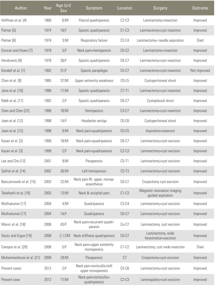
anteriorly located intradural arachnoid cyst are very rare in the cervical region and we could find only 24 examples reported previously. With taking to account two current cases, the number of the cases published so far in the lit- erature reach to 26 cases (Table 1).
Of the total these 26 reviewed cases, 15 occurred in males and the remaining 11 were female. Age ranged from 2 to 43 years with mean of 15/1 years. In 19 cases the cyst was detected in the first two decade of life [3- 7,11-13,17]. Four cases were diagnosed in the third de- cade [8,14,15,19]. Two patients in the forth decade and one in his fifth decade became symptomatic [1,9,18].
Most of these arachnoid cysts were considered con- genital, however minor or major trauma was suspected to play a role in five instances [3,4,17,22]. Trauma might take part in the pathology and semiology of intradural arachnoid cysts in two ways: either by producing a breach in the arachnoid membrane and subsequent development of a cyst or may trigger a silent preexisting arachnoid cyst into a symptomatic one.
The cyst occupied one or two vertebral segments in majority [3-6,8,11,13,16,17,21,22]. But it extended full cervical length from foramen magnum to lower cervical region in five occasions [9,12,18,19].
From clinical point of view, neck pain or wry neck are prominent symptom, but concurrence of weakness of upper or lower extremity of one or two weeks duration makes the patients or their parents to seek medical advice in our survey 38.5% of the cases had neck pain. Quadri- paresis was the most frequent sign seen in 50% of the
patients. Torticollis in our first case seems to be a com- pensatory event made for free passage of CSF.
In pre MRI era, myelography and computed tomog- raphy (CT) myelography were used to diagnose these lesions [1,3,7]. In conventional myelogram, displace- ment and compression of the spinal cord was visualized.
However, the arachnoid cyst could have been filled only in supine delayed myelogram. With regard to CT, CSF attenuation may be recognizable on plain CT scans using rapid high resolution machines, but intrathecal metriza- mide is almost always necessary to confirm and elucidate the details [3,7].
MRI is the modality of choice in diagnosis of the ante- rior cervical arachnoid cysts being demonstrated as a low signal round or oval lesions in T1 and hyperintense mass in T2 images compatible with CSF. Nowadays, with in- creased application of MRI, the numbers of the intradural arachnoid cysts are reported in increasing frequency and in earlier stage of cord compression. Twelve out of 26 re- viewed cases were diagnosed in post MRI era [13-21].
In differential diagnosis of such unusual cysts in such a rare location, arachnoid cysts secondary to arachnoiditis should be born in mind. Developments of such ventrally located cysts after bacterial or tuberculous meningitis have been reported in the cervical region quite resem- bling congenital arachnoid cysts [23]. Entrogenous cysts also known as edodermal cysts might express themselves with the same MRI features similar to arachnoid cysts and final diagnosis can be only made intraoperatively [24,25]. Intradural spinal parasitic cysts such as cysticer- cosis and hydatid cyst, although rare but should remain in differential diagnosis of arachnoid cysts specially in endemic areas [26,27].
Surgery is indicated once an intradural arachnoid cyst is suspected. The first step is laminectomy or lamino- plasty, Nowadays en block laminoplasty is suggested to avoid postoperative kyphosis. This procedure is mostly and particularly recommended in the patients with cer- vical and cervicothoracic intradural cysts and tumors.
Although children are more expected to develop post- laminectomy kyphosis but this deformity is not uncom- mon in adults. Subsequent to dural opening and minimal cord retraction, the cyst can be reached. In this stage, although complete surgical excision of the cyst seems desirable but it is not possible in all instances because of the scarring and adherence of arachnoid membrane to the cord. However, wide fenestration and partial removal Fig. 6. Postoperative T2-weighted magnetic resonance sagittal images
of the cervical spine showing near to complete resolution of the cyst.
Table 1. Ventral cervical arachnoid cyst
Author Year Age (yr)/Sex Symptom Location Surgery Outcome
Hoffman et al. [4] 1960 6/M Flaccid quadriparesis C2-C3 Laminectomy+resection Improved
Palmer [5] 1974 19/F Spastic quadriparesis C1-C3 Laminectomy+cyst resection Improved
Palmer [6] 1974 3/M Respiratory failure C2-C4 Laminectomy+ needle aspiration Died Duncan and Hoare [7] 1978 3/F Neck pain+hemiparesis C0-C2 Laminectomy+resection Improved Herskowitz [8] 1978 28/F Spastic quadriparesis C6-C7 Laminectomy+cyst resection Improved Kendall et al. [1] 1982 31/F Spastic paraplegia C5-C7 Laminectomy+cyst resection Not improved Chan et al. [9] 1985 37/M Upper extremity weakness C0-L5 Cystoperitoneal shunt Improved Jena et al. [10] 1990 17/M Spastic quadriparesis C7-T1 Laminectomy+cyst resection Improved
Rabb et al. [11] 1992 2/F Spastic quadriparesis C6-C7 Cystopleural shunt Improved
Chen and Chen [22] 1996 18/M Hemiparesis C3-C7 Laminectomy+cyst resection Improved
Jean et al. [12] 1998 14/F Headache vertigo C0-C6 Cystoperitoneal shunt Improved
Jean et al. [12] 1998 9/M Neck pain+quadriparesis C0-C5 Aspiration+reservoir Improved Kazan et al. [3] 1999 18/M Neck pain+quadriparesis C6-C7 Laminectomy+cyst excision Improved Kazan et al. [3] 1999 2/F Neck pain+quadriparesis C2-C3 Laminectomy+cyst excision Improved
Lee and Cho [13] 2001 9/M Paraparesis C5-T1 Laminectomy+cyst excision Improved
Safriel et al. [14] 2002 26/M Left hemiparesis C5-T3 Laminectomy+cyst excision Improved Banczerowski et al. [15] 2003 22/M Neck pain Rt. upper, monop-
araesthesia C6-C7 Corpectomy cyst excision Improved
Takahashi et al. [16] 2003 13/M Neck & occipital pain C1-C3 Magnetic resonance imaging
guided aspiration Improved
Muthukumar [17] 2004 4/M Quadriparesis C3-C4 Laminectomy+cyst excision Improved
Muthukumar [17] 2004 14/F Quadriparesis C6-C7 Laminectomy+cyst excision Improved
Maiuri et al. [18] 2006 43/F Neck pain+recurrent quadri-
paresis Co-C7 Laminectomy, cyst excision Improved Gezici and Ergun [19] 2008 2,1/2M Neck stiffness quadriparesis C0-C7 Laminectomy, wide
fenestration+excision Improved Campos et al. [20] 2008 5/F Neck pain+upper extremity
monoparesis C1-C2 Laminectomy, cyst wide resection Died
Muhammedrezai et al. [21] 2008 29/M Paraparesis C7 Corpectomy+cyst excision Improved
Present cases 2012 2/F Neck pain+torticollis+left
upper monoparesis C5-C6 Laminectomy+cyst excision Improved Present case 2012 17/M Neck pain+torticollis+
quadriparesis C2-C3 Laminopalsty+cyst excision Improved
of the cyst allowing maximal communication of the cyst with subarachnoid spaces is an accepted mode of surgery and has been applied in 69% of the reported cases with success. Recurrence might be expected if insufficient fenestration or aspiration alone is used [6,12]. Cystoperi- toneal or cystopleural shunt might be used as a primary mode of surgery or in recurrences [9,11,12]. Intermittent aspiration with application of percutaneous reservoir was use in one occasion [12]. Despite of previous reports on recurrence and even death with aspiration [5]. There is one report of image guided aspiration with good recovery [16]. However, we do not recommend aspiration as an ac- ceptable modality of treatment regarding the fact that the arachnoid cysts of different types and locations mostly recur after aspiration .
In order to obviate the need for cord retraction, ante- rior approach through cervical body vertebrectomy has been suggested and done in two separate reports [15,21].
But, since majority of these cysts can be easily accessed through the posterior approach without morbidity, the issue of anterior approach should be remained open for discussion.
Prognosis for complete recovery should be expected with excellent outcome, especially if the arachnoid cysts are diagnosed early.
Ultimately, this conclusion was made that anteriorly located arachnoid cyst of the cervical region should be considered in children and young adults with neck pain or torticollis specially if these are followed with motor weakness. Surgery should be done as soon as the diagno- sis is made, nowadays, complete recovery after surgery should be expected and despite of lack of any report on postoperative kyphosis in laminectomized cases, lamino- plasty is strongly recommended in children in order to avoid post-laminectomy deformity.
Conflict of Interest
No potential conflict of interest relevant to this article was reported.
References
1. Kendall BE, Valentine AR, Keis B. Spinal arachnoid cysts: clinical and radiological correlation with prog- nosis. Neuroradiology 1982;22:225-34.
2. Alvisi C, Cerisoli M, Giulioni M, Guerra L. Long-
term results of surgically treated congenital intradu- ral spinal arachnoid cysts. J Neurosurg 1987;67:333- 5.
3. Kazan S, Ozdemir O, Akyuz M, Tuncer R. Spinal in- tradural arachnoid cysts located anterior to the cervi- cal spinal cord: report of two cases and review of the literature. J Neurosurg 1999;91:211-5.
4. Hoffman EP, Garner JT, Johnson D, Shelden CH.
Traumatic arachnoidal diverticulum associated with paraplegia: case report. J Neurosurg 1973;38:81-5.
5. Palmer JJ. Spinal arachnoid cysts: report of six cases.
J Neurosurg 1974;41:728-35.
6. Palmer JJ. Cervical intradural arachnoid cyst in a 3-year-old child: report of a case. Arch Neurol 1974;31:214-5.
7. Duncan AW, Hoare RD. Spinal arachnoid cysts in children. Radiology 1978;126:423-9.
8. Herskowitz J, Bielawski MA, Venna N, Sabin TD. An- terior cervical arachnoid cyst simulating syringomy- elia: a case with preceding posterior arachnoid cysts.
Arch Neurol 1978;35:57-8.
9. Chan RC, Thompson GB, Bratty PJ. Symptomatic an- terior spinal arachnoid diverticulum. Neurosurgery 1985;16:663-5.
10. Jena A, Gupta RK, Sharma A, Prakesh VE, Khushu S. magnetic resonance diagnosis of spinal arach- noid cyst: a report of two cases. Childs Nerv Syst 1990;6:107-9.
11. Rabb CH, McComb JG, Raffel C, Kennedy JG. Spi- nal arachnoid cysts in the pediatric age group: an association with neural tube defects. J Neurosurg 1992;77:369-72.
12. Jean WC, Keene CD, Haines SJ. Cervical arachnoid cysts after craniocervical decompression for Chiari II malformations: report of three cases. Neurosurgery 1998;43:941-4.
13. Lee HJ, Cho DY. Symptomatic spinal intradural arachnoid cysts in the pediatric age group: descrip- tion of three new cases and review of the literature.
Pediatr Neurosurg 2001;35:181-7.
14. Safriel YI, Sanchez G, Jhaveri HS. Giant anterior cer- vicothoracic arachnoid cyst. Spine (Phila Pa 1976) 2002;27:E366-8.
15. Banczerowski P, Lipoth L, Vajda J, Veres R. Surgery of ventral intradural midline cervical spinal patholo- gies via anterior cervical approach: our experience.
Ideggyogy Sz 2003;56:115-8.
16. Takahashi S, Morikawa S, Egawa M, Saruhashi Y, Matsusue Y. Magnetic resonance imaging-guided percutaneous fenestration of a cervical intradural cyst. Case report. J Neurosurg 2003;99:313-5.
17. Muthukumar N. Anterior cervical arachnoid cyst presenting with traumatic quadriplegia. Childs Nerv Syst 2004;20:757-60.
18. Maiuri F, Iaconetta G, Esposito M. Neurological pic- ture. Recurrent episodes of sudden tetraplegia caused by an anterior cervical arachnoid cyst. J Neurol Neu- rosurg Psychiatry 2006;77:1185-6.
19. Gezici AR, Ergun R. Cervical anterior intradural arachnoid cyst in a child. Acta Neurochir (Wien) 2008;150:695-8.
20. Campos WK, Linhares MN, Brodbeck IM, Ruhland I. Anterior cervical arachnoid cyst with spinal cord compression. Arq Neuropsiquiatr 2008;66:272-3.
21. Muhammedrezai S, Ulu MO, Tanriover N, Moghad- dam AM, Akar Z. Cervical intradural ventral arachnoid cyst resected via anterior corpectomy with reconstruction: a case report. Turk Neurosurg 2008;18:241-4.
22. Chen HJ, Chen L. Traumatic interdural arachnoid cyst in the upper cervical spine. Case report. J Neu- rosurg 1996;85:351-3.
23. Lolge S, Chawla A, Shah J, Patkar D, Seth M. MRI of spinal intradural arachnoid cyst formation following tuberculous meningitis. Br J Radiol 2004;77:681-4.
24. Asazuma T, Sato M, Ichimura S, et al. Endodermal cyst of the cervical spine treated by an anterior ap- proach for resection and shunting. J Spinal Disord Tech 2002;15:258-60.
25. Sasani M, Ozer AF, Oktenoglu BT, Peker K, Bozkus MH, Sarioglu AC. Excision of an asymptomatic cer- vical intradural neurenteric cyst through the anterior approach: a study of two cases and a review of the literature. Spine J 2007;7:720-7.
26. Ciftci E, Diaz-Marchan PJ, Hayman LA. Intradural- extramedullary spinal cysticercosis: MR imaging findings. Comput Med Imaging Graph 1999;23:161- 4.
27. Pushparaj K, Sundararajan M, Madeswaran K, Am- balavanan S. Primary spinal intradural hydatid cyst:
a short report. Neurol India 2001;49:203-4.