Asian Spine Journal Vol. 3, No. 2, pp 73~79, 2009
Copyright � 2009 by Korean Society of Spine Surgery
This is an open-access article distributed under the terms of the Creative Commons Attribution License (http://creativecommons.org/licenses/by/3.0), Which permits unrestricted use, distribution, and reproduction in any medium, provided the original work is properly cited.
Asian Spine Journal�pISSN 1976-1902 eISSN 1976-7846
Received Aug 18, 2009; 1st revised Sep 9, 2009; accepted Sep 10, 2009 Corresponding author:Lee Kyu Yeol, MD
Department of Orthopedic Surgery, Dong-A University College of Medicine, Dongdaesin-dong 3-ga, Seo-gu, Busan 602-715, Korea
Tel: +82-51-240-2867, Fax: +82-51-243-9764, E-mail: [email protected]
Treatment Outcome of Cervical Tear Drop Fracture
Hyeon Jun Kim, Kyu Yeol Lee, Woo Chul Kim
Department of Orthopedic Surgery, Dong-A University College of Medicine, Busan, Korea S
Sttuuddyy DDeessiiggnn:: This is a retrospective study.
P
Puurrppoossee:: We wanted to evaluate the clinical results of surgical and conservative treatment for cervical tear drop fracture.
O
Ovveerrvviieeww ooff LLiitteerraattuurree:: The tear drop fracture of the lower cervical spine is generally associated with a high incidence of neurological deficits and surgery is needed to treat this injury. Tear drop fracture of C2 is usually a stable fracture that is amendable to conservative treatment.
M
Meetthhooddss:: We reviewed the outcomes of 25 patients. Cervical tear drop fracture was classified as the extension and flexion types according to the mechanism of injury. The neurologic symptoms were evaluated by the Frankel classification system, and the loss of lordosis and disc height, and the duration of bony union were analyzed.
R
Reessuullttss:: Twenty one patients had the flexion type injury and 4 patients had the extension type injury. All the patients with the flexion type were treated by anterior decompression and plate stabilization. All the patients with the extension type were treated conservatively. Ten patients with the flexion type had neurologic deficits. The nerve root injuries recovered fully and the incomplete injuries had an average 1.5 grade recovery. Radiologically, the extension type fracture showed bony union at an average of 12.8 weeks. For the patients with the flexion type fracture, the loss of lordosis was 2.6�and the loss of disc height was 2.1 mm. The period of bony union in 20 cases was 13.0 weeks.
C
Coonncclluussiioonnss:: Anterior plate stabilization was an effective treatment for the flexion type tear drop fracture. Conservative treatment is thought to be one of the good clinical methods for treating the extension type tear drop fracture.
Key WWords: Cervical spine, Tear drop fracture, Anterior plate stabilization
Introduction
Cervical spine injury is a severe injury that may lead to tetraplegia or death. Therefore, the importance of treatment and rehabilitation for cervical spine injury has been under- scored due to the severe social and economic losses. Satis- factory decompression has not been achieved for cases of cervical tear drop fracture that are treated with skeletal trac- tion by using skull tongs or halo-vest fixation. Despite ade- quate traction, there are some cases in which the fragment
of the vertebral body was displaced to the anterior aspect of the spinal canal on the posterior side of the vertebral body.
Cervical tear drop fracture is divided into two types based on the mechanism of injury, and most of which is flexion type injury. The cases of cervical tear drop fracture of the C2 vertebral body usually manifest as a hyperextension type injury. The anterior longitudinal ligament originates from the base of C2 (the axis). Accordingly, in cases of extension injury, the avulsion fracture occurs at the bottom of the anteroinferior side of the vertebral body. This can be differ- entiated from a tear drop fracture of the other lower cervical
The flexion type tear drop fractures were classified into four types according to the system of Korres et al.6, which is based on the size of the anterior inferior fragment and the displacement of the posterior part of the vertebral body into the spinal canal. In Type I, there was rupture of the posteri- or ligament and the characteristics of a small fracture (< 3
mm in size) on the anterior inferior angle of the vertebral body, half of the vertebral body lacked sagittal fracture at the posterior body and retrolisthesis was seen. In Type II, there were characteristics of the coronary fracture of the anterior inferior angle of the vertebral body, a lack of the retrolisthesis and there was sagittal fracture of the posterior Kim et al. Treatment Outcome of Cervical Tear Drop Fracture/ 75
Fig. 2. A 52 year old male patient with a C5 tear drop fracture (Case No.14). (A) The preoperative lateral roentgenogram shows anterior displacement of a bony fragment. (B) The CT shows a T-shape pattern with a sagittal fracture. (C) The postoperative radiograph shows the consolidation of the grafted bone. (D) The postoperative radi- ograph shows the consolidation of the grafted bone.
B
D A
C
accordance with Frankel’s classification.
On the simple lateral X-rays, the mean loss of lordosis was 2.0�(1.5�to 2.4�) in the cases of Type I, it was 2.3�
(1.6�to 3.1�) in the cases of Type II, 2.8�(2.5�to 3.0�) in the cases of Type IIIa, 3.1�(2.5�to 3.5�) in the cases of Type IIIb and 4.45�(4.2�to 4.7�) in the cases of Type IV.
The loss of disc height was 1.2 mm (0.5 to 1.8 mm) in the cases of Type I, 2.2 mm (1.1 to 3.5 mm) in the cases of Type II, 2.1 mm (1.8 to 2.3 mm) in the cases of Type IIIa, 2.5 mm (2.1 to 3.2 mm) in the cases of Type IIIb and 4.2 mm (4.1 to 4.2 mm) in the cases of Type IV. Overall, the mean loss of lordosis was 2.6�(1.5�to 4.7�) and the mean loss of disc height was 2.1 mm (0.5 mm to 4.2 mm). With excluding one case of Type IV, all the remaining cases had bone union with a high union rate of 92%. The duration for bony union was a mean of 13.0 weeks (11.4 to 16.4 weeks) (Table 2).
In regard to the complications, there were no cases of dis- placement of the graft or metal failure. There were no cases of complications such as postoperative infection or iatro- genic nerve injury. There was nonunion in one case of Type
IV. There was one case of the intermittent dysphagia. Fol- lowing the accident, in one case in which there was a com- plete spinal cord injury, death occurred due to respiratory complications at three months postoperatively.
Discussion
With the increased occurrence of traffic accidents and industrial disasters, cervical spinal cord injury due to cervi- cal spine fracture and dislocation may leave severe compli- cations such as quadriplegia where there is almost no chance for rehabilitation. For these patients, it is important that treatment and rehabilitation be stated early. According to Durbin8and Jacob9, spinal cord injury frequently occurs in young active male patients, and the major causes have been reported to be traffic and industrial accidents. Also in our series, 20 of the 25 patients were men. The mean age was 43.2 years and this is a relatively young age. The caus- es of injury included ten cases of traffic accident and this accounted for 40% of the total cases. The common sites of Kim et al. Treatment Outcome of Cervical Tear Drop Fracture/ 77
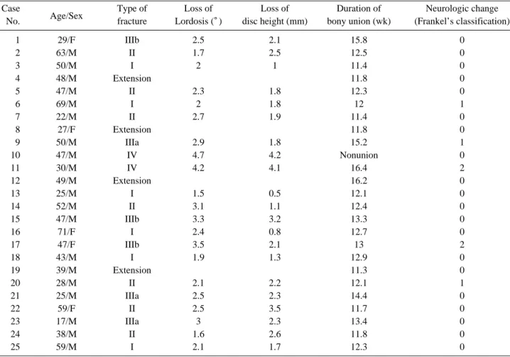
Table 2. Data on 25 patients with tear drop fracture of the cervical spine Case
Age/Sex Type of Loss of Loss of Duration of Neurologic change
No. fracture Lordosis (�) disc height (mm) bony union (wk) (Frankel’s classification)
1 29/F IIIb 2.5 2.1 15.8 0
2 63/M II 1.7 2.5 12.5 0
3 50/M I 2 1 11.4 0
4 48/M Extension 11.8 0
5 47/M II 2.3 1.8 12.3 0
6 69/M I 2 1.8 12 1
7 22/M II 2.7 1.9 11.4 0
8 27/F Extension 11.8 0
9 50/M IIIa 2.9 1.8 15.2 1
10 47/M IV 4.7 4.2 Nonunion 0
11 30/M IV 4.2 4.1 16.4 2
12 49/M Extension 16.2 0
13 25/M I 1.5 0.5 12.1 0
14 52/M II 3.1 1.1 12.4 0
15 47/M IIIb 3.3 3.2 13.3 0
16 71/F I 2.4 0.8 12.7 0
17 47/F IIIb 3.5 2.1 13 2
18 43/M I 1.9 1.3 12.9 0
19 39/M Extension 11.3 0
20 28/M II 2.1 2.2 12.1 1
21 25/M IIIa 2.5 2.3 14.4 0
22 59/F II 2.5 3.5 11.7 0
23 17/M IIIa 3 2.3 13.4 0
24 38/M II 1.6 2.6 11.8 0
25 59/M I 2.1 1.7 12.3 0
Kim et al. Treatment Outcome of Cervical Tear Drop Fracture/ 79
ple lateral X-rays and the dynamic flexion-extension X- rays. Accordingly, in all the cases, for conservative treat- ment, the patients were recommended to wear a Philadel- phia neck brace for 12 weeks. Medical treatment was per- formed for the management of clinical symptoms such as neck pain.
Conclusions
For the cases of the extension type cervical tear drop frac- ture, the findings suggestive of bone union confirmed that complications would not occur. For the cases of the exten- sion type cervical tear drop fracture, in most of the cases, with excluding the Type IV cases, the anterior plate stabi- lization using an anterior metal plate might be a useful modality. Yet posterior fixation might also be considered for the cases in which the stability cannot be maintained fol- lowing anterior interbody fusion or for those Type IV cases.
REFERENCES
01. Bohlman HH, Boada E: Fractures and dislocations of the lower cervical spine. (in The Cervical Spine Research Soci- ety eds. The cervical spine, 2nd ed. Philadelphia, Lippin- cott: 355, 1989)
02. Pierce DS: Acute treatment of spinal cord injuries. (in Pierce DS, Nickel VH eds. The total care of spinal cord injuries. Boston, Little Brown and Company: 1, 1977) 03. Bohler J, Gaudernak T: Anterior plate stabilization for
fracture-dislocations of the lower cervical spine. J Trauma 1980; 20: 203-205.
04. Bremer AM, Nguyen TQ: Internal metal plate fixation combined with anterior interbody fusion in cases of cervi- cal spine injury. Neurosurgery 1983; 12: 649-653.
05. Castaing J: Les traumatismes recents du Raquis cervical inferieur: traitement chirurgical par voie anterieure. Rev Chir Orthop 1984; 70: 519-522.
06. Korres DS, Stamos K, Andreakos A, Spyridonos S, Kavadias K: The anterior inferior angle fracture of a lower cervical vertebra. Eur Spine J 1994; 3: 202-205.
07. Allen BL Jr, Ferguson RL, Lehmann TR, O’Brien RP:
A mechanistic classification of closed, indirect fractures and dislocations of the lower cervical spine. Spine (Phila Pa 1976) 1982; 7: 1-27.
08. Durbin FC: Fracture-dislocations of the cervical spine. J Bone Joint Surg Br 1957; 39: 23-38.
09. Jacobs B: Cervical fractures and dislocations (C3-7). Clin Orthop Relat Res 1975; (109): 18-32.
10. Norrell H, Wilson CB: Early anterior fusion for injuries of the cervical portion of the spine. JAMA 1970; 214: 525- 530.
11. Penning L: Normal movements of the cervical spine. AJR Am J Roentgenol 1978; 130: 317-326.
12. Burke DC, Berryman D: The place of closed manipula- tion in the management of flexion-rotation dislocations of the cervical spine. J Bone Joint Surg Br 1971; 53: 165-182.
13. Castellano V, Bocconi FL: Injuries of the cervical spine with spinal cord involvement (myelic fractures): statistical considerations. Bull Hosp Joint Dis 1970; 31: 188-194.
14. Chung JY, Shin HC, Kim HS: Anterior plate fixation of the racture-dislocation of cervical spine. J Korean Orthop Assoc 1988; 23: 1541-1548.
15. Evans DK: Reduction of cervical dislocations. J Bone Joint Surg Br 1961; 43: 552-555.
16. Smith GW, Robinson RA: The treatment of certain cervi- cal-spine disorders by anterior removal of the intervertebral disc and interbody fusion. J Bone Joint Surg Am 1958; 40:
607-624.
17. Bailey RW, Badgley CE: Stabilization of the cervical spine by anterior fusion. J Bone Joint Surg Am 1960; 42:
565-594.
18. Capen DA, Garland DE, Waters RL: Surgical stabiliza- tion of the cervical spine: a comparative analysis of anteri- or and posterior spine fusions. Clin Orthop Relat Res 1985;
(196): 229-237.
19. Cabanela ME, Ebersold MJ: Anterior plate stabilization for bursting teardrop fractures of the cervical spine. Spine (Phila Pa 1976) 1988; 13: 888-891.
20. Stauffer ES: Fractures and dislocation of the spine. Part I:
The cervical spine. (in Rockwood CA Jr, Green DP eds.
Fractures in adults, 2nd ed. Philadelphia, Lippincott: 987, 1984).
21. Aebi M, Zuber K, Marchesi D: Treatment of cervical spine injuries with anterior plating. Indications, techniques, and results. Spine (Phila Pa 1976) 1991; 16: S38- S45.
22. Adams MS, Crawford NR, Chamberlain RH, et al: Bio- mechanical comparison of anterior cervical plating and combined anterior/lateral mass plating. Spine J 2001; 1:
166-170.