INTRODUCTION
Approximately 1--5% of the spinal schwannomas arise in the sacrum and frequently grow to a considerable size before be- coming symptomatic; hence, the term “giant sacral schwanno- ma” refers to tumors extending into the vertebral body and the extraspinal space (1). Most schwannomas are solid or heteroge- neous solid tumors, but they can rarely undergo cystic degener- ation, xanthomatous change, or hemorrhage (2). A completely cystic appearance with only thin peripheral wall enhancement is extremely rare.
A few cases of solitary cystic schwannomas arising from the spine have been previously reported (3-8). To the best of our knowledge, there is only one previous reported case of a giant solitary cystic schwannoma of the sacrum. Here, we describe a rare case of a totally cystic giant sacral schwannoma presenting as lumbar back pain. Institutional review board approval was
obtained.
CASE REPORT
A 69-year-old man presented with chronic lower lumbar back pain radiating to the lower limb. The back pain had progressed for 6 months, and the patient did not have any prior neurologi- cal symptoms or any history of trauma or surgery.
The computed tomography (CT) imaging showed a large os- teolytic mass involving the upper sacrum (Fig. 1). The mass was 5.6 × 5.4 × 4.5 cm in size, and marginal sclerosis was noted. The magnetic resonance imaging (MRI) revealed a well-defined uni- locular cystic mass in the sacrum, which was occupying most of the left upper sacrum. It partially extended to the retroperitone- um (Fig. 2A-D). The fluid-fluid level with intermediate signal intensity on T1- and T2-weighted images indicated a recent hemorrhage or cystic degeneration of the mass (Fig. 2A, D). The
J Korean Soc Radiol 2014;71(1):1-5 http://dx.doi.org/10.3348/jksr.2014.71.1.1
Received March 17, 2014; Accepted April 24, 2014 Corresponding author: Se Jeong Jeon, MD
Department of Radiology, Wonkwang University School of Medicine, 895 Muwang-ro, Iksan 570-711, Korea.
Tel. 82-63-859-1920 Fax. 82-63-851-4749 E-mail: [email protected]
This is an Open Access article distributed under the terms of the Creative Commons Attribution Non-Commercial License (http://creativecommons.org/licenses/by-nc/3.0) which permits unrestricted non-commercial use, distri- bution, and reproduction in any medium, provided the original work is properly cited.
Schwannomas are benign tumors arising from the myelinated nerve sheath. These tumors are generally solid, and degenerative changes can occur. However, cystic de- generation is usually noted only in parts of the tumor, and complete cystic change is rare; only a few such cases have been reported. To our knowledge, only one case of a completely cystic giant schwannoma in the lumbosacral area has been report- ed. Here, we report a similar case with the descriptions of the magnetic resonance imaging and pathological findings.
Index terms Bone Cysts Cysts
Neurilemmoma Neuroimaging Schwannoma Spinal Tumors
A Purely Cystic Giant Sacral Schwannoma Mimicking a Bone Cyst:
A Case Report
1완전 낭성변화를 보인 거대 신경초종: 증례 보고1
Han-A Lee, MD
1, Se Jeong Jeon, MD
1, See Sung Choi, MD
1, Hye-Won Kim, MD
1, Hun Soo Kim, MD
2Departments of 1Radiology, 2Pathology, Wonkwang University School of Medicine, Iksan, Korea
operative course, and the patient subsequently became free of symptoms.
The histopathologic examination showed a predominantly cystic component with extensive degeneration and hemorrhage (Fig. 3A). Within the few solid components of the tumor, wavy spindle cells with an interlacing, fascicular pattern were observed, whereas in other areas, palisading nuclei in whorled formations and arrangement in rows of eosinophilic processes (Verocay bod- ies) were noted. The solid components presented two distinct types of tissue patterns; Antoni A is where the spindle cells are arranged in a relatively compact palisading pattern, and Antoni B is comprised of loose reticular, often in cystic arrangements (Fig. 3B). The widespread areas of hemorrhagic, cystic, and mi- contrast-enhanced T1-weighted images showed thin rim en-
hancement of the mass, but there was no septum or nodular en- hancing in the solid portion (Fig. 2E). The preoperative differen- tial diagnosis were cystic spinal lesions including dermoid cysts, epidermoid cysts, arachnoid cysts, neurenteric cysts, cystic tera- tomas, meningoceles, primary bone cysts or cystic tumors, and perineural cysts.
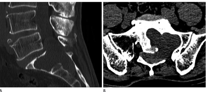
The tumor was excised using a posterior midline approach. In the operative fields, the large cystic tumor was associated with the extensive bony erosion involving the sacral body and was found to be firmly attached to the left S1 nerve root. The cystic component of the tumor was aspirated, and this was followed by tumor excision. There were no complications during the post- Fig. 1. Lumbar spine computed tomography imaging.
A. Sagittal image shows a well-defined osteolytic mass at the sacral region (L5--S2).
B. Axial image shows that the mass had a lobulated margin and extended to the intraosseous area of S1 and the epidural space.
A B
Fig. 2. Lumbar spine magnetic resonance imaging.
A, B. Sagittal T2-weighted images show a well-defined, unilocular cystic mass. This mass had a fluid-fluid level (or a fluid-debris level) (arrow) and extended to the presacral area (arrowheads).
C. Axial T2-weighted image shows that this mass was a purely cystic mass.
D. Sagittal T1-weighted image shows the fluid-fluid level (arrow).
E. Fat-suppressed contrast enhanced T1-weighted sagittal image shows thin rim enhancement of the cystic mass.
A B C D E
ally do not have specific symptoms; 2) intrasacral (osseous) tu- mors present with mild local pain, and neurological deficit is unusual; and 3) spinal tumors (dumb-bell tumors) are usually as- sociated with neurological deficit or other symptoms (7).
MRI is a valuable method for the diagnosis and characterization of the tumor, with regard to size, location, and relations to other organs. The depiction of the tumor originating from an adjacent nerve root is known to be a pathognomonic MRI finding of a neu- rogenic tumor. On MRI, the typical features of a schwannoma are as follows: T1-weighted images show iso- to hypointense signal intensity compared to the spinal cord, T2-weighted images show hyperintense signal intensity or heterogeneous signal intensity, and contrast enhanced T1-weighted images show good enhance- ment. The heterogeneous signal intensities on T2-weighted or contrast-enhanced T1-weighted images indicate heterogeneous cellularity and degenerative changes such as necrosis, cyst for- mation, hemorrhage, fibrosis, and calcification (3-7, 10). The fea- tures of typical spinal schwannomas and giant spinal schwanno- mas on MRI scans are not significantly different. However, the giant schwannomas show a tendency towards more heterogene- ity than the typical tumors (10).
The cyst formation is not an unusual finding, especially in large schwannomas, but this is most often partial and does not involve the entire lesion. The mechanism of cyst formation can be explained by 2 theories. First, the degeneration of the Antoni B cells could lead to cyst formation, which progresses in size over time. Second, tumor growth results in ischemic necrosis. A fluid crocystic degeneration were noted, but these lacked features of
degenerative nuclear pleomorphic changes indicative of ancient schwannoma.
On postoperative day 21, the patient was doing well with no complaints, although the follow-up MRI scan has not been per- formed.
DISCUSSION
Schwannomas are benign tumors that originate from Schwann cells in the myelinated nerve sheath. The sacral region is an un- common site for spinal schwannoma, compared to the thoracic or lumbar region (3, 5, 8).
These tumors are generally slow in growth, with common symptoms such as pain or paresthesia developing well before spreading into the extraspinal tissues. However, these tumors sometimes present as huge masses extending into the vertebral body and extraspinal tissues, and then, they are referred to as the giant invasive spinal schwannomas (5, 6, 9, 10). Although the term giant schwannoma is not clearly defined yet, the generally accepted definition is a tumor extending to more than 2 vertebral bodies while eroding the vertebral bodies and extending into myofascial planes, with the extraspinal extension of more than 2.5 cm (9). The giant spinal schwannoma is rare, especially in the sacral region. On the basis of the anatomic location, a giant sacral schwannoma can be classified into 3 types. Each type shows dif- ferent clinical features as follows: 1) retroperitoneal tumors usu- Fig. 3. Microphotography.
A. Microphotography (H&E stain, × 40) shows a predominantly cystic component with extensive degeneration and hemorrhage.
B. Microphotography (H&E stain, × 100) presents two distinct types of tissue patterns: Antoni A, where the spindle cells are arranged in a rela- tively compact palisading pattern, and Antoni B, comprising loose, reticular, often cystic arrangements.
A B
tion to the nerve root provided a decisive clue. The role of a ra- diologist is crucial in making the surgeons aware of different pa- thologies in the cases of sacral tumors, thereby helping them to decide on the extent of surgical removal, based on MRI findings in conjunction with intraoperative findings.
REFERENCES
1. Abdelwahab IF, Hermann G, Stollman A, Wolfe D, Lewis M, Zawin J. Case report 564: Giant intraosseous schwannoma.
Skeletal Radiol 1989;18:466-469
2. Conti P, Pansini G, Mouchaty H, Capuano C, Conti R. Spi- nal neurinomas: retrospective analysis and long-term out- come of 179 consecutively operated cases and review of the literature. Surg Neurol 2004;61:34-43; discussion 44 3. Ogose A, Hotta T, Sato S, Takano R, Higuchi T. Presacral
schwannoma with purely cystic form. Spine (Phila Pa 1976) 2001;26:1817-1819
4. Borges G, Bonilha L, Proa M Jr, Fernandes YB, Ramina R, Zanardi V, et al. Imaging features and treatment of an in- tradural lumbar cystic schwannoma. Arq Neuropsiquiatr 2005;63:681-684
5. Nyapathy V, Murthy UK, Chintamani J, Sridhar DY. A case report of a giant presacral cystic schwannoma with sig- moid megacolon. J Radiol Case Rep 2009;3:31-37
6. Albert AF, Kirkman MA, du Plessis D, Sacho R, Cowie R, Tzerakis NG. Giant solitary cystic schwannoma of the cer- vical spine: a case report. Clin Neurol Neurosurg 2012;114:
396-398
7. Pongsthorn C, Ozawa H, Aizawa T, Kusakabe T, Nakamura T, Itoi E. Giant sacral schwannoma: a report of six cases. Ups J Med Sci 2010;115:146-152
8. Wu D, Ba Z, Huang Y, Zhao W, Shen B, Kan H. Totally cystic schwannoma of the lumbar spine. Orthopedics 2013;36:
e679-e682
9. Sridhar K, Ramamurthi R, Vasudevan MC, Ramamurthi B.
Giant invasive spinal schwannomas: definition and surgi- cal management. J Neurosurg 2001;94(2 Suppl):210-215 10. Yu NH, Lee SE, Jahng TA, Chung CK. Giant invasive spinal
schwannoma: its clinical features and surgical manage- ment. Neurosurgery 2012;71:58-67
hemorrhage, leading to the fluid-fluid levels (6, 8). The nearly complete or complete cystic formation of the spinal schwanno- mas is rare. A few cases of giant schwannoma with complete cystic change have been reported, and most of these cases in- volved septation or a focal solid portion within the tumor mass.
The case reported here is one of a completely cystic mass. To our knowledge, only 3 such cases have been previously reported:
one involving the cervical region and the other two involving the lumbar region (4, 6, 8).
The diagnosis of purely cystic spinal schwannoma is difficult because of its rarity. Therefore, this tumor type needs to be con- sidered with differential diagnosis of cystic spinal lesions, in- cluding dermoid cysts, epidermoid cysts, arachnoid cysts, neur- enteric cysts, cystic teratomas, meningoceles, primary bone cysts or cystic tumors, and perineural cysts. MRI can be useful in the differentiation of cystic schwannomas from other spinal cystic masses (3, 6, 8).
The treatment strategies for cystic giant spinal schwannomas are not different from those for typical schwannomas. Further- more, the prognosis is good for the cystic giant spinal schwanno- mas and it is comparable to that of the solid spinal schwanno- mas (3, 7, 10).
The complete resection is the treatment of choice, as residual tumor is likely to regrow, increasing the risk of surgical compli- cations due to the repeat operations. However, in cases of the gi- ant invasive spinal schwannomas, a complete tumor resection is difficult because of the invasive tumor growth in all directions.
The risk of neurological deficit and surgery-related complica- tions is relatively high on this account. Therefore, the best op- tion is to attempt to achieve a complete resection, but if this is too difficult or risky, removal should be performed to the maxi- mum extent as possible. The measurement of the Ki-67 index and MRI should be performed as follow-up evaluations for the sequential assessment of tumor progression. The Ki-67 index is useful for predicting tumor regrowth or recurrence when com- plete resection is not achieved (10).
In conclusion, the giant spinal schwannomas with complete cystic degeneration are very rare, but this tumor type should be considered in the cases of cystic sacral masses. In these cases, careful evaluation of the anatomic relation of the tumor is im- portant. In our case, the distinction between bone cyst with
완전 낭성변화를 보인 거대 신경초종: 증례 보고1
이한아
1· 전세정
1· 최시성
1· 김혜원
1· 김헌수
2신경초종은 신경 수초에서 기원하는 양성 종양이다. 이 종양은 일반적으로 고형성 종양이며 가끔 퇴행성 변화를 나타낸다.
종양 일부에서 낭성변화가 일어날 수 있지만 완전한 낭성변화를 보이는 경우는 매우 드물며 이에 관하여 소수의 증례보고 가 있었다. 요천추부위에 발생한 완전 낭성변화를 보인 거대 신경초종에 대해서는 오직 한 증례만이 보고되었다. 우리는 이와 비슷한 증례를 경험하여 자기공명영상 소견과 병리학적 소견에 대해서 보고하고자 한다.
원광대학교 의과대학 1영상의학과학교실, 2병리과학교실