ABSTRACT
Spinal extradural arachnoid cysts (SEACs) are rare and usually asymptomatic, and they usually do not require surgical treatment. If symptoms manifest, however, surgical treatment is required. A 25-year-old male patient complained of impotence upon admission. Magnetic resonance images (MRIs) of his lumbar spine showed a SEAC located longitudinally from the T11 to L3, which was accompanied by thecal sac compression. Verifying the location of the dural defect is crucial for minimizing surgical treatments. Cystography, myelography, and lumbar spine MRI were conducted to locate the leak in real-time; however, it was not found.
Hence, the location of the cerebrospinal fluid leak was estimated based on cystography, computed tomography, myelography, and MRI findings. We suggest that the region with the earliest contrast-filling, as well as the middle and widest area of the cyst, may correspond to the location of the dural defect.
Keywords: Arachnoid cysts; Spine; Cerebrospinal fluid leak; Minimally invasive surgery
INTRODUCTION
A spinal extradural arachnoid cyst (SEAC) is a rare disease, accounting for about 1% of all primary spinal cord tumors.12) SEACs are known to arise from a congenital dural defect which allows the arachnoid membrane to herniate through the adjacent dura mater and communicate with the intraspinal subarachnoid space. Although SEACs occur with no symptoms in most case, a surgical excision should be performed in case a patient has syptoms.4) Cystography or myelography is the most commonly used methods to find the real-time leak site in order to perform minimally invasive surgery. Occasionally, the real-time leak site is not clearly observed in radiological images. In such cases, therefore, it is necessary to estimate the location of the dural defect site. In this study, our experiences of SEAC case would be reported and an alternative method in estimating the location of the dural defect of SEAC would be introduced.
Case Report
Received: Apr 2, 2020 Revised: Jul 27, 2020 Accepted: Aug 20, 2020 Address for correspondence:
Seung-Won Choi
Department of Neurosurgery, Chungnam National University Hospital, 282 Munhwa-ro, Jung-gu, Daejeon 35015, Korea.
E-mail: [email protected]
Copyright © 2020 Korean Neurotraumatology Society
This is an Open Access article distributed under the terms of the Creative Commons Attribution Non-Commercial License (https://
creativecommons.org/licenses/by-nc/4.0/) which permits unrestricted non-commercial use, distribution, and reproduction in any medium, provided the original work is properly cited.
ORCID iDs Seok-won Lee
https://orcid.org/0000-0001-8610-887X Jeongwook Lim
https://orcid.org/0000-0003-1395-3145 Jin-Young Youm
https://orcid.org/0000-0001-9609-0415 Hyon-Jo Kwon
https://orcid.org/0000-0003-1077-2461 Hyeon-Song Koh
https://orcid.org/0000-0003-2659-5535 Seon-Hwan Kim
https://orcid.org/0000-0002-5600-2801 Conflict of Interest
The authors have no financial conflicts of interest.
Seok-won Lee , Seung-Won Choi, Jeongwook Lim , Jin-Young Youm , Hyon-Jo Kwon , Hyeon-Song Koh , and Seon-Hwan Kim
Department of Neurosurgery, Chungnam National University Hospital, Daejeon, Korea
How to Find Dural Defect of Spinal
Extradural Arachnoid Cyst
CASE REPORT
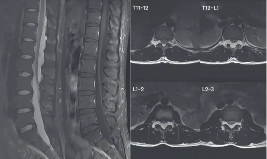
With complain of impotence, a 25-year-old male was admitted in our hospital. Physical and neurologic examinations of the patient have shown no signs of neurologic deficit. An L-spine magnetic resonance image (MRI) was conducted to find a spinal lesion, which has revealed a SEAC located from T11 to L3 level. In addition, thecal sac compression was also discovered (FIGURE 1).
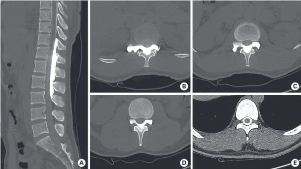
Initially, a cystography was conducted to determine the real-time cerebrospinal fluid (CSF) leak point; however, the real-time leak point has not been clearly detected through the examination (FIGURE 2). Because the post-cystography computed tomography (CT) showed
L1–2 L2–3
FIGURE 1. A spinal epidural arachnoid cyst locates from T11 to L3, along with thecal sac compression.
FIGURE 2. A cystography is conducted to verify the real-time cerebrospinal fluid leak point. The real-time leak is not clearly detected through this examination.
that the contrast had spread into the subarachnoid space, we attempted to find the dural defect (FIGURE 3) using myelography and myelo-CT by studying the CSF flow. However, these examinations did not show any signs of the real-time leak site, although they demonstrated that the contrast had spread into the cyst (FIGURES 4 & 5). Prior to surgery, it is essential to locate the dural defect site in order for the operation to be minimized. Thus, the location of the dural defect has been estimated based on the earliest contrast-filling region in myelography, as well as the mid-level and widest area of the cyst found on the MRI (FIGURE 6A-C, TABLE 1). Consequently, a partial laminectomy was performed at the level of T12. After removal of the ligament flavum, the dural defect was then be able to be detected, and it was discovered that the nerve root and arachnoid membrane were partially herniated through the defect (FIGURE 7A). Thus, we pushed the root and arachnoid membrane back inside the
A
B C
E D
FIGURE 3. Post cystography computed shows that contrast has spread into the subarachnoid space, indicating the presence of a dural defect site.
FIGURE 4. Although a myelography is conducted to locate the dural defect site by detecting the cerebrospinal fluid flow, the real-time leak site has not been localized.
defect and closed the defect by suture and patch with TachoComb® (CSL Behring, Tokyo, Japan) (FIGURE 7B). Follow-up MRI was performed two months after surgery, which has shown the SEAC was finally disappeared (FIGURE 8). Furthermore, the patient's symptoms have recently improved.
FIGURE 5. Myelo-computed tomography is conducted to locate the real-time leak site. Similar to the myelography findings, the precise real-time leak site has not been observed.
A B C
T12–L1 T12–L1
T12–L1 L1
FIGURE 6. We estimated the location the dural defect based on the earliest contrast-filling region in myelography (A), mid-level of the cyst (B), and widest area of the cyst in magnetic resonance image (C) in order to minimize the operation procedure.
TABLE 1. Information to suggest dural defect site
Cyst Site
First filling location T12-L1. (FIGURE 6A)
Mid-level of cyst L1. (FIGURE 6B)
Widest level of cyst T12-L1. (FIGURE 6C)
DISCUSSION
SEAC is a rare disease. It is commonly found in the thoracic level while it is uncommon to develop in the lumbosacral and thoracolumbar level. The exact mechanism of the formation of arachnoid cyst has not yet been identified, but several theories have been proposed to explain it. The cysts can be divided into 5 categories for 1) congenital reasons; 2) arachnoid adhesions that were caused by viral or bacterial infection; 3) secondary arachnoiditis caused by subarachnoid hemorrhage, usage of contrast media, application of spinal anesthetics, and meningitis; 4) traumatic injuries to the vertebral column caused by lumbar punctures used in diagnostic procedures, or anesthetic and intradural surgery; and 5) idiopathic causes.5,6,11) It was previously documented that congenital asymptomatic cysts could be enlarged due to trauma and can become symptomatic.7,8)
Cyst expansion may be caused by active secretion from internal linings of cell that would lead to an abnormal osmotic gradient between the subarachnoid space and cyst, pulsatile CSF dynamics and one-way valve mechanism.2)
A B
FIGURE 7. A partial laminectomy at T12 has been performed. (A) After removal of ligamentum flavum, the dural defect site has been detected and it was discovered that a nerve root and the arachnoid membrane were partially herniated through the defect. (B) The root and arachnoid membrane has intentionally pushed back inside the defect and the defect has been closed with sutures and patches of TachoComb®.
FIGURE 8. Magnetic resonance image performed 2 months after surgery has revealed the disappearance of the spinal extradural arachnoid cyst.
and closure of the communication. If the size of cyst is large, conventional laminectomy and resection of cyst may cause a risk of postoperative kyphotic deformity (33–100%).1) Moreover, longer operating time and technical difficulty might continuously challenges the surgeons.
Closure of the dural defect without cyst resection via selective laminectomy is reported to achieve good neurological outcome without recurrence and with lower rates of complication.
Hence, verifying the communication site and closure is the treatment of choice for large SEACs.
To verify the communicating site, MRI, CT-myelography, and cystography are valuable diagnostic tools. Among these tools, MRI would be useful to detect CSF flow to localize a defective site. A study has reported that the CSF flow in MRI can identify the pulsating turbulent flow void of a defective site.3) Cystography and CT-myelography are also useful diagnostic tools in the light of the fact that they are more reliable tools which can detect the anatomical location of the cyst, and measure the severity of compression of the spinal cord and nerve roots.3) Some reports indicate that CT-myelography can help locate the dural defect site between the spinal subarachnoid space and the cyst cavity (i.e. the real-time leak site).3,10) Therefore, CSF communicating sites have been verified through MRI, cystography, CT-myelography. If the real-time leak site is not clearly observed, however, it is suggested the site be detected through the finding s of the method that we performed as described farther on. First, we assumed that the communicating site would be at the level of the earliest contrast-filling location. Second, we guessed it would be at the mid-level of the cyst. Third, we supposed it would be around the widest portion of the cyst. In our case introduced above, the earliest contrast-filling site on myelography was located at the level of T12-L1 (FIGURE 6A-C). The mid-level of the cyst was at the level of L1 and the widest area of cyst was at the level of T12-L1 on MRI and CT-myelography. The three estimated locations were identical to T12-L1, and therefore a stepped approach has been planned. That is, as first step, detection and closure of the dural defect would be implemented through the selective laminectomy at the estimated level, and if failed, second step including extended laminectomy and total resection of the cyst would be proceeded with. With such a plan, a selective partial laminectomy was carried out at T12, and the dural defect site has been ultimately confirmed to be located at that same level. Without extended multilevel laminectomy, eventually, the dural defect has been closed and a successful outcome has been granted.
CONCLUSION
Identifying the exact location of dural defects in case of a SEAC is an important step in the surgical planning in order to minimize the extent of laminectomy and reduce the complication rate. For a location undetected, we suggest estimating the communication site based on the earliest contrast-filling region, mid-level of the cyst, and widest area of the cyst. We also recommend attempting a stepped approach, i.e. the first step of selective laminectomy at the estimated site, and the second step for extended laminectomy and total resection of the cyst in case of failure of the first step. In conclusion, closure of the
communication site alone through selective laminectomy would be an effective and less- invasive treatment option.
REFERENCES
1. Amhaz HH, Fox BD, Johnson KK, Whitehead WE, Curry DJ, Luerssen TG, et al. Postlaminoplasty kyphotic deformity in the thoracic spine: case report and review of the literature. Pediatr Neurosurg 45:151-154, 2009 PUBMED | CROSSREF
2. Choi JY, Kim SH, Lee WS, Sung KH. Spinal extradural arachnoid cyst. Acta Neurochir (Wien) 148:579- 585, 2006
PUBMED | CROSSREF
3. Choi SW, Seong HY, Roh SW. Spinal extradural arachnoid cyst. J Korean Neurosurg Soc 54:355-358, 2013 PUBMED | CROSSREF
4. Hughes G, Ugokwe K, Benzel EC. A review of spinal arachnoid cysts. Cleve Clin J Med 75:311-315, 2008 PUBMED | CROSSREF
5. Kazan S, Özdemir O, Akyüz M, Tuncer R. Spinal intradural arachnoid cysts located anterior to the cervical spinal cord. Report of two cases and review of the literature. J Neurosurg 91:211-215, 1999
PUBMED | CROSSREF
6. Lee TT, Alameda GJ, Gromelski EB, Green BA. Outcome after surgical treatment of progressive posttraumatic cystic myelopathy. J Neurosurg 92:149-154, 2000
PUBMED | CROSSREF
7. Myles LM, Gupta N, Armstrong D, Rutka JT. Multiple extradural arachnoid cysts as a cause of spinal cord compression in a child. Case report. J Neurosurg 91:116-120, 1999
PUBMED | CROSSREF
8. Marbacher S, Barth A, Arnold M, Seiler RW. Multiple spinal extradural meningeal cysts presenting as acute paraplegia. Case report and review of the literature. J Neurosurg Spine 6:465-472, 2007 PUBMED | CROSSREF
9. Nabors MW, Pait TG, Byrd EB, Karim NO, Davis DO, Kobrine AI, et al. Updated assessment and current classification of spinal meningeal cysts. J Neurosurg 68:366-377, 1988
PUBMED | CROSSREF
10. Panigrahi S, Mishra SS, Dhir MK, Parida DK. Giant thoracolumbar extradural arachnoid cyst: an uncommon cause of spine compression. Neurol India 60:540-542, 2012
PUBMED | CROSSREF
11. Suryaningtyas W, Arifin M. Multiple spinal extradural arachnoid cysts occurring in a child. Case report. J Neurosurg 106:158-161, 2007
PUBMED | CROSSREF
12. Yabuki S, Kikuchi S. Multiple extradural arachnoid cysts: report of two operated cousin cases. Spine 32:E585-E588, 2007
PUBMED | CROSSREF